http://rodriguesia.jbrj.gov.br
Nota Científica / Short Communication:
Nota Científica / Short Communication:
Nota Científica / Short Communication:
Nota Científica / Short Communication:
Nota Científica / Short Communication:
The formation of the stigmatic surface in
The formation of the stigmatic surface in
The formation of the stigmatic surface in
The formation of the stigmatic surface in
The formation of the stigmatic surface in
Passiflora elegans
Passiflora elegans
Passiflora elegans
Passiflora elegans
Passiflora elegans (Passifloraceae)
(Passifloraceae)
(Passifloraceae)
(Passifloraceae)
(Passifloraceae)
11111A formação da superfície estigmática em Passiflora elegans (Passifloraceae)
Adriano Silvério
2*&
Jorge Ernesto de Araujo Mariath
2,31Part of the PhD thesis of the first author. Programa de Pós-Graduação em Botânica, Universidade Federal do Rio Grande do Sul.
2Universidade Federal do Rio Grande do Sul, Depto. Botânica, Lab. Anatomia Vegetal. Av. Bento Gonçalves 9500, Prédio 43423, s/206, 91501-970, Porto Alegre, RS, Brasil. 3CNPq Productivity in Research Scholarship recipient.
*Corresponding author: jorge.mariath@ufrgs.br
Abstract
The stigma surface is a complex multicellular structure where the development of the pollen tube begins. This
development is necessary for sucess in fertilization and depends on recognition processes that involve the anatomy of
the stigma.
Passiflora
is an economically important genus because of its edible fruits. Many authors have described the
stigma of
Passiflora
but nothing is known about the ontogenesis of this structure. This work aimed to describe the
formation of the stigmatic surface of
Passiflora elegans
. Results showed that, in bud, the stigmatic surface of this species
is flat with small cells. The cells in the subdermal layer have large vacuoles and the nucleus, near to the external
periclinal walls. During its development the stigma surface becomes uneven due to the elongation of cells in the
subdermal layer. Elongation results in an increase of external secretory surface area of the stigmas, and probably plays
an important role in pollen recognition. The polysaccharide content found in the inner walls of these structures might
be involved in the signal process for pollen tube growth during its early development. The morphological evidence
presented here shows that, as the stigma of
Passiflora
is formed by dermal and subdermal cells, it should not be
characterized as colleters or papillae and, therefore, it is defined here as stigma emergences.
Key-words
: anatomy, stigma development, stigma emergence, pollination.
Resumo
A superfície estigmática é uma estrutura multicelular complexa, onde o tubo polínico inicia o seu desenvolvimento,
necessária para a fecundação. Este desenvolvimento depende de condições favoráveis que envolvem a anatomia do
estigma durante o processo de reconhecimento.
Passiflora
é um gênero economicamente importante devido aos seus
frutos comestíveis. O estigma de
Passiflora
tem sido descrito por vários autores, mas o seu processo de formação é
desconhecido. Esse trabalho tem por objetivo descrever o processo de formação da superfície estigmática de
Passiflora
elegans
. Os resultados demonstram que durante a fase de botão jovem, a superfície estigmática é composta por
pequenas células e apresenta superfície plana. As células da camada subdepidérmica apresentam grandes vacúolos e
núcleo, próximo da parede periclinal externa. Durante o seu desenvolvimento, a superfície estigmática torna-se
irregular devido ao alongamento de células da camada subdepidérmica. Essas modificações resultam em um acréscimo
da superfície secretora externa do estigma, e provavelmente desempenham um importante papel no reconhecimento
do pólen. Os conteúdos polissacarídicos encontrados na superfície interna dessas estruturas podem estar envolvidos
com os processos de sinalização do tubo polínico durante seu desenvolvimento inicial. As evidências morfológicas
observadas nesse trabalho demonstram que as estruturas presentes na superfície do estigma de
Passiflora
são constituídas
por células de origem dérmica e subdérmica, e não devem ser caracterizadas como coléteres ou papilas, sendo assim,
caracterizadas nesse trabalho como emergências estigmáticas.
Palavras-chave
: anatomia, desenvolvimento do estifma, emergência estigmática, polinização.
Species of Passiflora L. are characterized by having a sporophytic and gametophytic self-incompatibility system (Rêgo et al. 1999, 2000;
1% OsO4, washed in 0.1 M sodium phosphate buffer at pH 7.2 (Weber 1992), dehydrated in acetone, critical point dried (Gersterberger & Leins 1978), sputter-coated with gold using a Balzers SCD 050, and examined using a Jeol 6060 SEM.
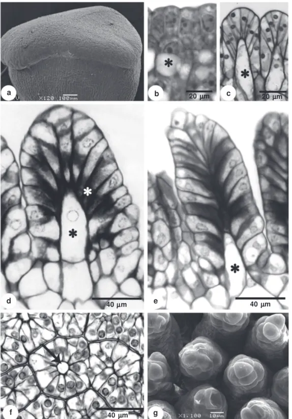
It was found, during the initial stages of development, that the apical portion of the stigma has a slightly sinuous surface (Fig. 1a), the dermal layer has cells with an evident nucleus and portions of condensed chromatin (Fig. 1b), and the subdermal layer has cells with large vacuoles and respective nucleus displaced near the external periclinal walls (Fig. 1b).
Subsequent to the initial developmental phase, the external surface of the stigma begins to form multiple dome-shaped projections, as a result of anticlinal divisions followed by anticlinal and radial elongation of the dermal and some of the subdermal cells (Fig. 1c). The height of each emergence on the stigmatic surface continues to increase, while the expanding subdermal cells divide transversally giving rise to projections that have an apical and a basal cell (Fig. 1d). The apical cells remain in direct contact with epidermal cells, in which the internal periclinal wall and the proximal portion of the anticlinal wall accumulate compounds of pectic nature (Fig. 1d). At the end of development, a specialized structure is formed on the stigma, comprised of cells from the dermal and subdermal layer (Fig. 1e). Once these structures have formed the stigma surface, it appears papillate, but in cross-section it can be seen that each projection has a multicellular organization around a central axis formed by the subdermal cell (Fig. 1f). Scanning electron microscopy revealed numerous multicellular projections on the stigmatic surface (Fig. 1g).
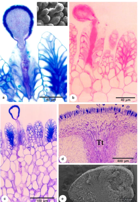
During pollination, the pollen germinates on this pappilate surface (Fig. 2a) and the pollen tube path follows the central region of the structure, which is rich in pectic compounds that have accumulated along the anticlinal and periclinal walls (Fig. 2b-c). Beyond the stigma, the pollen tubes grow into parenchyma (Fig. 2c-e) and the transmitting tissue that have cells with similar chemical properties.
The surface of the stigma is crucial during pollination, because pollen recognition depends on the lipids stored in the stigmatic cells and on the glycoproteins secreted from them onto the outer surface (Tilton et al. 1984). After hydration, the pollen grain germinates and the pollen tube emerges and grows over the stigma. During this time, specific enzymes loosen the cell wall of the papillae Passifloraceae has been described in previous
studies. Puri (1947) described the flower anatomy of this genus, and considered the stigma to be large, with massive structures; however, no further anatomical comments were provided. Raju (1956) classified these structures as projections that facilitate pollen grain retention during pollination events, and also as a site for the growth of pollen tubes during their passage towards the transmitting tissue. In one study, the stigma of Passifloraceae was classified as dry with unicellular papillae (Heslop-Harrison & Shivanna 1977), and in another study Passiflora racemosa Brot. and other
Passiflora species and genera in the family were reported to have multicellular papillae (Bernhard 1999). The classification used by Bernhard (1999) was also used by Souza et al. (2006) for P. edulis f.
flavicarpa Degener. These authors noted that the papillate structures had cells with large vacuoles and thin walls. However these characterizations of the pappilate stigmatic surface of Passiflora were based only on the final stages of stigma development. There is still no consensus on the origin of this structure, which is probably induced by the lack of specific ontogenetic studies. The goal of this work is to analyze the ontogenetic process of these structures at the stigmatic surface of Passiflora elegans Mast., an endemic species of southern Brazil.
Stigmas of 50 floral buds, measuring 0.3 to 2 cm, and 20 buds in pre-anthesis were collected from plants found on the Campus do Vale, at Rio Grande do Sul Federal University. A voucher specimen was deposited in the ICN Herbarium (ICN 52108).
Figure 1 –
Development of the
Passiflora elegans
stigma – a. young stigma under scanning electron microscopy; b. longitudinal
section of the young stigma with the cells of the subdermal layer with large vacuoles (*); c. longitudinal section of the stigma
showing epidermal cells pushed by cells from the subdermal layer (*); d. cross-sectional of the dermal cell with pectins walls
(white asterisk) and division of subdermal cell (*); e. longitudinal section of the stigma emergence in the final phase of
development; f. cross-section of the stigma emergence region in the final phase of development, showing the cell of the subdermal
layer positioned in the central region; g. electromicrography of the stigma surface showing the stigma emergences.
g f
e
c
d
b a
40 µµµµµm 40 µµµµµm
20 µµµµµm 20 µµµµµm
Figure 2 –
Stigma and style of the
Passiflora elegans
flower – a. stigma surface with a pollen tube on the stigma
emergence. Detail under scanning electron microscopy; b. histochemical test for the presence of pectins; c. longitudinal
section of the stigma surface and the way of the pollen tube penetration through the stigma emergences and
parenchymatous tissue; d. longitudinal section of the stigma and style showing the transmitting tissue at the apical
center of the style (Tt); e. stigma and style under scanning electron microscopy.
c
b a
e d
100 µµµµµm
40 µµµµµm
500 µµµµµm
400 µµµµµm
preparing it for the penetration of the pollen tube (Micheli 2001). Calcium is probably a messenger during this process, inducing enzyme secretion and the consequential loosening of the cell wall (Elleman & Dickinson 1986; Hiscock et al. 2002).
In this study, a large amount of pectin was observed on the dermal and subdermal cell walls of the stigma, which coincides at the cellular level with the pollen tube path during its germination. Pectins probably stimulate the pollen tube growth of P. elegans, and calcium is made available to this structure along its course. Calcium (Ca2+) is a key
element in this process, regulating elongation and orientation of the pollen tube during its development (Malhó et al. 2006).
Pectins are synthesized in dictyosomes, in a methyl-sterified form. The methyl-sterification of carboxylic groups prevents Ca2+ binding, making the
cell wall less rigid. As methyl-sterification increases, the fluidity of the pectin gel also increases, allowing the cell to expand while the integrity of its structure is maintained, due to the hydrophilic properties of pectins (Micheli 2001; Taylor & Hepler 1997).
Braum (2008) observed in style cells, adjacent to a growing pollen tube, the accumulation of pectic material in the vacuoles and, near the cell walls. These traits are important because they promote changes in the cell walls of the transmitting tissue, allowing for the passage of the pollen tube. In P. edulis, there are reports of the occurrence of pectic compounds, mainly along the inner periclinal walls of the cells that constitute the dermal layer structures (Souza et al. 2006). It is possible that the same mechanism described by Braum (2008) occurs in the stigma of P. elegans.
The stigmatic surface cells of Passiflora are structurally and ontogenetically similar to colleters (Paiva & Machado 2006), which are usually associated with the secretion of mucilaginous compounds. Thomas (1991) cites the occurrence of colleters in approximately 60 families of angiosperms, mainly on stipules and sepals. In Passifloraceae, these structures are known to occur on leaf surfaces (Solereder 1908), and are abundant on young plant parts, especially along the borders of foliar primordia and stipules (González 1998).
Colleters secrete a viscous material on the external surface (Thomas 1991; Klein et al. 2004; Barreiro & Machado 2007). This process differs from what was observed in this study, as the stigmatic emergences found on P. elegans have pectic compounds in their inner walls.
The morphological characteristics of the stigmatic projections revealed in this work do not agree with the previous descriptions of the literature, that used the term papilla to define these “projections of epidermal cells.” In addition, previous studies did not classify these structures as colleters. For this reason, we conclude that “stigma emergence” is a better term to classify the structures found on the stigmas of Passiflora, as they are formed from the dermal and subdermal layers and they do not secrete mucilage.
Bernhard (1999) considered the characteristic stigma of Passifloraceae to be largely distributed among the genera of this family, but rare in the other families of angiosperms. From a taxonomic perspective, the stigmatic surface also appears to be an important trait that could be used to help describing Passifloraceae. Additional studies on other taxa in the family are needed to confirm this.
References
Barreiro, D.P. & Machado, S.R. 2007. Coléteres dendróides em Alibertia sessilis (Vell.) K. Schum.,uma espécie não-nodulada de Rubiaceae. Revista Brasileira de Botânica 30: 387-399. Bernhard, A. 1999. Flower Structure, development and
systematics in Passifloraceae and in Abatia
(Flacourtiaceae). International Journal of Plant Science 160: 135-150.
Braum, A.F. 2008. Morfologia, anatomia e imunocitoquímica da interação entre pólen e estigma em duas espécies
de Passiflora (Passifloraceae). Dissertação de
Mestrado. Universidade Federal do Rio Grande do Sul, Porto Alegre. 116p.
Elleman, C.J. & Dickinson, H.G. 1986. Pollen-stigma interaction in Brassica, structural reorganisation in the pollen grains during hydration. Journal Cell Science 80: 141-157.
Feder, N. & O´Brien, T. P. 1968. Plant microtechnique, some principles and new methods. American Journal of Botany 55: 123-142.
Gerrits, P.O. & Smid, L. 1983. A new, less toxic polymerisation system for the embedding of soft tissue in glycol methacrylate and subsequent preparing of serial sections. Journal of Microscopy 132: 81-85. Gersterberger, P. & Leins, P. 1978.
Rasterelektronen-mikroskopische Untersuchungen an Blütenknospen
von Physalis philadelphica (Solanaceae).
Anwendung einer neuen Präparationsmethode. Berichte der Deutschen Botanischen Gesellschaft 91: 381-387.
González, A.M. 1998. Colleters in Turnera and Piriqueta
Heslop-Harrison, Y. & Shivanna, K.R. 1977. The receptive surface of Angiosperm stigma. Annals of Botany 41: 1233-1258.
Hiscock, S.J.; Hoedemaekers, K.; Friedman, W.E. & Dickinson, H.G. 2002. The stigma surface and pollen-stigma interactions in Senecio squalidus
(Asteraceae) following cross (compatible) and self (incompatible) pollinations. International Journal of Plant Science 163: 1-16.
Johansen, D.A. 1940. Plant microtechnique. 3 ed. Paul B. Hoeber, Inc., New York. 790p.
Klein, D.E.; Gomes, V.M.; Silva-Neto, S.J. & Cunha, M. 2004. The structure of colleters in several species
of Simira (Rubiaceae). Annals of Botany 94: 733-740.
Malhó, R.; Liu, Q.; Monteiro, D.; Rato, C.; Camacho, L. & Dinis, A. 2006. Signalling pathways in pollen germination and tube growth. Protoplasma 228: 21-30. Micheli, F. 2001. Pectin methylesterases: cell wall
enzymes with important roles in plant physiology. Trends in Plant Science 6 (9): 414-419.
Paiva, E.A.S. & Machado, S.R. 2006. Colleters in
Caryocar brasiliense (Caryocaraceae) ontogenesis,
ultrastructure and secretion. Brazilian Journal of Biology 66: 301-308.
Puri, V. 1947. Studies in floral anatomy VI. Vascular anatomy of the flower of certain species of the Passifloraceae. American Journal of Botany 34: 562-573. Raju, M.V.S. 1956. Embryology of the Passifloraceae. I.
Gametogenesis and seed development of Passiflora
calcarata Mast. Journal of the Indian Botanical
Society 35: 126-138.
Rêgo, M.M.R.; Bruckner, C. H.; Silva, E.A.M.; Finger, F.L.: Siqueira,, D.L. & Fernandes, A.A. 1999.
Self-incompatibility in passion fruit: evidence of two locus genetic control. Theoretical and Applied Genetics 98: 564-568.
Rêgo, M. M., Rêgo, E. R., Bruckner, C. H., da Silva, E. A. M., Finger, F. L. & Pereira, K. J. C. 2000. Pollen
tube behavior in yellow passion fruit following compatible and incompatible crosses. Theoretical and Applied Genetics 101: 685-689.
Roland, J.C. & Vian, B. 1991. General preparation and staining of thin sections. In: Hall, J.L & Hawes, C. (eds.). Electron microscopy of plant cells. Academic Press, London. Pp. 1-66.
Sass, J.E. 1951. Botanical microtechnique. 2ed. Iowa State College Press, Iowa. 228p.
Solereder, H. 1908. Systematic anatomy of the dicotyledons. 2º vol. Clarendon Press, Oxford. 1182p.
Souza, M.M.; Pereira, T.N.S.; Dias, A.J.B.; Ribeiro B.F. & Viana, A.P. 2006. Structural, hystochemical and cytochemical characteristics of the stigma and style
in Passiflora edulis f. flavicarpa (Passifloraceae).
Brazilian Archives and Biotechnology 49: 93-98. Suassuna, T.M.F.; Bruckner, C.H.; Carvalho, C.R. &
Borém, A. 2003. Self-incompatibility in passion fruit: evidence of gametophytic-sporophytic control. Theoretical and Applied. Genetics 106: 298-302. Taylor, L.P. & Hepler, P.K. 1997. Pollen germination
and tube growth. Annual Review of Plant Physiology and Plant Molecular Biology 48:461-491. Thomas, V. 1991. Structural, functional and phylogenetic
aspects of the colleter. Annals of Botany 68: 287-305.
Tilton, V.R.; Wilcox, L.W. & Palmer, R.G. 1984. Postfertilization wandlabrinthe formation and function in the central cell of soybean, Glycine max
(L.) Merr. (Leguminosae). Botanical Gazette 145: 334-339.
Weber, M. 1992. The formation of pollenkitt in
Apium nodiflorum (Apiaceae). Annals of Botany
70: 573-577.