ABSTRACT
ORIGINAL AR
Ademar Lopes
Maria do Rosário Dias
recurrence of carcinoma of the
uterine cervix in stages IB and IIA
Treatment and Research Center of Hospital do Câncer A. C. Camargo,
São Paulo, Brazil
CONTEXT AND OBJECTIVE: Infl ammatory reac-tion intensity has been indicated as a possible recurrence risk factor in carcinoma of the uterine cervix. Some authors observed greater risk with weak infl ammatory reaction, while others described the opposite. This study aimed to evaluate risk factors for initial-stage uterine cervix carcinoma recurrence (IB and IIA), considering infl ammatory reaction intensity.
DESIGN AND SETTING: Retrospective cohort at Hospital do Câncer A. C. Camargo.
METHODS: 289 patients with diagnosed uterine cervix carcinoma (stages IB and IIA) who underwent radical surgery between 1980 and 1999 were studied. Data were col-lected from medical records. Histological sections from tumors and lymph nodes could be reviewed in 247 cases. Five-year disease-free survival rates were calculated using the Kaplan-Meier method and curves were compared using the log-rank test. Cox’s proportional-hazards model was used for multivariate analysis. Recurrence risk was estimated using hazard ratios (HR).
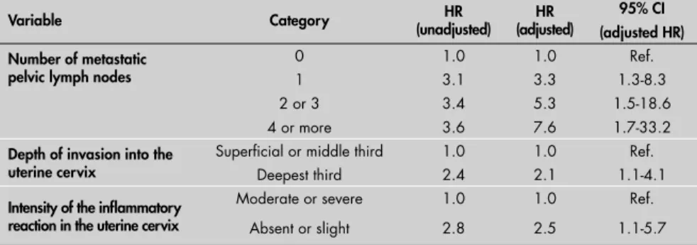
RESULTS: Forty-three recurrences were found. Multivariate analysis identifi ed the following independent recurrence risk factors: number of metastatic pelvic lymph nodes (one lymph node: HR = 3.3 [1.3-8.3]; two or three: HR = 5.3 [1.5-18.6]; four or more: HR = 7.6 [1.7-33.2]), tumor invasion depth (deepest third: HR = 2.1 [1.1-4.1]) and infl ammatory reaction intensity in the uterine cervix (absent or slight: HR = 2.5 [1.1-5.7]).
CONCLUSION: This study identifi ed that absent or slight infl ammatory reaction was an independent risk factor for recurrence. The other risk factors were the number of metastatic pelvic lymph nodes and invasion of the deepest third of the uterine cervix.
KEY WORDS: Uterine cervical neoplasms. Recurrence. Survival. Infl ammation. Lymphatic metastasis. Neoplasm invasiveness.
INTRODUCTION In developed countries, carcinoma of the uterine cervix represents around 4% of all tumors among women and is the sixth most frequent type of cancer in the population. In developing countries, it is the most common or second most common type of cancer among women, corresponding to 15% of tumors.1
The treatment is dictated by the clinical staging that is recommended by the Interna-tional Federation of Gynecology and Obstetrics (Fédération Internationale de Gynécologie et d’Obstétrique, FIGO).2 The initial stages IB and
IIA can be treated by means of Piver-Rutledge class III radical hysterectomy,3 in association
with pelvic lymphnodectomy, or by means of radiotherapy alone.4 In the more advanced
stages (IIB to IVB), radiotherapy presents better results than does surgery.5 Around 20% of the
patients with carcinoma of the uterine cervix at its initial stages (IB and IIA) who have been adequately treated by radical surgery develop disease recurrence.6 In 90% of such cases, the
recurrence occurs within two years.7 The reason
for this recurrence is persistence of the disease at microscopic scale locoregionally (pelvis) and/or at a distance, in an organ (for example, in the lungs, liver or bones).
The literature describes a variety of risk factors for recurrence of carcinoma of the uterine cervix in its initial stages, such as the presence of metastases in lymph nodes, tumor size, depth of invasion into the cervical stroma, invasion of the parametrium, histological type, degree of cell differentiation, blood capillary embolization and lymphatic capillary emboli-zation, among others.8 The intensity of the
infl ammatory reaction in the uterine cervix consequent to carcinoma has been indicated as a risk factor for recurrence. If on the one hand some authors have observed increased risk of recurrence when the infl ammatory reaction was weak,9-21 others have described
discordant results.22-25
de Oliveira Latorre
Reinthaller et al. evaluated 158 women with carcinoma of the uterine cervix in stages IA to IIB and observed that the women with a severe infl ammatory reaction in the uter-ine cervix presented lower recurrence rates, independent of the histological type.14 Kainz et
al. identifi ed the intensity of the cervical infl am-matory reaction as a risk factor for recurrence, in multivariate analysis. Absent or moderate infl ammatory reaction implied a reduction in disease-free survival.15 In a study on 73 cases
of carcinoma of the uterine cervix in stage IB, Bethwaite et al. noted a signifi cant association between low density of T lymphocytes in the uterine cervix and disease recurrence.16 Chao
et al. evaluated 83 patients with carcinoma of the uterine cervix (stage IIB) who underwent radical hysterectomy and found that, in the cases in which the intensity of the infl amma-tory reaction in the cervix was slight, recurrence was signifi cantly more frequent (50.0% versus 16.9%).18 Horn et al. evaluated patients with
carcinoma of the uterine cervix in its initial stage who underwent surgical treatment and found that absence of peritumoral infl amma-tory reaction was associated with greater risk of recurrence.20
The intensity of the infl ammatory reac-tion in the uterine cervix is considered in some scoring systems to be a risk factor for the recur-rence of carcinoma of the uterine cervix, as in the Malignancy Grading System10 and the
Par-tial Index.11 In these scoring systems, and also
in others that are derived from them,12,13,17,19
the absence of infl ammatory reaction or a slight reaction is implicated in greater recur-rence risk. However, neither of these scoring systems is based on multivariate analysis to determine the intensity of the infl ammatory reaction as a risk factor for recurrence.
mechanisms, thereby promoting depression of the local cell immunity. A variety of mediators involved in immunodepression are produced by neoplastic cells, and the following are prominent: transforming growth factor-beta (TGF-β), interleukin IL-4, interleukin IL-6, interleukin IL-10, macrophage chemotactic protein-1 (MCP-1), macrophage deactivating factor (MDF), suppressive E-receptor (SER), immuno-suppressive acidic protein (IAP), prostaglandin PGE2 and protein p15E, among others.26,27
Recently, it has been reported that certain metal-loproteinases not only degrade the extracellular matrix but also are implicated in the immuno-suppression promoted by the tumor.28
OBJECTIVE To evaluate the risk factors for recurrence of carcinoma of the uterine cervix in its initial stages (IB and IIA), considering the intensity of the infl ammatory reaction in the uterine cervix in this analysis.
METHODS This was a retrospective cohort study in which patients diagnosed with carcinoma of the uterine cervix who underwent radical surgical treatment at the Treatment and Research Center of Hospital do Câncer A. C. Camargo between 1980 and 1999 were evaluated. The inclusion criteria were: 1) Histological proof of invasive carcinoma of the uterine cervix; 2) Clinical staging IB and IIA, according to the criteria established by FIGO; 3) Absence of any previous treatment; 4) Surgical treatment performed by means of Piver-Rutledge class II or III radical hysterectomy3 and pelvic lymphnodectomy.
The medical records were consulted and information was collected on a specifi c form
for sociodemographic, clinical, histopatho-logical, therapeutic and follow-up data. The histological sections from the tumors and lymph nodes (stained using hematoxylin-eosin, HE) were reviewed in 247 cases. In 42 cases, it was not possible to conduct the review because of the poor quality of the slides or because the slides could not be located in the fi les of the Department of Pathology.
The sample was composed of 289 women, whose ages ranged from 18 to 72 years (mean = 44.4 years; standard deviation, SD = 10.0 years; median = 44.0 years). Most of them were white (75.8%) or brown-skinned (16.6%), married (76.2%) and of low educa-tional level (17.6% were illiterate and 71.3% had only done elementary education). Most of the patients were staged clinically as IB (90.7%) and the others as IIA.
The radical hysterectomy method most frequently utilized was type III (97.9%) and the remaining hysterectomies were type II. The mean number of lymph nodes dissected during the surgery was 19.4 (SD = 8.6; median = 19.0), with a range from 1 to 60. Involved surgical margins were observed in 3.8% of the patients (11 cases).
Postoperative radiotherapy was adminis-tered to 118 patients (40.8%). Of these, in 39 cases the indication for complementary treatment was because of metastases in the pel-vic lymph nodes. In the other cases, although the lymph nodes did not present metastases shown by the HE method, at least one of the following events was observed: tumor lesions larger than 4 cm; moderately or poorly dif-ferentiated tumors; presence of deep invasion into the cervical stroma; lymphatic capillary embolization; blood capillary
emboliza-tion; involved or narrow surgical margins; and involvement of the parametrium.
Teletherapy was utilized in association with vaginal brachytherapy in 89 cases, vaginal brachytherapy alone was utilized in 20 cases, and teletherapy alone was utilized in nine cases. In two cases, the adjuvant radiotherapy was utilized in association with chemotherapy (cisplatin 40 mg/m2), both in the year 1999.
The adjuvant teletherapy dose utilized ranged from 540 to 6040 cGy, with a median of 4500 cGy (mean = 4612 cGy; SD = 831 cGy).
The patients were followed up for a mean of 103.7 months, ranging from seven days to 259.5 months (SD = 63.2 months; median = 102.7 months). Sixty-three patients (21.8%) were lost to follow-up, and their mean fol-low-up was 47.8 months (SD = 44.2 months; median = 38.6 months), ranging from 15 days to 208.9 months.
The information collected was stored in a computerized database and analyzed by means of the Statistical Package for the Social Sciences (SPSS) software, version 10.0. Five-year
dis-ease-free survival rates were calculated using the Kaplan-Meier method, and the survival curves were compared using the log-rank test. Multivariate analysis was performed using Cox’s proportional-hazards model, and the recurrence risk was estimated using hazard ratios (HR). The modeling technique was the stepwise forward selection method, and the signifi cance level was set at 5.0%.
This study formed part of a larger research project within the postgraduate program at Fundação Antônio Prudente. It was submitted in advance to, and approved by, the Research Ethics Committee of the Treatment and Research Center of Hospital do Câncer A. C. Camargo (approval no. 354/01). Because this was a retrospective study, there was no need to draw up an informed consent statement.
RESULTS Over the study period, 43 recurrences (14.9%) were recorded, distributed thus: 22 within the pelvic region, 12 at a distance, seven simultaneously pelvic and at a distance and two at unknown locations. The mean length of time until recurrence was 29.3 months (SD = 24.9 months; median = 21.9 months), ranging from 2.4 to 99.0 months. The fi ve-year disease-free survival rate for the population studied was 86.3%.
Univariate analysis showed that, among the sociodemographic and clinical variables, only a number of previous gestations greater than four (p = 0.020) was associated with recurrence (Table 1). Among the
histopatho-Table 1. Five-year disease-free survival rate according to sociodemographic and clinical variables. Hospital A. C. Camargo (Hospital do Câncer), 1980-1999
Variable Category n* Five-year disease-free survival (%) p
Age Up to 45 years 159 87.5 0.252
More than 45 years 128 84.8
Skin color White 217 87.3 0.429
Non-white 70 83.2
Education level Up to completion of elementary
education† 230 86.2 0.929
High school or university-level 29 85.5
Number of previous gestations Up to 4 140 90.1 0.020
More than 4 144 82.4
Menopause No 209 86.7 0.247
Yes 75 84.7
Clinical stage IB 200 86.8 0.917
IIA 63 87.9
logical variables, associations were observed between recurrence and the number of meta-static pelvic lymph nodes (p = 0.001); the depth of invasion of the tumor, stratifi ed into thirds (p = 0.008); and the intensity of the infl ammatory reaction in the uterine cervix (p = 0.009). Among the treatment-related variables, none showed any association with recurrence (Table 2).
The multivariate model identifi ed the fol-lowing independent risk factors for recurrence: number of metastatic pelvic lymph nodes (one lymph node: hazard ratio, HR = 3.3; two or three lymph nodes: HR = 5.3; four or more lymph nodes: HR = 7.6); depth of tumor invasion into the uterine cervix (deepest third: HR = 2.1); and intensity of the infl ammatory reaction in the uterine cervix (absent or slight: HR = 2.5) (Table 3).
DISCUSSION In the present sample, the independent variables associated with recurrence were the number of pelvic lymph nodes involved in metastases, the depth of invasion into the cervical stroma and the intensity of the in-fl ammatory response in the uterine cervix. Of these, only the latter is not habitually cited in the literature as a risk factor for recurrence. However, there are authors who have been unable to demonstrate any difference in the risk of recurrence, in relation to the intensity of the infl ammatory reaction in the uterine cervix.22-24 Others have found
re-sults that are the opposite of what is present above.25 Studying prognostic factors in 196
cases of carcinoma of the uterine cervix in stages IB and IIA without metastases in the lymph nodes, Samlal et al. found that the risk of recurrence was around 2.5 times greater in cases with severe infl ammatory reaction in the uterine cervix.25
Lack of standardization of the criteria utilized for defi ning the intensity of the in-fl ammatory reaction is probably responsible for this difference in results. In the present study, the fact that a single pathologist reviewed the tumor slides reduced the bias produced by variation between observers. Nonetheless, the reproducibility of the re-sults found here is debatable, because what may correspond to slight inflammatory reaction in one pathologist’s opinion may represent a moderate reaction according to another pathologist. There is a need to develop criteria for assessing the intensity of the infl ammatory reaction that are less sub-jective, thereby enabling better reproduction of the results in future studies.
Table 3. Independent variables associated with recurrence that were identifi ed using Cox’s proportional-hazards model (adjusted for adjuvant radiotherapy and decade of treatment)
Variable Category (unadjusted)HR (adjusted)HR 95% CI
(adjusted HR) Number of metastatic
pelvic lymph nodes
0 1.0 1.0 Ref.
1 3.1 3.3 1.3-8.3
2 or 3 3.4 5.3 1.5-18.6
4 or more 3.6 7.6 1.7-33.2
Depth of invasion into the uterine cervix
Superfi cial or middle third 1.0 1.0 Ref.
Deepest third 2.4 2.1 1.1-4.1
Intensity of the infl ammatory reaction in the uterine cervix
Moderate or severe 1.0 1.0 Ref.
Absent or slight 2.8 2.5 1.1-5.7
HR = hazard ratio; 95% CI = 95% confi dence interval. Ref= reference categry.
Table 2. Five-year disease-free survival rate according to histopathological and treat-ment-related variables. Hospital A. C. Camargo (Hospital do Câncer), 1980-1999
Variable Category n* Five-year disease-free survival (%) p
Histological type Epidermoid 204 85.9 0.714
Adenocarcinoma 35 87.9
Adenoscamous 5 74.0
Histological grade 1 44 92.4 0.161
2 124 82.6
3 78 83.9
Mitotic index Up to 10 cga 60 87.1 0.174
11 to 20 cga 74 79.9
More than 20 cga 110 89.7
Tumor size Up to 2 cm 58 78.6 0.238
2.1 to 4.0 cm 96 88.8
More than 4.0 cm 33 86.5
Depth of invasion into the uterine cervix
Superfi cial and middle thirds 159 89.2 0.008
Deepest third 85 80.3
Capillary embolization
(blood and/or lymphatic) NoYes 125120 87.384.7 0.271
Number of metastatic pelvic lymph nodes
0 240 88.8 0.001
1 22 68.8
2 or 3 17 79.9
4 or more 8 71.4
Perineural invasion No 215 86.6 0.190
Yes 30 82.5
Invasion of the parametrium
No 112 83.8 0.234
Yes 7 100.0
Invasion of the uterine
body No 218 84.9 0.493
Yes 34 87.1
Intensity of the infl ammatory reaction in the uterine cervix
Absent or slight 141 81.4 0.009
Moderate or severe 105 92.6
Tumor necrosis Absent or slight 190 85.8 0.804
Moderate or severe 56 87.0
Decade of treatment 1980s 157 81.9 0.163
1990s 130 91.1
Type of radical hysterectomy II 6 83.3 0.847
III 281 86.4
Number of lymph nodes dissected
Less than 20 152 89.1 0.187
20 or more 134 83.0
Surgical margins Free 247 85.7 0.799
Involved 11 90.9
Postoperative radiotherapy No 165 83.2 0.311
Yes 118 90.2
The presence of metastases in pelvic lymph nodes is the most important histopathologi-cal factor in carcinoma of the uterine cervix for overall, cancer-specifi c and disease-free survival.19,29 Nevertheless, the simple presence
or absence of metastases in the lymph nodes is not the only factor implicated in recurrence. The number of lymph nodes involved has also been described as a risk factor for recur-rence, and progressively shorter disease-free survival with increasing numbers of lymph node metastases has been noted.17,30-34 In the
present sample, the recurrence risk became progressively greater with increasing numbers of metastatic pelvic lymph nodes.
The depth of tumor invasion into the uterine cervical stroma showed a close rela-tionship with the recurrence of carcinoma of the uterine cervix, in the present study. This result is in agreement with published fi nd-ings.17,35-37 Samlal et al. demonstrated that
the depth of tumor invasion is a risk factor for recurrence even in patients without lymph node metastases.25
Differing from what is described in the literature, some variables in the present study did not show any association with recurrence risk, not even in the univariate analysis. This was, for example, found with regard to capil-lary embolization, which has been strongly associated with the presence of regional lymph
node metastases in cases of carcinoma of the uterine cervix, and also with recurrence.29
Identifi cation of emboli inside capillaries is not always an easy task, since the retraction of the peritumoral stroma that takes place in histological sections may make it diffi cult to identify capillary invasion in slides stained using hematoxylin-eosin.29 It is possible that
this technical difficulty may have created a bias. Another variable that did not show any association with recurrence was tumor size, which has traditionally been described as a risk factor for recurrence. Since surgical specimens are not stored in the Department of Pathology of the Treatment and Research Center of Hospital do Câncer A. C. Camargo, the tumor measurements were obtained from the anatomopathological reports that were available in the medical records. Since these anatomopathological examinations were not performed by the same person, the tumor measurement technique probably varied, which created the bias that is responsible for this result.
The classical criteria for indicating postoperative radiotherapy in cases of carci-noma of the uterine cervix are the presence of lymph node metastases, tumor lesions larger than 4 cm, moderately or poorly dif-ferentiated tumors, presence of deep stromal invasion, lymphatic capillary embolization,
blood capillary embolization, involved or narrow surgical margins, and involvement of the lateral cervical ligaments (parametri-um).38 It needs to be considered at this stage
whether patients who had not presented any of these risk factors for recurrence but whose anatomopathological examination demonstrated a weak infl ammatory reaction in the uterine cervix should undergo post-operative radiotherapy. The answer is that they probably should receive radiotherapy, but this procedure should not be adopted as the routine in cancer treatment centers until the matter has been duly assessed by means of prospective studies, considering that adjuvant radiotherapy is not a harmless procedure.38 It will only be possible to assess
the real benefi t of including infl ammatory reaction in the indications for postoperative radiotherapy through prospective studies that are appropriately set up for this purpose.
1. Parkin DM, Bray FI, Devesa SS. Cancer burden in the year 2000. The global picture. Eur J Cancer. 2001;37(Suppl 8):S4-66. 2. Benedet JL, Odicino F, Maisonneuve P, et al. Carcinoma of the
cervix uteri. Int J Gynaecol Obstet. 2003;83(Suppl 1):41-78. 3. Piver MS, Rutledge F, Smith JP. Five classes of extended
hys-terectomy for women with cervical cancer. Obstet Gynecol. 1974;44(2):265-72.
4. Wilkinson EJ, Malik S. National Institutes of Health Consensus Development Conference statement on cervical cancer. April 1-3, 1996. J Womens Health. 1998;7(5):604-5. 5. Chi DS, Gemignani ML, Curtin JP, Hoskins WJ. Long-term
experience in the surgical management of cancer of the uterine cervix. Semin Surg Oncol. 1999;17(3):161-7.
6. Wang CJ, Lai CH, Huang HJ, et al. Recurrent cervical car-cinoma after primary radical surgery. Am J Obstet Gynecol. 1999;181(3):518-24.
7. Hart K, Han I, Deppe G, et al. Postoperative radiation for cervical cancer with pathologic risk factors. Int J Radiat Oncol Biol Phys. 1997;37(4):833-8.
8. Fregnani JHTG. Doença micrometastática nos linfonodos pélvicos no carcinoma do colo do útero: diagnóstico e risco de recorrência. [thesis]. São Paulo: Fundação Antônio Prudente — Hospital A. C. Camargo; 2005.
9. van Nagell JR Jr, Donaldson ES, Wood EG, Parker JC Jr. The signifi cance of vascular invasion and lymphocytic infi ltration in invasive cervical cancer. Cancer. 1978;41(1):228-34. 10. Stendahl U, Willen H, Willen R. Classifi cation and grading of
invasive squamous cell carcinoma of the uterine cervix. Acta Radiol Oncol Radiat Phys Biol. 1979;18(6):481-96. 11. Stendahl U, Eklund G, Willen R. Invasive squamous cell
car-cinoma of the uterine cervix. IV. Analysis of a histopathologic malignancy grading system and construction of a partial index. Acta Radiol Oncol. 1981;20(5):289-4.
12. Pagnini CA, Della Palma P, De Laurentiis G. Malignancy grading in squamous carcinoma of uterine cervix treated by surgery. Br J Cancer. 1980;41(3):415-21.
13. Willen H, Eklund G. Invasive squamous cell carcinoma of the uterine cervix. IX. Construction of a partial index in a histopathologic malignancy grading system. Acta Radiol Oncol. 1985;24(2):133-8.
14. Reinthaller A, Tatra G, Breitenecker G, Janisch H. Prognosefak-toren beim invasiven Zervixkarzinom der Stadien Ia-IIb nach radikaler Hysterektomie unter besonderer Berücksichtigung der Stromareaktion. [Prognostic factors in stage Ia-IIb invasive cervix cancer after radical hysterectomy with special reference to stroma reaction]. Geburtshilfe Frauenheilkd. 1991;51(10):809-13. 15. Kainz C, Gitsch G, Tempfer C, et al. Vascular space invasion
and infl ammatory stromal reaction as prognostic factors in patients with surgically treated cervical cancer stage IB to IIB. Anticancer Res. 1994;14(5B):2245-8.
16. Bethwaite PB, Holloway LJ, Thornton A, Delahunt B. In-fi ltration by immunocompetent cells in early stage invasive carcinoma of the uterine cervix: a prognostic study. Pathology. 1996;28(4):321-7.
17. Kristensen GB, Abeler VM, Risberg B, Trop C, Bryne M. Tumor size, depth of invasion, and grading of the invasive tumor front are the main prognostic factors in early squamous cell cervical carcinoma. Gynecol Oncol. 1999;74(2):245-51.
18. Chao HT, Wang PH, Tseng JY, Lai CR, Chiang SC, Yuan CC. Lymphocyte-infi ltrated FIGO Stage IIB squamous cell carcinoma of the cervix is a prominent factor for disease-free survival. Eur J Gynaecol Oncol. 1999;20(2):136-40. 19. Grafl und M, Sorbe B, Hussein A, Bryne M, Karlsson M.
The prognostic value of histopathologic grading parameters and microvessel density in patients with early squamous cell carcinoma of the uterine cervix. Int J Gynecol Cancer. 2002;12(1):32-41.
20. Horn LC, Raptis G, Nenning H. DNA cytometric analysis of surgically treated squamous cell cancer of the uterine cervix, stage pT1b1-pT2b. Anal Quant Cytol Histol. 2002;24(1):23-9. 21. Yamazawa K, Matsui H, Ishikura H, Seki K, Mitsuhashi A,
Sekiya S. Signifi cance of perivascular lymphocytic infi ltrates on survival of patients with invasive cervical cancer. J Immunother (1997). 2003;26(2):149-55.
22. Gauthier P, Gore I, Shingleton HM, Soong SJ, Orr JW Jr, Hatch KD. Identifi cation of histopathologic risk groups in stage IB squamous cell carcinoma of the cervix. Obstet Gynecol. 1985;66(4):569-74.
23. Berman ML, Bergen S, Salazar H. Infl uence of histological features and treatment on the prognosis of patients with cervi-cal cancer metastatic to pelvic lymph nodes. Gynecol Oncol. 1990;39(2):127-31.
24. Zaino RJ, Ward S, Delgado G, et al. Histopathologic predic-tors of the behavior of surgically treated stage IB squamous cell carcinoma of the cervix. A Gynecologic Oncology Group study. Cancer. 1992;69(7):1750-8.
25. Samlal RA, van der Velden J, Ten Kate FJ, Schilthuis MS, Hart AA, Lammes FB. Surgical pathologic factors that predict recurrence in stage IB and IIA cervical carcinoma patients with negative pelvic lymph nodes. Cancer. 1997;80(7):1234-40. 26. Botti C, Seregni E, Ferrari L, Martinetti A, Bombardieri E.
Immunosuppressive factors: role in cancer development and progression. Int J Biol Markers. 1998;13(2):51-69. 27. Elgert KD, Alleva DG, Mullins DW. Tumor-induced immune
dysfunction: the macrophage connection. J Leukoc Biol. 1998;64(3):275-90.
28. Melo FHM, Junqueira MS, Chammas R. Mecanismos de invasão e metástases. In: Brentani MM, Coelho FRG, Kowalski LP, editors. Bases da oncologia. 2nd ed. São Paulo: Lemar; 2003.
p. 202-26.
29. Singh N, Arif S. Histopathologic parameters of prognosis in cervi-cal cancer--a review. Int J Gynecol Cancer. 2004;14(5):741-50. 30. Sevin BU, Lu Y, Bloch DA, Nadji M, Koechli OR, Averette
HE. Surgically defi ned prognostic parameters in patients with early cervical carcinoma. A multivariate survival tree analysis. Cancer. 1996;78(7):1438-46.
31. Tsai CS, Lai CH, Wang CC, et al. The prognostic factors for patients with early cervical cancer treated by radical hys-terectomy and postoperative radiotherapy. Gynecol Oncol. 1999;75(3):328-33.
32. Ishikawa H, Nakanishi T, Inoue T, Kuzuya K. Prognostic fac-tors of adenocarcinoma of the uterine cervix. Gynecol Oncol. 1999;73(1):42-6.
33. Aoki Y, Sasaki M, Watanabe M, et al. High-risk group in node-positive patients with stage IB, IIA, and IIB cervical carcinoma after radical hysterectomy and postoperative pelvic irradiation. Gynecol Oncol. 2000;77(2):305-9.
34. Fregnani JHTG. Proposta de escore preditivo de recorrência em pacientes submetidas a tratamento cirúrgico radical do carcinoma do colo do útero estádios IB e IIA. [Proposal of a predictive recurrence score in patients submitted to radical surgical treatment of stage IB and IIA carcinoma of the cervix uteri]. Rev Bras Ginecol Obstet. 2003;25(9):694-5. 35. Delgado G, Bundy B, Zaino R, Sevin BU, Creasman WT,
Major F. Prospective surgical-pathological study of disease-free interval in patients with stage IB squamous cell carcinoma of the cervix: a Gynecologic Oncology Group study. Gynecol Oncol. 1990;38(3):352-7.
36. Kamura T, Tsukamoto N, Tsuruchi N, et al. Multivariate analysis of the histopathologic prognostic factors of cervical cancer in patients undergoing radical hysterectomy. Cancer. 1992;69(1):181-6.
37. Lai CH, Hong JH, Hsueh S, et al. Preoperative prognostic variables and the impact of postoperative adjuvant therapy on the outcomes of Stage IB or II cervical carcinoma patients with or without pelvic lymph node metastases: an analysis of 891 cases. Cancer. 1999;85(7):1537-46.
38. Novaes PE, Cruz EA. Tumores ginecológicos. In: Pellizon ACA, Salvajoli JV, Maia MAC, Novaes PERS, Fogaroli RC, Ferrigno R, editors. Rotinas e condutas em radioterapia. 2nd ed. São Paulo:
Lemar; 2002. p. 57-63.
Sources of funding: This paper is part of a larger study carried out within the postgraduate program at the Treatment and Re-search Center of Hospital do Cancer A. C. Camargo, which was funded by Fundação de Amparo à Pesquisa do Estado de São Paulo (Fapesp). Grant number 2001/11939-9
Confl ict of interest:None
Date of fi rst submission:December 4, 2006
Last received: January 31, 2007
Accepted: June 19, 2007
AUTHOR INFORMATION José Humberto Tavares Guerreiro Fregnani, MD, PhD.
As-sistant professor, Department of Morphology, Faculdade de Ciências Médicas da Santa Casa de São Paulo. PhD from Fundação Antônio Prudente, Treatment and Research Center, Hospital A. C. Camargo (Hospital do Câncer), São Paulo, Brazil.
Fernando Augusto Soares, MD, PhD. Department of Pathology, Treatment and Research Center, Hospital A. C. Camargo (Hospital do Câncer). Full Professorship from Universidade de São Paulo, São Paulo, Brazil.
Pablo Roberto Novik, MD, MSc. Department of Gynecology, Treatment and Research Center, Hospital do Câncer A. C. Camargo. MSc from Fundação Antônio Prudente, Treatment and Research Center, Hospital A. C. Camargo (Hospital do Câncer), São Paulo, Brazil.
Ademar Lopes, MD, PhD. Department of Pelvic Surgery, Treatment and Research Center, Hospital A. C. Camargo (Hospital do Câncer). Full Professorship from Universidade de São Paulo, São Paulo, Brazil.
Maria do Rosário Dias de Oliveira Latorre, MD, PhD. De-partment of Epidemiology, Faculdade de Saúde Pública da Universidade de São Paulo. Full Professorship from Universidade de São Paulo, São Paulo, Brazil.
Address for correspondence: José Humberto Tavares Guerreiro Fregnani
Departamento de Morfologia, Faculdade de Ciências Médicas da Santa Casa de São Paulo
Rua Dr. Rua Cesário Motta Júnior, 61 São Paulo (SP) — Brasil — CEP 01221-020 Tel./Fax. (+55 11) 2176-7000, ramal 5509 E-mail: [email protected]
Copyright © 2007, Associação Paulista de Medicina
RESUMO
Intensidade da reação infl amatória cervical como fator de risco para recorrência do carcinoma do colo do útero nos estádios IB e IIA
CONTEXTO E OBJETIVO: A intensidade da reação infl amatória é citada como possível fator de risco para recorrência do carcinoma do colo do útero (CCU). Alguns autores observaram aumento do risco com reação infl amatória fraca, enquanto outros descreveram o oposto. Este estudo procurou avaliar os fatores de risco para recorrência do CCU em estádios iniciais (IB e IIA), considerando na análise a reação infl amatória.
TIPO DE ESTUDO E LOCAL: Coorte retrospectiva, no Centro de Tratamento e Pesquisa Hospital A. C. Camargo (Hospital do Câncer).
MÉTODOS: Estudaram-se 289 pacientes com diagnóstico de CCU (estádios IB e IIA) submetidas à cirurgia radical entre 1980 e 1999. Realizou-se a coleta de dados nos prontuários. Os cortes histológicos dos tumores e dos linfonodos foram revistos em 247 casos. As taxas de sobrevida livre de doença em cinco anos foram calculadas pelo método de Kaplan-Meier e as curvas comparadas pelo teste de log-rank. Para a análise multivariada empregou-se o modelo de riscos proporcionais de Cox. A estimativa do risco de recorrência foi o hazard ratio (HR).
RESULTADOS:Registraram-se 43 recorrências. A análise multivariada identifi cou os seguintes fatores de risco independentes para recorrência: número de linfonodos pélvicos metastáticos (1 linfonodo: HR = 3,3 [1,3-8,3]; 2 ou 3 linfonodos: HR = 5,3 [1,5-18,6]; 4 ou mais linfonodos: HR=7,6 [1,7 – 33,2]), profundi-dade de invasão do tumor (terço profundo: HR = 2,1 [1,1-4,1]) e intensiprofundi-dade da reação infl amatória no colo do útero (ausente ou leve: HR = 2,5 [1,1-5,7]).
CONCLUSÃO:Este estudo identifi cou a reação infl amatória ausente ou de intensidade leve como fatores de risco independentes para recorrência. Os outros fatores de risco foram o número de linfonodos pélvicos metastáticos e a invasão do terço profundo do colo do útero.