Mid-term follow-up of acetabular reconstruction using bovine
Mid-term follow-up of acetabular reconstruction using bovine
Mid-term follow-up of acetabular reconstruction using bovine
Mid-term follow-up of acetabular reconstruction using bovine
Mid-term follow-up of acetabular reconstruction using bovine
freeze-dried bone graft and reinforcement device
freeze-dried bone graft and reinforcement device
freeze-dried bone graft and reinforcement device
freeze-dried bone graft and reinforcement device
freeze-dried bone graft and reinforcement device
Acompanhamento a médio prazo da reconstrução acetabular com enxerto ósseo
Acompanhamento a médio prazo da reconstrução acetabular com enxerto ósseo
Acompanhamento a médio prazo da reconstrução acetabular com enxerto ósseo
Acompanhamento a médio prazo da reconstrução acetabular com enxerto ósseo
Acompanhamento a médio prazo da reconstrução acetabular com enxerto ósseo
liofilizado bovino e dispositivo de reforço
liofilizado bovino e dispositivo de reforço
liofilizado bovino e dispositivo de reforço
liofilizado bovino e dispositivo de reforço
liofilizado bovino e dispositivo de reforço
RICARDO ROSITO2; CARLOS ROBERTO GALIA1; CARLOS ALBERTODE SOUZA MACEDO1; LOURDES MARIA ARAÚJO CAMARGO QUARESMA3;
LUIS FERNANDO MOREIRA, TCBC-RS4
A B S T R A C T A B S T R A C T A B S T R A C T A B S T R A C T A B S T R A C T
Objective Objective Objective Objective
Objective: To report clinical and radiographic graft incorporation capability of bovine freeze-dried bone grafts. MethodsMethodsMethodsMethodsMethods: Twenty five patients were enrolled. The mean follow-up was eight years. Grafts were purified and freeze-dried. Clinical analysis was based on the score of Merle d’Aubigné and Postel and an established score criteria for radiographic bone incorporation was used for radiographic analyses. ResultsResultsResultsResultsResults: Good clinical and radiographic results were found in 80% and 72% of the cases, respectively. ConclusionConclusionConclusionConclusionConclusion: Bovine freeze-dried grafts can be safely and adequately used in acetabular revision in total hip arthroplasty.
Key words: Key words: Key words: Key words:
Key words: Prostheses and Implants. Animals. Bones and bones. Freeze Drying. Acetabulum/surgery. Arthroplasty.
From the Orthopedics Department - Tissue Bank, Hospital de Clínicas de Porto Alegre, School of Medicine, Universidade Federal do Rio Grande do Sul (UFRGS), Porto Alegre, RS, Brasil.
1. Surgeon, Orthopedics Department - Tissue Bank, Hospital de Clínicas de Porto Alegre, School of Medicine, Universidade Federal do Rio Grande do Sul (UFRGS), Porto Alegre, RS, Brasil; 2. Assistant Professor of Surgery, School of Medicine, UFGRS, Porto Alegre, RS, Brasil; 3. Professor of Surgery Postgraduate Program, School of Medicine, UFGRS, Porto Alegre, RS, Brasil; 4. Biologist - Empresa Brasileira de Pesquisa Agropecuária (EMBRAPA).
INTRODUCTION
INTRODUCTION
INTRODUCTION
INTRODUCTION
INTRODUCTION
A
cetabular bone loss is one of the main problems in revision total hip arthroplasty1. Treatment of type I andII deficiency1 with impacted cancellous morselized bone
has been shown to provide good results2. Therefore,
treatment of severe defects, type III and IV, is more challenging. A valuable option in those cases is the use of bulk allograft, but the rate of failure of structural grafts, not supported by a reinforcement device, has been shown to increase in the past years3. As an alternative, some
authors have advocated the use of an acetabular reinforcement ring instead. This device seems to protect grafts from overstress, helping to settle the reconstructed acetabulum until graft is integrated4, 5.
However, choosing the reconstruction technique for an acetabular defect is not the only concern. The use of bone graft is essential too, and autografts are of limited amount to replace these losses. Moreover, tissue requirements are far greater than the real availability when considering the use of allografts6. This situation led us to
search for an alternative tissue processing to submit the grafts to some method of disinfection and sterilization, as well as to attempt the use of xenografts from bovine source. For this reason, a lyophilization process was developed; and after initially confirmed by experimental studies in animals7
and followed by use in other general orthopedic procedures, this cohort was started.
Therefore, the aim of this study was to demonstrate clinical and radiographic bone integration capability of bovine freeze-dried bone grafts, produced at our Tissue Bank in Hospital de Clínicas de Porto Alegre University Hospital (HCPA, TBHCPA), in acetabular reconstruction procedures.
METHODS
METHODS
METHODS
METHODS
METHODS
From May 1997 to February 2005, 25 patients with severe acetabular defects, type III and IV of D’Antonio et al.1 classification, were consecutively submitted to
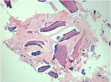
the production of bone grafts which kept the main characteristics (proteins and minerals) almost unchanged (Figure 1). The preparation method could not be fully disclosed to protect intellectual property. In the process, protein denaturation with 20% hydrogen peroxide is followed by alcohol extraction of lipids. The end product is composed of minerals (65%) and proteins (27%). It is definitively sterilized in autoclave. The failure of the arthroplasty was determined to be aseptic in all patients. The type and extent of the acetabular defects had been determined from preoperative radiographs and intraoperative assessments. The clinicopathologic characteristics of patients are shown in table 1.
All patients were operated on by the same team and same reconstruction technique. The posterior approach was performed in all cases.
Technique Technique Technique Technique Technique
The loose prosthesis was removed and all cement, debris, granulomata and fibrous membrane were completely cleared. Carefully reaming of the acetabulum cavity was performed in order to achieve the vascular bone bed, and then the acetabulum was reconstructed by cancellous morselized (chip size, approximately 8 mm3) freeze-dried
bone. The chips were pressed into acetabulum defects and were carefully condensed. The flanges of the reinforcement device (MDTÒ, São Paulo, Brazil) were bent into shape to fit the specific anatomy of the acetabular region. The hook of the device was placed under de teardrop portion. The upper flange of the metal ring was screwed to the ilium. This should result in a stable composite (consisting of the load-bearing host bone, grafts and implant) with an impacted bone graft located beneath the ring. Afterwards, a polyethylene cup was cemented into the acetabular reinforcement device. The amount of bone graft used ranged from 40 g to 60 g in all cases.
Patient analysis was based on the clinical and radiographic evaluation. The clinical analysis was based on functional criteria established by Merle d’Aubigné and Postel8.
For the radiographic analyses, several subjectively established features such as radiolucency, density, trabecular bone formation and component migration were used. Thus, a radiographic analysis of bone integration, based on Conn’s et al.9 criteria was developed to establish bone incorporation
of the grafts in the two groups. Each criterion, except migration, received an independent score ranging from 0 to 2 for each of the three zones of De Lee and Chanrley10
in the acetabulum, where 0 was the worst and 2 the best result11. The sum of the scores was then, multiplied by three
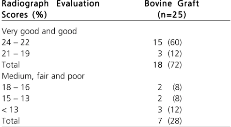
for the acetabulum. For migration, a score of 2, 4 or 6 was established when there was more than 6 mm, 3 to 5 mm, or less than 3 mm of prosthesis displacement. Therefore, a total of 24 points could be achieved for acetabular assessment. Adequate results were considered those with a sum of 19 or greater.
RESULTS
RESULTS
RESULTS
RESULTS
RESULTS
No severe complication occurred in the early post-operative period. Only two deaths two and six years after the procedure were recorded and both were unrelated to RTHA. No more patients were lost to follow-up.
Overall, no minor events were clinically observed. However, a case of superficial infection (cellulites) occurred six months following the procedure and was successfully treated with antibiotics. Clinical and radiographic (Figures 2-4) outcomes are shown in tables 2 and 3, respectively.
DISCUSSION
DISCUSSION
DISCUSSION
DISCUSSION
DISCUSSION
The goals of reconstruction of severe acetabular defects in revision arthroplasty of the hip are to restore the bone stock, to repair the hip mechanics and to obtain stability, and the use of bone graft is imperative to achieve them. Autografts are excellent, but their amount is usually limited and thus allografts have been frequently used. Therefore, a bone bank under strict quality control is necessary to minimize the risk of disease transmission.
Table 1 -Table 1 -Table 1
-Table 1 -Table 1 - Clinicopathologic characteristics of the patients. F e a t u r e s
F e a t u r e sF e a t u r e s
F e a t u r e sF e a t u r e s Bovine GraftBovine GraftBovine GraftBovine GraftBovine Graft n = 25 (%) n = 25 (%)n = 25 (%) n = 25 (%) n = 25 (%)
Female 20 (80,0)
Male 5 (20,0)
Mean (SD) Age (years) 57,7 (12,3) Mean (SD) follow-up (Mo) 96,1 (24,49) Acetabular deficiency
Type III 14 (56)
Type IV 11 (44)
sd= standard desviation. Figure 1 -Figure 1 -Figure 1
Nevertheless, there is not one single technique that provides a solution to all deficiencies. Reconstruction with impacted cancellous morselized bone has been providing good results2,12, but there is some skepticism about its use in hostile
acetabula, especially those of type III and IV deficiencies1,4,5,13.
Additionally, reconstruction of structural allografts is still controversial3. Like others, these authors also consider that
the occurrence of a severe acetabular defect is an indication for reconstruction by reinforcement device combined with bone grafts. This method provides initial stability, prevents grafts from mechanic stress until graft integration is achieved4,5.
For hip procedure, it´s believed that the most suitable graft would be the one slightly changed by any process. In the physical and chemistry analyses, the freeze-dried bones produced at TBHCPA kept most of the mineral and protein characteristics virtually unchanged and both grafts from bovine and human sources are strongly alike, though not keeping the same texture and malleability as their deep-frozen counterparts14. This enabled proper
handling of freeze-dried bones, both from a technical and mechanical viewpoint, after re-hydration15.
By the Merle d’Aubigné and Postel8 criteria, the
average results obtained for bovine grafts were considered good and very good in 80% of the cases. Although the follow-up may be considered not long enough for a more reliable clinical evaluation, it is indicative that the use of bovine freeze-dried grafts during that period did not cause any harm to the patients or significant differences between both. By comparing these results with those in the literature with similar follow-up - but using allogeneic deep-frozen grafts instead -, no considerable differences that could be attributed to the use of freeze-dried bovine grafts were observed4,5,13.
Several studies have clinically and radiographically evaluated, in a number of bone diseases, the use of human and bovine freeze-dried grafts demonstrating very good results. However, few indexed articles concerning the use
Figure 2 -Figure 2 -Figure 2 Figure 2
-Figure 2 - Anteroposterior radiography of the acetabular loosening, type IV deficiency by D’Antonio et al.1
Figure 3 -Figure 3 -Figure 3 Figure 3
-Figure 3 - Immediate post-operative radiography showing the acetabular reconstruction with lyophilized bovine graft and reinforcement device.
Figure 4 Figure 4 Figure 4 Figure 4
of human or bovine freeze-dried grafts in RTHA have been reported. This reluctance of hip surgeons to use freeze-dried grafts may be partially related to a number of available grafts with different steps in the production process for distinct purposes and indications as well. As a result, different mechanical and biological responses may be obtained, leading to an unjustified concern to use this type of graft16.
Levai and Boisgard14 reported thirty revision cases performed
on loose total hip replacements with a specific technique for acetabular reconstruction combining the use of a bovine bone substitute and a support ring. No migration of the acetabular implant or osteolysis of the heterograft was seen in 27 (90%) cases within three years after procedure. Radiologically, the heterograft gradually condensed, and its appearance was similar to that observed with allografts. In that series, the two failures with implant migration and heterograft osteolysis were considered as technical bias related to the use of the Muller ring, and in both cases, it was supported by the cancellous heterograft and not by the host bone14. De Roeck and Drabu17 reported on 32 patients
who underwent RTHA using cemented components and allograft impaction of processed freeze-dried bones. The overall endurance with this type of graft at a mean follow-up of four years was 91%. There were no femoral component failures, although revision was required in three
patients due to failure of the acetabulum. Freeze-dried graft can require longer re-hydration for adequate impaction. The results of impaction bone grafting with freeze-dried bone alone have been satisfactory, although these authors do not feel entirely secure with its use alone in cases of hostile acetabulum17. Thien et al.18 reported an overall survival rate
for acetabular reconstructions of 86% in seven cases using impacted freeze-dried cancellous bone chips and a cemented cup, with a follow-up average of 7 years (range, 5-9 years). At this median follow-up period, no aseptic loosening was observed and the results for freeze-dried allograft bone chips were acceptable. Charalambides et al.19 published a paper addressing their poor results in 27
RTHA cases, followed up for an average of 2.5 years, after the use of autograft and xenograft (Surgibone) bones combined. Seventeen (62%) out of the 27 patients showed apparent bone incorporation within three months. In three (11%) patients, however, there was no incorporation. Three other cases (11%) appeared to have what they considered a pseudoinfection (with no agent identified); and one patient, who had the revision procedure revised again has suffered from deep MRSA infection. Disregarding the case of unequivocal infection, unrelated to graft, and, even considering the three cases of pseudoinfection, that may not be related to the grafts, one finds 77% of good results, which is similar to those obtained by other methods, and therefore, their poor results may be argued. Moreover, the authors also showed a histologic sample from that patient who required, despite apparently radiographic incorporation, revision of the replacement due to acetabular loosening. New bone formation from the grafted area and residual necrotic bony spicules from the graft material were observed, which clearly demonstrates graft incorporation20.
Considering the radiographic criteria, despite biases, the results obtained with bovine freeze-dried bone grafts from TBHCPA, in this series, were comparable to each other and to those reported in the literature, even with those reports of the use of deep-frozen allografts. Using a similar technique of impacting deep-frozen graft and cement, and with a similar follow-up, Kerboull et al.4
reported a similar rate of 92% of graft incorporation. Therefore, the achievements obtained in RTHA seem to be more related to surgeon skills, inherent limitations of the techniques and the severity of the case rather than to the type of graft used.
The use of freeze-dried bone grafts provides a decrease in the risk of transmission of infectious diseases and tumors, since chemical reagents are used in its process to inactivate bacteria, viruses and, probably, prions due to exposure to sodium hypochloride21. After the whole process,
the bone is also submitted to some kind of sterilization22,23
that in our tissue bank virtually achieves 100% of effectiveness. This way, concerns related to prion transmission (EEB), attributed to the use of freeze-dried bovine bone graft seems to be inappropriate. Also, careful selection and country of origin of herd, especially Brazil, that has been always a risk-free country for EEB, should be considered24.
Table 2 -Table 2 -Table 2
-Table 2 -Table 2 - Clinical outcomes.
ClInical Evaluation ClInical EvaluationClInical Evaluation
ClInical EvaluationClInical Evaluation Bovine GraftBovine GraftBovine GraftBovine GraftBovine Graft Merle d’Aubigné and Postel
Merle d’Aubigné and PostelMerle d’Aubigné and Postel
Merle d’Aubigné and PostelMerle d’Aubigné and Postel (n=25) n (%)(n=25) n (%)(n=25) n (%)(n=25) n (%)(n=25) n (%) Very good and good
12 8 (20)
11 9 (35)
10 2 (12)
Total 19 (76) Medium, fair and poor
9 3 (12)
8 1 (4)
7 or < 7 2 (8)
Total 6 (24)
Table 3 -Table 3 -Table 3
-Table 3 -Table 3 - Radiograph results.
Radiograph Evaluation Radiograph EvaluationRadiograph Evaluation
Radiograph EvaluationRadiograph Evaluation Bovine GraftBovine GraftBovine GraftBovine GraftBovine Graft Scores (%)
Scores (%)Scores (%)
Scores (%)Scores (%) ( n = 2 5 )( n = 2 5 )( n = 2 5 )( n = 2 5 )( n = 2 5 ) Very good and good
24 – 22 15 (60)
21 – 19 3 (12)
Total 18 (72)
Medium, fair and poor
18 – 16 2 (8)
15 – 13 2 (8)
< 13 3 (12)
From the mechanical point of view, some studies that use non-decalcified freeze-dried bone concluded that there is no mechanical difference between freeze-dried and deep-frozen bone, and if there is one, this favors the freeze-dried ones, since they lack fat, blood and marrow cells15,25.
Although there is a shortage of data regarding xenograft use in RTHA, clinical complications have not been observed, except for those complications expected with the
use of allografts or xenografts in general, since physical and chemistry characterizations have confirmed both bones are alike26,27. The obtained results, therefore, have shown
that the use of freeze-dried bovine bone grafts does not cause adverse reactions of any kind, further supporting their safety.
In conclusion, the lyophilization process of bones from bovine, accomplished in tissue bank, is of suitable quality to be used in RTHA.
R E S U M O R E S U M O R E S U M O R E S U M O R E S U M O
Objetivo: Objetivo: Objetivo: Objetivo:
Objetivo: Relatar a capacidade clínica e radiográfica de integração de enxertos ósseos liofilizados bovinos. Método: Método: Método: Método: Vinte e cincoMétodo: pacientes foram incluídos. O período médio de seguimento foi de oito anos. Os enxertos foram purificados e liofilizados. O tipo e extensão dos defeitos acetabulares foram determinados por estudo radiográfico pré-operatório e visualização per-operatória. A análise clínica baseou-se no escore de Merle d’Aubigné e Postel1, e critérios de pontuação estabelecidos para a osteointegração
radiográfica foram usados para as análises radiográficas. ResultadosResultadosResultadosResultados: Não houve complicações no pós-operatório imediato. DuasResultados mortes ocorreram em dois e seis anos depois do procedimento não relacionadas ao processo cirúrgico. Os resultados clínicos e radiográficos foram considerados como bons em 80% e 72% dos casos, respectivamente. ConclusãoConclusãoConclusãoConclusãoConclusão: Enxertos liofilizados bovinos podem ser usados com segurança na revisão acetabular da artroplastia total de quadril e o processo de liofilização de ossos bovinos é adequado para uso cirúrgico.
Descritores: Descritores: Descritores: Descritores:
Descritores: Próteses e Implantes. Animais. Ossos e ossos. Liofilização. Acetábulo/cirurgia. Artroplastia.
REFERENCES
REFERENCES
REFERENCES
REFERENCES
REFERENCES
1. D’Antonio JA, Capello WN, Borden LS, Bargar WL, Bierbaun BF, Boettcher WG et al. Classification and management of acetabular abnormalities in total hip arthroplasty. Clin Orthop. 1989; 243:126-37.
2. Schreurs BW, Slooff TJ, Buma P, Gardeniers JW, Huiskes R. Acetabular reconstruction with impacted morsellised cancelous bone graft and cement. A10-to- 15 year follow-up of 60 revision arthroplasties. J Bone Joint Surg Br. 1998; 80B(3): 391-5. 3. Shinar AA, Harris WH. Bulk structural autogenous grafts and
allografts for reconstruction of the acetabulum in total hip arthroplasty. Sixteen-year-average-follow-up. J Bone Joint Surg Am.1997; 79(2):159-68.
4. Kerboull M. Hamadouche M, Kerboull L. The Kerboull acetabular reinforcement device in major acetabular reconstructions. Clin Orthop. 2000; 378:155-68.
5. Winter E. Piert M. Volkmann R. Maurer F. Eingartner C. Weise K. Weller S. Allogeneic cancellous bone graft and a Burch-Schneider ring for acetabular reconstruction in revision hip arthroplasty. J Bone Joint Surg Am. 2001; 83A(6):862-7.
6. Finkemeier CG. Bone-grafting and bone-graft substitutes. J Bone Joint Surg Am. 2002; 84A(3):454-64.
7. Galia CR, Rosito R, Mello TM, Macedo C. Uso de enxerto ósseo homólogo e heterólogo em diáfise femoral de ratos: comparação entre enxerto ósseo congelado e liofilizado. Rev Bras Ortop. 2005; 40(3):141-6.
8. d’Aubigné RM, Postel M. Functional results of hip arthroplasty with acrylic prosthesis. J Bone Joint Surg Am. 1954; 36A(3):351-47. 9. Conn RA, Peterson LFA, Stauffer RN, Ilstrup D. Management of
acetabular deficiency: Long-term results of bone grafting the acetabulum in total hip arthroplasty. Orthopaedics Trans. 1985; 9:451-4.
10. De Lee JG, Charnley J. Radiological demarcation of cemented sockets in total hip replacement. Clin Orthop. 1976; 121:20-32. 11. Cohen J. Statistical power analysis for behavioural sciences. London:
London Academic Press; 1969.
12. Slooff TJ, Huiskes R, van Horn J, Lemmens AJ. Bone grafting in total hip replacement for acetabular protrusion. Acta Orthop Scand. 1984; 55(6):593-6.
13. Boldt JG, Dilawari P, Agarwal S, Drabu KJ. Revision total hip arthroplasty using impaction bone grafting with cemented nonpolished stems and charnley cups. J Arthroplasty. 2001; 16(8):943-52.
14. Levai JP, Boisgard S. Acetabular reconstruction in total hip revision using a bone graft substitute. Early clinical and radiographic results. Clin Orthop. 1996; 330:108-14.
15. Macedo CAS, Galia CR, Silva ALB, César PC, Sanches PRS, Duarte LS, Muller LM. Comparação da resistência à compressão do osso bovino congelado e liofilizado. Rev Bras Ortop. 1999; 34(9/10):529-34. 16. Zasacki W. The efficacy of application of lyophilized,
radiation-sterilized bone graft in orthopedic surgery. Clin Orthop Relat Res. 1991; 272:82-7.
17. de Roeck NJ, Drabu KJ. Impaction bone grafting using freeze-dried allograft in revision hip arthroplasty. J Arthroplasty. 2001; 16(2):201-6.
18. Thien TM, Welten ML, Verdonschot N, Buma P, Yong P, Schreurs BW. Acetabular revision with impacted freeze-dried cancellous bone chips and a cemented cup: a report of 7 cases at 5 to 9 years’ follow-up. J Arthroplasty. 2001; 16(5):666-70.
19. Charalambides C, Beer M, Cobb AG. Poor results after augmenting autograft with xenograft (Surgibone) in hip revision surgery: a report of 27 cases. Acta Orthop. 2005; 76(4):544-9.
20. Donk S, Buma P, Slooff TJ, Gardeniers J, Schreurs BW. Incorporation of morsellised cancelous bone grafts: a study of 24 acetabular biopsy specimes. Clin Orthop. 2002; 396:131-41.
21. Taylor D. Inactivation of the BSE agent. Comptes Rendus de I’Academie des Scinces Serie III. Sciences de la Vie. 2002; 325:75. 22. Mbithi JN, Springthorpe VS, Sattar AS. Chemical disinfection of hepatitis A virus on environmental surfaces. Appl Environ Microbiol. 1990; 56(11):3601-4.
23. Aranda-Anzaldo A, Viza D, Busnel RG. Chemical inactivation of human immunodeficiency virus in vitro. J Virol Methods. 1992; 37(1):71-81.
24. Wenz B, Oesch B, Horst M. Analysis of the risk of transmitting bovine spongiform encephalopathy through bone grafts derived from bovine bone. Biomaterials. 2001; 22(12):1599-606. 25. Cornu O, Bavadekar A, Godts B, Van Tomme J, Delloye C, Banse X.
26. Galia CR, Macedo CA, Rosito R, de Mello TM, Camargo LM, Moreira LF. In vitro and in vivo evaluation of lyophilized bovine bone biocompatibility. Clinics (São Paulo). 2008; 63(6):801-6.
27. Rosito R, Galia CR, Macedo CA, Moreira LF, Quaresma LM, Palma HM. Acetabular reconstruction with human and bovine freeze-dried bone grafts and a reinforcement device. Clinics(São Paulo). 2008; 63(4):509-14.
Received in 20/09/2008
Accepted for publication in 15/12/2008 Conflict of interest: None
Financial source: None
How to cite: How to cite:How to cite: How to cite:How to cite:
Rosito R, Galia CR, Macedo CAS, Quaresma LMAC, Moreira LF. Mid-term follow-up of acetabular reconstruction using bovine freeze-dried bone graft and reinforcement device. Rev Col Bras Cir. [periódico na Internet] 2009; 36(3). Disponível em URL: http://www.scielo.br/rcbc
Correspondence address: Correspondence address:Correspondence address: Correspondence address:Correspondence address: Ricardo Rosito