Cop
yright
© ABE&M t
odos os dir
eit
os r
eser
vados
.
Soft tissue metastases from
differentiated thyroid cancer
diagnosed by
18F FDG PET-CT
Metástases em tecidos moles de câncer de tireoide diagnosticadas por 18F FDG PET-CT
Inés Califano1, Sergio Quildrian2, Martín Coduti3, Erica Rojas Bilbao4, José Otero2, Leonardo Califano3
SUMMARY
Distant metastases of differentiated thyroid cancer are unusual; lung and bones are the most frequently affected sites. Soft tissue metastases (STM) are extremely rare. We describe two ca-ses of patients with differentiated thyroid cancer metastasizing to soft tissues. Both patients had widespread metastatic disease; clinically asymptomatic soft tissue metastases were found by 18-Fluordeoxyglucose positron emission tomography/computed tomography (18F FDG PET-CT), and conirmed by cytological and/or histopathological studies. These indings underscore the ability of 18F FDG PET-CT in accurately assessing the extent of the disease, as well as the utility of the method to evaluate regions of the body that are not routinely explored. Arq Bras Endocrinol Metab. 2013;57(4):317-21
SUMÁRIO
As metástases a distância em carcinoma diferenciado de tireoide são raras. Pulmão e ossos são os lugares mais frequentemente atingidos. As metástases em tecidos moles são extremamen-te infrequenextremamen-tes. Nesextremamen-te artigo, descrevemos dois casos de pacienextremamen-tes com câncer diferenciado de tireoide com metástases em tecidos moles. Ambos os pacientes padeciam de enfermida-de avançada. As metástases em partes moles foram assintomáticas e enfermida-detectadas mediante
18-Fluordeoxyglucose positron emission tomography/computed tomography (18F FDG PET-CT), sendo conirmadas por citologia e/ou histopatologia. Esses achados ressaltam o valor do 18F FDG PET-CT para a avaliação correta da extensão da enfermidade e a utilidade do método para avaliar regiões do corpo que não são exploradas nos estudos de rotina. Arq Bras Endocrinol Metab. 2013;57(4):317-21
1 Service of Endocrinology,
Instituto de Oncología “Angel H. Roffo”, Universidad de Buenos Aires, Argentina 2 Department of Soft Tissues
Surgery, Instituto de Oncología “Angel H. Roffo”, Universidad de Buenos Aires, Argentina 3 Department of Head and Neck
Surgery, Instituto de Oncología “Angel H. Roffo”, Universidad de Buenos Aires, Argentina 4 Department of Pathology,
Instituto de Oncología “Angel H. Roffo”, Universidad de Buenos Aires, Argentina
Correspondence to:
Inés Califano
Service of Endocrinology, Instituto de Oncología “Angel H. Roffo”,
San Martín, 5481 1407, Caba, Argentina [email protected]
Received on Nov/5/2012 Accepted on Nov/28/2012
INTRODUCTION
S
oft tissues comprise over 40%-50% of the totalbody weight; however, hematogenous metastases to these areas are uncommon. It has been hypothesized that muscle and subcutaneous soft tissue are hostile en-vironments for the survival of cancer cells (1).
Although occasionally soft tissue metastases (STM) may present themselves as painful masses, they are usu-ally asymptomatic. Therefore, they may be an unex-pected inding in imaging studies (2).
Differentiated thyroid cancer (DTC) is character-ized by excellent prognosis and long-term survival. Metastases occur in about 5%-10% of patients. The ma-jor sites of metastases are the lungs and bones. Meta-static disease to skeletal muscle is extremely rare and tends to be diagnosed in patients with advanced stage neoplasms (3).
18-Fluordeoxyglucose positron emission
tomogra-phy/computed tomography (18F FDG PET-CT) is a
Cop
yright
© ABE&M t
odos os dir
eit
os r
eser
vados
.
patients with elevated thyroglobulin and negative whole body radioiodine scan, after thyroidectomy and radio-iodine ablation. Also, the inding of FDG-avid lesions
usually implies in poor prognosis. As 18F FDG PET-CT
yields whole-body imaging, it is not unusual to ind foci of metastatic tissue in previously unsuspected sites; this may lead to the modiication of treatment strategies in up to 28% of patients (4).
Two cases of unsuspected STM from DTC that
were found by 18F FDG PET-CT are described in this
report.
CASE REPORT 1
A 26 year-old male without history of exposure to ra-diation or familial thyroid disease was diagnosed with papillary thyroid cancer (PTC) with peripheral thyroid soft tissues invasion [T3NxM0 EI; risk of recurrence was intermediate according to the ATA guidelines (5) and high according to LATS (6)] in 2002. He was tre-ated elsewhere with total thyroidectomy,
lymphadenec-tomy, and 150 mCi131I. In May 2005, four metastatic
left cervical lymph nodes were removed. He received an
additional 150 mCi131I dose, with a negative post-dose
scan. Serum thyroglobulin levels during these procedu-res were not available. Three months later, he was re-ferred to our hospital due to suspicious cervical lymph nodes in the cervical ultrasound. Fine-needle aspiration biopsy (FNAB) conirmed metastatic PTC. The patient underwent a thoroughly surgical revision of the thyroid lodge and lymph node dissection (levels III-IV), which yielded two metastatic lymph nodes out of two ted, the largest measuring 2 cm; the remaining resec-ted material was granulomatous tissue. Subsequently, he had a negative neck ultrasonography. However, six months later, he presented a palpable ixed left cervi-cal mass. FNAB was positive for PTC metastases. Tra-cheal invasion was suspected on CAT scan, and intra-luminal invasion was conirmed by bronchoscopy. In March 2007, asegmental tracheal resection with level VII lymphadenectomy was performed. He received off--label treatment with rosiglitazone as a redifferentiating agent, aiming to restore radioiodine uptake (7) and a new dose of 200 mCi of radioiodine. A post-dose scan showed central neck uptake; stimulated thyroglobulin was 13 ng/mL with negative antibodies. The patient was lost to follow up until a year later; a mass in the upper mediastinum was observed in MRI; he received another dose of 100 mCi of radioiodine, with a
nega-tive post-dose scan, and his stimulated thyroglobulin
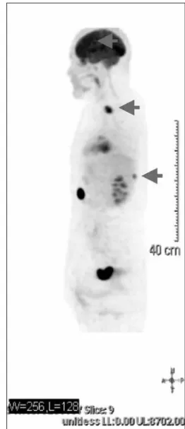
rose to 96 ng/mL. A18F FDG PET-CT (Figure 1)
sho-wed hypermetabolic foci in the mediastinum and lo-wer neck (SUVm 11,2-14,3), in central nervous system (left frontal lobe, SUVm 10,5, which was conirmed by MRI), and paravertebral dorsal soft tissues (SUVm 6,4). FNAB of this lesion was compatible with PTC metastases (Figure 2). Bilateral pulmonary micronodu-les were observed. Gamma knife surgery of the cerebral metastases was performed, as well as 3D external beam radiotherapy (6000 Cgy) to the neck and mediastinum; both lesions remained stable subsequently. The para-vertebral metastases increased its volume. The patient refused surgery and was offered off-label treatment with sunitinib (8).
Cop
yright
© ABE&M t
odos os dir
eit
os r
eser
vados
.
CASE REPORT 2
A 64 year-old female with a 30 year history of goiter was diagnosed with follicular variant of papillary thyroid cancer (FVPTC), (T3N0M0 EIII; risk of recurrence was intermediate according to the ATA guidelines, and high according to LATS) in 2003. She was treated el-sewhere with total thyroidectomy and ablation with 100 mCi of radioiodine. She was subsequently lost to follow up until 2006, when she was seen because of dyspnea. Extensive invasion of the larynx through the mucosa was observed; total laryngectomy was perfor-med. Histopathological diagnosis conirmed FVPTC. She was then referred to our hospital on 2007. She was given 200 mCi of radioiodine, the post-dose scan showing faint uptake in the neck. Stimulated thyro-globulin was 860 ng/mL, with negative antibodies. Neck sonogram and bone scan were negative for me-tastases. On the CAT scan, lung micro metastases were observed, as well as a 12-mm image in the pancreas. Levels of CA 19.9 and CEA were normal at that time, sonogram was irrelevant, and close follow-up was de-cided. A new treatment with 200 mCi of radioiodine was given, with a negative post-dose scan and rising
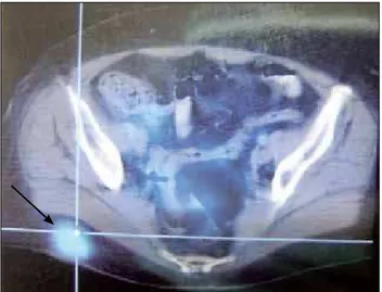
thyroglobulin (1,390 ng/mL). A 18F FDG PET-CT
scan was performed identifying hypermetabolic foci in the pancreas (a 28 mm mass with SUVmax 3,9), lower abdominal wall (SUVmax 2,4), and right gluteus (30 x 25 mm SUVmax 5,1, Figure 3); stimulated thyroglo-bulin was 3,259 ng/mL, and antibodies were negative. On the MRI the gluteal lesion was solid and hetero-geneous; FNAB showed an adenocarcinoma. A biopsy was performed, conirming PTC metastases (Figure 4). Immunohistological staining revealed positivity for TTF1 and thyroglobulin. FNAB of the abdominal wall
lesion was positive for ibrosis, and FNAB of the pan-creas was unsatisfactory.
Figure 2. FNAB of dorsal soft tissue lesion showing papillary thyroid cancer metastases.
Figure 3. 18 F FDG PET-CT showing a subcutaneous gluteal FDG-avid lesion.
Figure 4. Hematoxilin-eosin showing metastases of papillary thyroid cancer in subcutaneous tissue.
DISCUSSION
Cop
yright
© ABE&M t
odos os dir
eit
os r
eser
vados
.
Subcutaneous tissue and muscle metastases of DTC are extremely rare events. It is, however, dificult to know the real incidence of this presentation, because reports of cases are scarce (3,10,11-14). According to Song and cols. (15), up to 2011 there were 10 cases of muscle metastases in the English literature.
STM may be an unexpected inding in imaging studies, due to the fact that they are asymptomatic in the majority of the cases. Autopsy series in cancer patients report a prevalence of up to 17% of skeletal muscle metastases (2); therefore, it is likely that most STM remain undetected clinically and radiographically. Besides the possibility of underdiagnosis, several fac-tors have been implicated in the rare occurrence of STM, such as muscle motion, mechanical tumor destruction, muscle ability to remove tumor-produced lactic acid, changes in pH, accumulation of metabolites, and local temperature of the soft tissue sites. In addition, blood low is variable, inluenced by adrenergic receptors, and subject to variations in tissue pressure affecting cancer implantation. Whether traumatic injury to soft tissue is a risk factor for STM remains undetermined (1).
The most commonly reported malignancies that result in STM are lung, kidney and colon carcinoma (1); STM from DTC are extremely unusual. They tend to be found in patients with advanced disease, such as the cases we are reporting. Both patients had aggressive disease, showing widespread metastatic dissemination with concomitant loss of radioiodine uptake ability.
The 18F FDG PET-CT is a sensitive, noninvasive
method that allows simultaneous identiication and anatomic localization of metastases in patients with elevated thyroglobulin levels and negative radioiodine whole body scans. Additionally, it also provides prog-nostic information (16). Patients with large tumor volume and elevated SUVm values (as observed in the two cases we report here) are considered to have worse
prognosis and shorter survival (17). As 18F FDG
PET--CT yields whole-body imaging, it can identify foci of non-radioiodine avid disease in areas that are not rou-tinely explored. As increasing number of PET-CT scans are performed, it is likely that distant metastases in un-usual sites will be more frequently detected. Bae and cols. (18) reported one unsuspected skeletal muscle metastases from papillary thyroid cancer diagnosed by
18F FDG PET TC. The patient also had recurrent
dis-ease in the mediastinum.
STM as the presenting feature of DTC are excep-tional (11). In this event, distinction between a
met-astatic neoplasm and a primary soft tissue sarcoma is critical because treatment and prognosis are markedly different (1); cytological or histological conirmation of diagnosis is mandatory. In such case, the histologi-cal feature of epithelioid neoplasm with tubules and papillary structures is suspicious of synovial sarcoma. In this sarcoma, the immunohistochemical study reveals positivity with CK7, CK19, EMA, BCL2, and CD99, but TTF-1 and thyroglobulin are negative. The exhaus-tive study of primary thyroid neoplasm is relevant in the identiication of vascular invasion, dedifferentiated areas, extracapsular extension, and evaluation of other component (anaplastic, medullary and tall-cell variant). In both of our patients, extrathyroid extension was noted at diagnosis.
In conclusion, we have reported two cases of skin
and muscle metastases of DTC diagnosed by 18F FDG
PET CT. Our report underscores the utility of the method in selected cases to conirm the accurate ex-tension of the disease, and the need for cytological or histological conirmation due to the rarity of the diag-nosis.
Disclosure: no potential conlict of interest relevant to this article was reported.
REFERENCES
1. Plaza J, Perez-Montiel D, Mayerson J, Morrison C, Suster S. Me-tastases to soft tissue. A review of 118 cases over a 30-year pe-riod. Cancer. 2008;112:193-203.
2. Surov A, Hainz M, Holzhausen H, Arnold D, Katzer M, Schmidt J, et al. Skeletal muscle metastases: primary tumors, prevalence and radiological features. Eur Radiol. 2010;20:649-58.
3. Qiu ZL, Luo QY. Erector spinae metastases from differentia-ted thyroid cancer identiied by I-131 SPECT/CT. Clin Nucl Med. 2009;34:137-40.
4. Razfar A, Branstetter B, Christopoulos A, Lebeau S, Hodak S, He-ron D, et al. Clinical usefulness of positHe-ron emission tomography--computed tomography in recurrent thyroid carcinoma. Arch Oto-laryngol Head Neck Surg. 2010;136(2):120-5.
5. Cooper D, Doherty G, Haugen B, Kloos R, Lee S, Mandel S, et al. Revised American Thyroid Associaton management guidelines for patients with thyroid nodules and differentiated thyroid can-cer. Thyroid. 2009;19(11):1167-214.
6. Pitoia F, Ward L, Wohllk N, Friguglietti C, Tomimori E, Gauna A, et al. Recommendations of the Latin American Thyroid Society on diagnosis and management of differentiated thyroid cancer. Arq Bras Endocrinol Metab. 2009;53(7):884-97.
7. Kebebew E, Peng M, Reiff E, Treseler P, Woeber K, Clark OH, et al. A phase II trial of rosiglitazone in patients with thyroglobulin--positive and radioiodine-negative differentiated thyroid cancer. Surgery. 2006;140:960-6.
Cop
yright
© ABE&M t
odos os dir
eit
os r
eser
vados
.
carcinoma of the thyroid with functional imaging correlation. Clin Cancer Res. 2010;16(21):5260-8.
9. Benbassat C, Mechlis-Frish S, Hirsch D. Clinicopathological characteristics and long-term outcome in patients with distant metastases from differentiated thyroid cancer. World J Surg. 2006;30:1088-95.
10. Bruglia M, Palmonella G, Silvetti F, Rutigliano P, Criante P, Mar-morale C, et al. Skin and thigh muscle metastasis from papillary thyroid cancer. Singapore Med J. 2009;50 (2):e61-4.
11. Pucci A, Suppo M, Lucchesi G, Celeste A, Viberti L, Pelleritto R, et al. Papillary thyroid cancer presenting as a solitary soft tissue metastasis in an elderly hyperthyroid patient. Case report and re-view of the literature. Virchows Arch. 2006;48:857-61.
12. Zhao LX, Li L, Li FL, Zhao Z. Rectus abdominis muscle metastasis from papillary thyroid cancer identiied by I-131 SPECT/CT. Clin Nucl Med. 2010;35:360-1.
13. Iwai H, Ohno Y, Ito H, Kiyokawa T, Aoki N. Renal rupture asso-ciated with a poorly differentiated follicular thyroid carcinoma
metastasizing to the thigh muscle, lung and kidney. Intern Med. 2005;44:848-52.
14. Sevinc A, Buyukberber S, Sari R, Baysal T, Mizrak B. Follicular thyroid cancer presenting initially with soft tissue metastasis. Jpn J Clin Oncol. 2000;30(1):27-9.
15. Song HJ, Xue YL, Xu YH, Qiu ZL, Luo QY. Rare metastases of diffe-rentiated thyroid carcinoma: pictorial review. Endocr Relat Can-cer. 2011;18:R165-74.
16. Robbins R, Quiang W, Grewal R, Reibke R, Gonen M, Strauss H, et al. Real-time prognosis for metastatic thyroid carcinoma based on 2-[18f] luoro-2-deoxy-glucose-positron emission tomography scanning. J Clin Endocrinol Metab. 2006;91(2):498-505. 17. Pace L, Nicolai E, Klain M, Salvatore M. Diagnostic value of FDG/
PET TC imaging. Q J Nucl Med Mol Imaging. 2009;53:503-12. 18. Bae S, Lee S, Koo M, Hur S, Choi M, Cho D, et al. Distant, solitary