Effect of BjcuL, a lectin isolated from
Bothrops jararacussu
, on human
peripheral blood mononuclear cells
Weverson Luciano Pires
a,1, Onassis Boeri de Castro
a,1, Anderson Makoto Kayano
a,c,1,
Sulamita da Silva Setúbal
a, Adriana Silva Pontes
a, Neriane Monteiro Nery
a, Mauro Valentino Paloschi
a,
Soraya dos Santos Pereira
b, Rodrigo Guerino Stábeli
c, Carla Freire Celedônio Fernandes
b,d,
Andreimar Martins Soares
c, Juliana Pavan Zuliani
a,c,⁎
aLaboratório de Imunologia Celular Aplicada à Saúde, Fundação Oswaldo Cruz, FIOCRUZ Rondônia, Porto Velho, RO, Brazil bLaboratório de Engenharia de Anticorpos, Fundação Oswaldo Cruz, FIOCRUZ Rondônia, Porto Velho, RO, Brazil
cCentro de Estudos de Biomoléculas Aplicadas à Saúde (CEBio), Fundação Oswaldo Cruz, FIOCRUZ Rondônia e Departamento de Medicina, Universidade Federal de Rondônia, UNIR, Porto Velho,
RO, Brazil
dCentro de Pesquisa em Medicina Tropical, Porto Velho, RO, Brazil
a b s t r a c t
a r t i c l e
i n f o
Article history:
Received 11 November 2016
Received in revised form 13 January 2017 Accepted 2 February 2017
Available online 8 February 2017
BjcuL is a C-type lectin with specificity for the binding ofβ-D-galactose units isolated fromBothrops jararacussu venom. It triggers cellular infiltration in post capillary venules, increases edema and vascular permeability in mu-rine models, contributes toin vitroneutrophil activation and modulates macrophage functional activation to-wards an M1 state. The purpose of this study was to investigate the effect of BjcuL on human peripheral blood mononuclear cells (PBMCs) activation with a focus on PBMCs proliferation and inflammatory mediators release. Results showed that BjcuL is not toxic to PBMCs, that BjcuL inhibits PBMCs proliferation and that it stimulates PBMCs to produce superoxide anion and hydrogen peroxide, primarily via lymphocyte stimulation, but does not stimulate the production of nitric oxide and PGE2. These results demonstrate that BjcuL has an
immunomod-ulatory effect on PBMCs. Further studies are needed to confirm the immunomodulatory effect of BjcuL, to eluci-date the molecular mechanisms of action responsible for its effects and to determine its potential application as an immunopharmacological and biotechnological tool.
© 2017 Elsevier Ltd. All rights reserved. Keywords:
Snake venom Lectin PBMCs Mediators
1. Introduction
Bothrops jararacussuis one of Brazil's most dreaded snakes. This im-portant pit-viper may grow to a length of 2.2 m and its venom glands can deliver up to 1000 mg (dry weight) of highly-lethal venom in a sin-gle bite. It is responsible for 0.8% to 10% of serious snakebites in São Paulo State, Brazil (Milani Júnior et al., 1997).Bothrops jararacussu venom is composed primarily of enzymatic proteins such as phospholi-pases A2(PLA2), serine and metalloproteases, and non-enzymatic lectin proteins.
Snake venom lectins comprise a class of toxins that bind, non-cova-lently and with high specificity, to terminal galactoside residues of gly-cans. The recognition of specific glycans by lectins is an event that plays a key role in a variety of biological phenomena (Kishore et al., 1997).
The lectin BjcuL was isolated fromBothrops jararacussuvenom. This lec-tin is a C-type leclec-tin with specificity for the binding ofβ-D-galactose units (de Carvalho et al., 2002).
BjcuL has been shown to bind to lactose moieties and induce agglu-tination of erythrocytes. The literature shows that BjcuL weakly adheres to MDAMB-435, a human metastatic breast cancer cell line, and to OVCAR-5, a human ovarian carcinoma cell line (de Carvalho et al., 2001). Moreover, BjcuL inhibits growth in several cancer cell lines (Damasio et al., 2014; de Carvalho et al., 2001; Nolte et al., 2012; Pereira-Bittencourt et al., 1999). However, BjcuL does not inhibit adhe-sion of these cancer cells to the extracellular matrix proteinsfibronectin, laminin and type I collagen (de Carvalho et al., 2001).
BjcuL triggers cellular infiltration in post capillary venules (Elifi o-Esposito et al., 2007), increases edema and vascular permeability in mu-rine models (Panunto et al., 2006) and contributes to in vitro neutrophil activation (Elifio-Esposito et al., 2011). The literature on the infl amma-tory effects of BjcuL indicates that BjcuL modulates macrophage func-tional activation towards an M1 state (Dias-Netipanvi et al., 2016); the aim of this study was to investigate the effect of BjcuL on human PBMCs activation with a focus on PBMCs proliferation and inflammatory mediators release.
⁎ Corresponding author: Fundação Oswaldo Cruz (FIOCRUZ-Rondônia), Laboratório de Imunologia Celular Aplicada à Saúde, Rua da Beira, 7671 BR364, Km 3.5, CEP 76812-245, Porto Velho, RO, Brazil.
E-mail addresses:juliana.zuliani@fiocruz.br,[email protected],
[email protected],[email protected](J.P. Zuliani). 1 These authors contributed equally to this work.
http://dx.doi.org/10.1016/j.tiv.2017.02.003
0887-2333/© 2017 Elsevier Ltd. All rights reserved.
Contents lists available atScienceDirect
Toxicology in Vitro
2. Material and methods
2.1. Chemicals and reagents
MTT, RPMI-1640,L-glutamine, gentamicin, lipopolysaccharide (LPS), concanavalin A (Con-A), phorbol myristate acetate (PMA), Histopaque 1077, DMSO, OPD (o-1,2-phenylenediamine dihydrochloride), horse-radish peroxidase, nitroblue tetrazolium (NBT), carboxyfluorescein diacetate succinimidyl ester (CFDA-SE) and anti-iNOS antibody were purchased from Sigma (MO, USA). Cell Viability Kit, PE anti-human CD14, PE anti-human CD4, PE anti-human CD8 and PerCP anti-human CD3 were purchased from BD Pharmingen (CA, USA). 2′,7′ -dichlorodihydrofluorescein diacetate (DCFDA) was purchased from Molecular Probes (OR, USA). PGE2specific enzymatic immunoassay (EIA), anti-COX-1 and anti-COX-2 antibodies were purchased from Cay-man Chemicals (MI, USA). Fetal bovine serum (FBS) was obtained from Cultilab (São Paulo, Brazil). All salts and reagents were obtained from Merck (Darmstadt, Germany).
2.2. Ethical statement
This study was approved by the Brazilian Institutional Review Board of the Centre for Research in Tropical Medicine (CEPEM-RO), under pro-tocol numbered 067/08. All subjects were adult volunteers and all signed a Statement of Informed Consent.
2.3. Venom
TheBothrops jararacussusnake venom was provided by Serpentário Proteínas Bioativas (Batatais–SP) and maintained refrigerated (8 °C) in the Bank of Amazon Venoms at the Center of Biomolecular Studies Ap-plied to Health, CEBIO-UNIR-FIOCRUZ-RO, (authorization: CGEN/CNPq 010627/2011-1 and IBAMA 27131-1).
2.4. Affinity column preparation
Initially, 20 mL of Sepharose 6B resin was transferred to a Buchner funnel and washed with 30 mL of distilled water. After the complete elimination of water, the resin was suspended in 15 mL of NaOH 1 M containing 30 mg of sodium borohydrate. The contents were transferred to a plastic beaker and 5 mL of bisepoxi was added. The activation was processed for 5 h at room temperature under gentle agitation. The acti-vated resin was transferred to a Buchner funnel and washed with 300 mL of distilled water. Lactose coupling proceeded for 24 h at room temperature in bicarbonate buffer 0.5 M (pH 10.0), using an equal vol-ume of ligand solution and activated resin. After lactose coupling, the re-maining epoxi groups were blocked withβ-mercaptoethanol for 2 h at room temperature. The coupled resin was washed with 200 mL of sodi-um bicarbonate 0.5 M, 50 mL of sodisodi-um acetate 0.2 M (pH 4.0), and 50 mL of saline, respectively. The coupled resin (Sepharose 6B-CL-lac-tose) was maintained at 4 °C until use.
2.5. Purification of a lectin from Bothrops jararacussu venom
Bothrops jararacussuvenom (100 mg) was dissolved in CaTBS buffer (0.02 M Tris, 0.15 M NaCl and 0.01 CaCl2) (pH 7.4), and clarified by ul-tracentrifugation at 3500 ×gfor 5 min. The supernatant was applied and fractionated on a column (3 cm × 1 cm) of Sepharose 6B-CL-lactose which was pre-equilibrated with the same buffer used to dissolve the venom. The resin was washed until the optical density measured 280 nm returned to basal levels. The retained material was eluted using 0.1 M lactose in CaTBS. The resin fraction wasfiltered using Sephadex G-25 to remove the lactose. The purity of the eluted protein was evaluated with RP-HPLC using a C18 column (Discovery 0.45 cm × 25 cm; Supelco–USA) under a linear gradient 0–60% of ace-tonitrile in 0.1% trifluoroacetic acid (v/v). All chromatographic steps
were done using an Akta Purifier 10 system (GE Lifescience Healthcare –USA). SDS-PAGE 12.5%, performed as described byLaemmli (1970), confirmed the apparent weight and molecular homogeneity of the resin fraction. The protein concentration was estimated using Bradford reagent with a solution of BSA as the protein standard.
2.6. Molecular mass determination
Molecular mass was determined by mass spectrometry performed with MALDI equipment (matrix-assisted laser desorption ionization) using TOF analyzers (AXIMA TOF2Shimadzu Biotech) and a saturated solution of sinapinic acid as the ionization matrix. The sample was co-crystallized with the ionization matrix (1:3 ratio, respectively) and an-alyzed in linear mode.
2.7. Hemagglutinanting activity
Hemagglutinanting activity was performed using a microagglut ination technique to determine the presence of BjcuL. This technique used human erythrocytes previously collected with heparin (50 U/mL), and microtiter V-well plates. Each well contained 50μL of 3% erythrocytes suspension and CaTBS with 50μL samples in the same buffer. The nega-tive control contained 50μL of cell suspension and 50μL of CaTBS. Follow-ing the addition of erythrocytes, the plates were shaken briefly and incubated at room temperature. The agglutination was monitored visual-ly after 2 h.
2.8. Isolation of human peripheral blood mononuclear cells (PBMC)
Blood was donated by healthy volunteers who had not used medica-tion in the last 48 h. Blood was collected in vacuum tubes containing heparin, and diluted in phosphate buffered saline (PBS - 14 mM NaCl, 2 mM NaH2PO4·H2O, 7 mM Na2HPO4·12H2O) (pH 7.4). PBMCs were isolated by density gradient method—according toLacouth-Silva et al. (2015)—using Histopaque 10,771 (Sigma; MO, USA), as per the manufacturer's instructions. Briefly, the blood diluted in PBS was lay-ered on Histopaque at a 1:1 ratio and subjected to centrifugation at 400 ×gfor 30 min. The white layer representing PBMCs was gently as-pirated out and aseptically transferred into sterile centrifuge tubes. After centrifugation, the cell suspension containing the PBMCs was washed 3 times with PBS and cultured in sterile RPMI assay medium [RPMI-1640 medium supplemented with 100μg/mL of gentamicin, 2 mM ofL-glutamine and 10% of fetal bovine serum (FBS)]. Aliquots of the isolated PBMCs were used to determine the total number of cells in a Neubauer's chamber following cell staining (1:20, v/v) with Turk solution (0.2% violet crystal in 30% acetic acid). The purity of the isolated cell populations was determined by Panotic staining of cytospin prepa-rations and byflow cytometry analysis (FACscan). The mean purity was 99% for PBMC preparation. The number of cells was adjusted to 105or 106, relative to the amount of cells necessary for each experiment.
2.9. Cell viability assay by MTT reduction
The cytotoxicity of BjcuL on PBMCs was determined by an MTT assay (Reilly et al., 1998; Lacouth-Silva et al., 2015), which made it possible to analyze the capacity of living cells to reduce the 3-(4,5-dimethyl-2-thiazolyl)-2,5-diphenyl-2H-tetrazolium bromide (MTT, Sigma; MO, USA) to a purple formazan product. Cells were plated in 96-well plates (2 × 10⁵/well) and maintained in RPMI assay medium. PBMCs were in-cubated with RPMI (negative control) or BjcuL (5 and 10μg/mL diluted in RPMI; experimental group) for 96 h at 37 °C, under an atmosphere of 5% CO2. After incubation, the plate was centrifuged at 400 ×gfor 5 min and the supernatant was replaced by fresh RPMI containing MTT (0.5μg/mL). Four hours later, the plates were washed three times with PBS. The formazan crystals were dissolved in 100μL DMSO over 90 min and were evaluated by spectrophotometer at 540 nm (Bio-Tek
Synergy HT Multi-Detection, Winooski, VT). The results were expressed by optical density relative to the control.
2.10. Cell viability assay byflow cytometry (BD™cell viability kit)
PBMCs (1 × 10⁶cells/well) were suspended in RPMI assay medium and incubated, in duplicate, in 96-well plates with RPMI (negative con-trol) or BjcuL (5 and 10μg/mL; experimental group) for 1, 2, 8, 24, 48 and 96 h at 37 °C, in a humidified atmosphere of 5% CO2. Following that, 0.1μL of propidium iodide (PI) and 0.2μL of thiazole orange (TO) was added to each sample, stirred by vortexing and then incubated for 5 min at room temperature. Acquisition and analysis was then per-formed in FACScan.
2.11. Cell proliferation assay by MTT reduction
Cell proliferation was tested using a 3-(4,5-dimethylthiazol-2-yl)-2, 5-diphenyl tetrazolium bromide (MTT) assay. PBMCs were freshly iso-lated and piso-lated in 96-wellflat bottom tissue culture plates at a concen-tration of 2 × 10⁵cells/well containing 200
μL of RPMI assay medium. PBMCs were cultured with RPMI (negative control), Concanavalin A (5μg/mL; positive control), or BjcuL (5 and 10μg/mL; experimental group) for 96 h at 37 °C, under an atmosphere of 5% CO2. After incuba-tion, 10μL of MTT solution (5 mg/mL in PBS) was added to each well. After 4 h, the plates were centrifuged at 400 ×gfor 5 min and the super-natant was removed. After washing with PBS, the formazan crystals were dissolved with 100μL of DMSO and the plates were kept on an or-bital shaker for 90 min. The optical density (O.D.) was measured in a spectrophotometer at 570 nm (BioTek Synergy HT Multi-Detection, Wi-nooski, VT). The results were expressed by optical density relative to the controls.
2.12. Cell proliferation assay byflow cytometry
Cell proliferation was tested using carboxyfluorescein diacetate succinimidyl ester (CFDA-SE). In brief, isolated PBMCs were stained with CFDA-SE and plated in 96-wellflat bottom tissue culture plates at a concentration of 1 × 10⁶cells/well containing 100
μL of RPMI assay medium. After that, PBMCs were incubated with RPMI (negative control), Concanavalin A (5μg/mL; positive control), or BjcuL (5 and 10μg/mL; experimental group) for 72 h at 37 °C, under an atmosphere of 5% CO2. After incubation, cell surface labeling was performed with monoclonal antibody PerCP anti-human CD3, PE anti-human CD14, PE anti-human CD4, and PE anti-human CD8 incubated for 30 min on ice and protected from light. PBMCs were washed with PBS to remove un-bound antibodies. Acquisition and analysis was performed in FACScan.
2.13. Superoxide anion production assay
In this assay, the generation of superoxide was estimated by reduc-ing nitroblue tetrazolium (NBT, Sigma; MO, USA), a yellow liposoluble compound that becomes insoluble and blue in its reduced form (Pontes et al., 2014). In brief, 2 × 10⁵PBMCs/100
μL were incubated with 100μL of RPMI containing 0.1% NBT (negative control), 100μL of PMA containing 0.1% NBT (500 ng/mL; positive control), or BjcuL (5 and 10μg/mL diluted in RPMI containing 0.1% NBT; experimental group) and incubated for 2 h at 37 °C, in a humidified atmosphere of 5% CO2. At the end of the incubation period, the supernatant was re-moved and the cells were centrifuged at 800 ×gfor 5 min. After washing with PBS, the reduced NBT deposited inside the cells was dissolved,first by adding 120μL of 2 M KOH to solubilize the cell membranes, and then by adding 140μL of DMSO to dissolve the blue formazan via gentle shak-ing for 10 min at room temperature. The dissolved NBT solution was then transferred to a 96-well plate and absorbance was measured on a spectrophotometer at 620 nm (Bio-Tek Synergy HT Multi-Detection, Winooski, VT). Data were expressed as absorbance.
2.14. Determination of hydrogen peroxide (H2O2) production by human PBMCs
The technique used was described byPick and Keisari (1980), and adapted for microassay byPick and Mizel (1981), with modifications proposed byRusso et al. (1989). In brief, PBMCs (2 × 10⁵/100
μL) were resuspended in 1 mL of phenol red solution (140 mM NaCl, 10 mM potassium phosphate buffer (pH 7.0), 0.56 mM phenol red) con-taining 0.05 mg/mL of horseradish peroxidase. The cells were then incu-bated with RPMI (negative control) or BjcuL (5 and 10μg/mL) for 3 h at 37 °C, under a humidified atmosphere of 5% CO2. Cells were maintained simultaneously with and without stimulation by phorbol myristate ace-tate (PMA, 500 ng/mL). After incubation, the reaction was stopped by the addition of 1 N sodium hydroxide (10 mL). The absorbance was measured spectrophotometrically (Bio-Tek Synergy HT Multi-Detec-tion, Winooski, VT), at 620 nm, against the blank which consisted of phenol red medium. The data generated were compared to a standard curve conducted for each test. The results were expressed in terms of mM of H2O2produced.
2.15. Measurement of intracellular ROS levels
A peroxide-sensitivefluorescent probe 2′,7′-dichlorodihydrofluoresce in diacetate (DCFDA) was used to measure intracellular levels of ROS. DCFDA is converted by intracellular esterases to 2′,7′-dichlorodihy drofluorescin, which is then oxidized by H2O2to the highlyfluorescent 2′,7′-dichlorodihydrofluorescein (DCF). For this assay, 2 × 10⁶PBMCs were resuspended in RPMI without phenol red, and the cells were incu-bated with RPMI without phenol red (negative control), PMA (500 ng/ mL; positive control), or BjcuL (5 and 10μg/mL). After 3 h of incubation, 100μL of DCFDA, diluted with 10μM of PBS, was added and incubated for 30 min at 37 °C, under constant dark conditions (Nakajima et al., 2009). After incubation, cell surface labeling was performed with mono-clonal antibody PerCP anti-human CD3 or PE anti-human CD14, incubat-ed for 30 min on ice and protectincubat-ed from light. PBMCs were washincubat-ed with PBS to remove unbound antibodies. Acquisition and analysis was per-formed in FACScan.
2.16. Nitric oxide (NO) production assay
NO production by PBMC cultures, incubated with and without BjcuL, was determined in the supernatant. PBMCs, at a density of 2 × 10⁵/ 250μL in RPMI assay medium, were plated in 96-well plates and incu-bated with RPMI (negative control), LPS (1μg/mL; positive control), or BjcuL (5 and 10μg/mL; experimental group) for 96 h at 37 °C, in a hu-midified atmosphere of 5% CO2. After incubation, the supernatants of the cells were transferred to a reading plate, and Griess reagent was added (in the ratio of 1:1, v/v) for the determination of nitrite (Ding and Nathan, 1988). The absorbance was measured spectrophotometri-cally (Bio-Tek Synergy HT Multi-Detection, Winooski, VT), at 550 nm, and data were compared to a standard curve prepared with NaNO2 (2.5 to 80μM) and expressed in terms ofμM nitrite released by 2 × 10⁵cells.
2.17. Determination of prostaglandin E2(PGE2) production
sample absorbances were registered at 412 nm in a spectrophotometer (Bio-Tek Synergy HT Multi-Detection, Winooski, VT), and eicosanoids concentrations were estimated from standard curves expressed in terms of pg/mL of PGE2.
2.18. Immunoblotting
For this assay, 2 × 10⁶PBMC suspensions resuspended in assay me-dium were plated with RPMI (negative control), PMA (500 ng/mL; pos-itive control), or different concentrations of BjcuL (5 and 10μg/mL) and incubated at 37 °C, in a humidified atmosphere of 5% CO2,over a period of 2 h for COX-1 and COX-2, and a period of 24 h for iNOS determination. After incubation, PBMCs were lysed with RIPA buffer containing
phosphatase and protease inhibitors (1/100, Sigma), and were homog-enized. For COX-1, COX-2 and iNOS determination, total protein extracts (20μg) were prepared, resolved by 15% SDS/PAGE and transferred onto a PVDF membrane (Hybond, Amersham Pharmacia Biotech). Immuno-blotting was performed using monoclonal antibodies targeting COX-1, COX-2 (Cayman Chemical, MI, USA), or iNOS (Sigma, MO, USA). Blots were developed with 3,3′-diaminobenzidine tablets and hydrogen per-oxide (Sigma-Fast).
2.19. RT-PCR
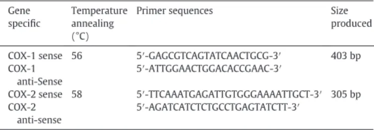
COX-1 and COX-2 gene expression was evaluated by Reverse tran-scription polymerase chain reaction (RT-PCR). For this assay, 1 × 10⁶/ 300μL PBMCs were incubated with RPMI (negative control), PMA (500 ng/mL; positive control), LPS (1 μg/mL; positive control), or BjcuL (5 and 10μg/mL; experimental group) for 6 h at 37 °C, in a humid-ified atmosphere of 5% CO2. Total RNA was extracted by TRIzol® LS Re-agent (Ambion, Life Technologies, USA). Synthesis of cDNA was performed using a SuperScriptTM III kit (InvitrogenTM, Life Technolo-gies, USA); a total of 500 ng of RNA was used in all experimental condi-tions. For the amplification of COX-1 and COX-2 specific genes via PCR, 500 ng of cDNA was added to a reaction mixture containing 1 × PCR buffer, 200μM dNTP, 0.2μM of gene specific primers (Invitrogen, Carls-bad, CA) (Table 1), 1.25μL of Taqpolymerase(HotMaster™, 5Prime) and water, for afinal volume of 25μL. The amplification product of GAPDH Table 1
Primer sequences for gene specific amplification of COX-1 and COX-2.
Gene specific
Temperature annealing (°C)
Primer sequences Size
produced
COX-1 sense 56 5′-GAGCGTCAGTATCAACTGCG-3′ 403 bp COX-1
anti-Sense
5′-ATTGGAACTGGACACCGAAC-3′
COX-2 sense 58 5′-TTCAAATGAGATTGTGGGAAAATTGCT-3′ 305 bp COX-2
anti-sense
5′-AGATCATCTCTGCCTGAGTATCTT-3′
Fig. 1.Isolation of BjcuL. (A) Samples of 100 mg were applied on Sepharose 6B-CL-lactose (1 cm × 3 cm) equilibrated with CaTBS. The fraction bound to resin was eluted using 0.1 M lactose solubilized in the same buffer, under aflow rate of 1.0 mL/min. (B) Sample purity degree was evaluated by RP-HPLC using a C-18 column (0,45 cm × 25 cm - Supelco), equilibrated with TFA 0,1% and eluted under a gradient of 0–70% acetonitrile in 0.1% trifluoroacetic acid (v/v). Protein elution was monitored at 280 nm. (C) Isolated protein (BjcuL) under non-reducing (lane 01) and reducing conditions (lane 02), andB. jararacussucrude venom were analyzed by SDS-PAGE, and the apparent molecular mass was determined by comparison with the migration of molecular mass marker (lane 04). (D) Mass spectrum showing a main peak of 32,830m/zcorresponding to the isolated protein (BjcuL) and a second peak of 16,332 m/z2 (double charge). The spectrum represents the average of the laser pulses.
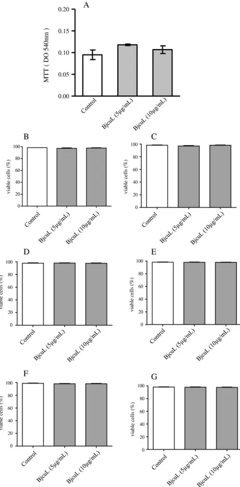
Fig. 2.Effect of BjcuL on PBMCs viability. Human PBMCs were isolated from buffy coats of healthy adult blood donors through a density gradient method and were analyzed in FACScan. (A) 2 × 10⁵cells were incubated with RPMI (control) or with different concentrations of BjcuL (5 and 10μg/mL; experimental group) for 96 h at 37 °C, in a humidified atmosphere of 5% CO
(glyceraldehyde 3-phosphate dehydrogenase) was used as cDNA con-trol. For fragment amplification, the reaction mixtures were submitted to initial denaturation at 94 °C for 5 min; followed by 30 cycles of 30 s at 94 °C for denaturation; 30 cycles at 58 °C (COX-2), and 30 cycles at 56 °C (COX-1) for annealing; 40–60 s at 72 °C for extension, depending on the amplicon size produced; and an additional extension at 72 °C for 10 min. The RT-PCR products were analyzed by 1.5% agarose gel electro-phoresis, stained with ethidium bromide and photo documented.
2.20. Statistical analysis
The means and standard error of the mean (S.E.M.) of all data were obtained. Data were tested for homogeneity of variances using Bartlett's test followed by Brow-Forsythe test and compared by using one-way
ANOVA, followed by Tukey's test in Graph-Pad PRISM 5.0 with signifi -cance probability levelsb0.05.
3. Results
3.1. Isolation, characterization and purification of BjucL
Fractionation ofB. jararacussucrude venom on Sepharose 6B CL lac-tose gave two peaks (Fig. 1A); elution with 0.1 M Lactose preceded the second peak and the second peak showed haemagglutination activity, suggesting the presence of C-type lectin. The purity degree was deter-mined by RP-HPLC using a C18 column (Fig. 1B).
The apparent molecular mass determined by SDS-PAGE under non-reducing conditions showed a single spot of approximately 25 kDa. Under reducing conditions, a single band of about 15 kDa was observed (Fig. 1C). On the other hand, the mass spectrometry procedure showed the molecular mass of isolated protein (BjcuL) to be 32,830 Da, with a second peak of about 16,332 Da corresponding to the double charge of the same protein.
3.2. Haemagglutination activity
The haemagglutinin activity was used to evaluate the presence of C-type lectins and was carried out using human erythrocytes. The results showed that this protein exhibits high haemagglutinin activity. More-over, the inhibition of this activity by EDTA confirms that BjcuL is a C-type lectin (data not shown).
3.3. Effect of BjcuL on human PBMCs viability
To further investigate BjcuL's effect on PBMCs function, we isolated these cells by density gradient, according to Lacouth-Silva et al. (2015). The purity of the isolated PBMCs was determined byflow cy-tometry using PE anti-human CD14 (Supplementary Fig. 1A and B) and PerCP anti-human CD3 (Supplementary Fig. 1C and D). To test the Fig. 3.Effect of BjcuL on PBMCs proliferation. PBMCs were isolated from buffy coats of
healthy adult blood donors through a density gradient method. 2 × 10⁵cells were incubated with RPMI (negative control), ConA (5μg/mL, positive control), or BjcuL (5 and 10μg/mL) for 96 h at 37 °C, in humidified atmosphere of 5% CO2. PBMCs proliferation was then assessed by MTT [3-(4.5-dimethylthiazol-2-yl)-2,5-diphenyltetrazolium bromide] metabolization method. Values from 4 to 5 donors represent the D.O. and the mean ± S.E.M. *Pb0.05 compared with control (ANOVA).
Fig. 5.Effect of BjcuL on hydrogen peroxide production. PBMCs (2 × 10⁵) were incubated with RPMI (negative control), PMA (500 ng/mL, positive control), or BjcuL (5 and 10μg/ mL) in phenol red solution for 3 h at 37 °C, in a humidified atmosphere of 5% CO2. The absorbance was measured by spectrophotometer at 620 nm. The results were expressed asμM of H2O2produced, and represent the mean ± SEM of four donors. *Pb0.05
compared to control (ANOVA).
Fig. 4.Effect of BjcuL on superoxide anion production by PBMCs. PBMCs (2 × 10⁵) were incubated with RPMI (negative control), PMA (500 ng/mL, positive control), and BjcuL (5 and 10μg/mL) for 2 h; mixtures contained 0.1% NBT to measure the formazan crystal formation resulting from NBT reduction by superoxide. The crystals were solubilized and the absorbance of the supernatant was determined at 620 nm in a spectrophotometer. Values from 4 to 5 donors represent the D.O. and the mean ± S.E.M. *Pb0.05 compared with control (ANOVA).
Fig. 6.Effect of BjcuL on ROS production. PBMCs (1 × 10⁶) were incubated with RPMI (negative control; A➔D), PMA (500 ng/mL, positive control, E➔G) or BjcuL (5 and 10μg/mL; H➔J and K➔M) in RPMI without phenol red for 3 h at 37 °C, in a humidified atmosphere of 5% CO2. Following the addition of DCFDA (10μM), DCFfluorescence was determined in FL1 in FACScan.
toxicity of BjcuL in isolated human PBMCs, we used the MTT assay, and investigated the effect of 96 h of BjcuL incubation. As shown inFig. 2A, the incubation of PBMCs with RPMI (negative control) or BjcuL (5 and 10μg/mL) did not affect cell viability at the time intervals and concen-trations tested. Cell viability was also evaluated by staining at different time intervals with propidium iodide (PI) and thiazole orange (TO), and analysis by FACScan.Fig. 2B, C, D, E, F and G show that incubation of PBMCs with RPMI (negative control) or BjcuL (5 and 10μg/mL) at dif-ferent time intervals did not affect cell viability (Supplementary Fig. 2).
3.4. Effect of BjcuL on PBMCs proliferation
In order to verify the effect of BjcuL on PBMCs functions, the prolifer-ation of mononuclear cells was investigated. InFig. 3, ConA (positive control) induced a significant proliferation of PBMCs when compared to RPMI (negative control). On the other hand, BjcuL inhibited PBMCs proliferation when used in concentrations of 5 and 10μg/mL. These data were confirmed using CFSE. This method relies on the ability of CFSE to covalently label long-lived intracellular molecules with the highlyfluorescent dye, carboxyfluorescein. Analysis of heterogeneous cell populations identified the presence and proportion of various pop-ulations of interest: Supplementary Fig. 3A and B show monocytes incu-bated with CFSE (negative control), and Supplementary Fig. 3C and D shows lymphocytes incubated with CFSE (positive control). Supple-mentary Fig. 4A, B, C, D, E and F show the different peaks that represent lymphocyte proliferation incubated with ConA (positive control). The data also shows that the proliferating cells in areas R2, R3 and R4 were CD8+. Supplementary Fig. 5A, B, C and D, and Supplementary Fig. 6A, B, C and D both show one peak, indicating that BjcuL (5 and 10μg/mL) did not stimulate lymphocyte to proliferate. Moreover, it can be seen that the main populations in area R3 were mainly CD4+ and CD8+.
3.5. Effect of BjcuL on ROS production by PBMCs
In order to investigate the interference of BjcuL in the respiratory burst and induction of superoxide anion and hydrogen peroxide by PBMCs, the cells were incubated with RPMI (negative control), PMA (positive control), or BjcuL (5 and 10μg/mL) in the presence of NBT 0.1% for superoxide anion production, horseradish peroxidase for hy-drogen peroxide production, and DCF-DA for ROS production.
As shown inFig. 4, BjcuL induced an increase in superoxide produc-tion as compared to the negative control (RPMI). Our results reveal that BjcuL activates human PBMCs to produce superoxide anion.Fig. 5shows that incubation of PBMCs with PMA induced a significant increase in hy-drogen peroxide production as compared to the control cells (incubated with phenol red solution). BjcuL, at concentrations of 5 and 10μg/mL, resulted in a significant increase in hydrogen peroxide production as compared to the control cells. Thesefindings demonstrate the ability of BjcuL to stimulate human PBMCs to produce hydrogen peroxide.
In order to identify the cell populations that BjcuL stimulates to pro-duce ROS, the cells were subjected to immunophenotyping using CD14-PE and CD3-PerCP, incubated with DFC, and analyzed by FASCan. As shown inFig. 6. (A➔D), in the negative control, 0.23% of CD3 and 0.22% of CD14 cells produced ROS. However, panel F and G show that, in the positive control, 56.05% of CD3 and 11.27% of CD14 cells produced ROS. When PBMCs were incubated with BjcuL at concentrations of 5 and 10μg/mL production of ROS increased by 59.9% and 64.5% respectively for CD3 cells, and by 15.57% and 12.4% respectively for CD14 cells. These results indicate that lymphocytes are the predominant cell type that produce ROS induced by BjcuL.
3.6. Effect of BjcuL on nitric oxide production by PBMCs
In order to investigate the abiltiy of BjcuL to induce the production of nitric oxide by human PBMCs, the cells were incubated with RPMI
(negative control), LPS (positive control), or BjcuL (5 and 10μg/mL). As shown inFig. 7A, incubation of PBMCs with LPS induced a significant increase in nitric oxide production as compared to the control cells. On the other hand, BjcuL at concentrations of 5 and 10μg/mL did not induce nitric oxide production in compariosn to the control cells, which sug-gests that this lectin does not stimulate PBMCs to produce nitric oxide. To investigate the effect of BjcuL on the protein expression profiles of iNOS in human PBMCs, the cells were incubated with RPMI (negative control), LPS (positive control), or BjcuL (5 and 10μg/mL).Fig. 7C shows that BjcuL induced slight iNOS expression, but this did not differ significantly from the control cells.
To verify the ability of BjcuL to induce the release of PGE2,and to in-duce COX-1 and COX-2 protein and gene expression in human PBMCs, the cells were incubated with RPMI (negative control), LPS (positive control), or BjcuL (5 and 10μg/mL). As shown inFig. 7B, incubation of PBMCs with LPS induced a significant liberation of PGE2as compared to the control cells. However, at the concentrations tested (5 and 10μg/mL), BjcuL did not induce PGE2liberation as compared to the con-trol cells; this suggests that BjcuL does not stimulate PBMCs to induce PGE2liberation. BjcuL did not induce an increase in either COX-1 or COX-2 protein expression as compared to the control (Fig. 7C). The same results were obtained for COX-1 and COX-2 mRNA gene expres-sion (Fig. 7D) as compared to the control. Thesefindings suggest that BjcuL does not stimulate COX protein and gene expression in PBMCs.
4. Discussion
The literature shows that BjcuL triggers inflammation with cellular infiltration in post capillary venules (Elifio-Esposito et al., 2007), in-creases edema and vascular permeability in murine models (Panunto et al., 2006), contributes to in vitro neutrophil activation (Elifi o-Esposito et al., 2011) and modulates macrophage functional activation towards an M1 state (Dias-Netipanvi et al., 2016).
First of all, BjcuL was isolated fromBothrops jararacussuvenom, as described byGartner and Ogilvie (1984),Guimarães-Gomes et al. (2004)andPereira-Bittencourt et al. (1999), using an affinity chroma-tography procedure with an immobilized carbohydrate matrix, which made it possible to obtain the lectin with a high degree of purity. In agreement with our results, several C-type lectins have been isolated from snake venoms of the genus Bothrops, includingB. atrox(Gartner et al., 1980), B. godmani (Lomonte et al., 1990), B. insularis (Guimarães-Gomes et al., 2004),B. jararacussu(de Carvalho et al., 1998),B. pirajai (Havt et al., 2005), andB. leucurus(Nunes et al., 2011), among others.
SDS-PAGE showed that, under reducing conditions, BjcuL migrated as a single protein with a molecular apparent mass corresponding to 14 kDa. In non-reducing conditions, a single band was observed (Fig. 3), suggesting that BjcuL forms dimmers. Moreover, the molecular mass determined by mass spectrometry showed a main peak of about 32,830 Da. These results are consistent with those obtained in previous studies dealing with BjcuL isolation (Elifio-Esposito et al., 2007), and in studies of other lectins (de Carvalho et al., 2002; Guimarães-Gomes et al., 2004; Havt et al., 2005).
Despite the importance of lectins, we still have a limited understand-ing of the mechanisms of action by which BjcuL effects the function of PBMCs. Since monocytes and lymphocytes play an important role in the immune response, this study aimed to evaluate the effects of Bjcul on isolated human PBMCs. Bjcul was isolated fromBothrops jararacussu venom and its effects on cell viability were examined. Our data show that BjcuL does not affect the viability of PBMCs, which indicates that BjcuL exhibits very low toxicity on this cell type. These results are in agreement withDias-Netipanyj et al. (2016).
gastric carcinoma cells MKN45 and AGS, due to its direct interaction with specific glycans on the cell surface. These authors also observed cell viability decrease caused by the disorganization of actinfilaments and apoptosis.
This study also investigated the effect of BjcuL on PBMCs prolifera-tion. Lectins are widely known for their mitogenic actions, which may be inhibited by low concentrations of saccharide (Sharon, 2008); how-ever, numerous lectins with antimitogenic actions have been described (Sharon, 2008). The induction of proliferation seems to be associated
with the binding of mitogenic lectins to the T cell receptor complex, which emits signals that lead to IL-2 synthesis. On the other hand, non-mitogenic lectins do not bind to the T cell receptor complex, nor do they bind to other accessory molecules that are essential for the ac-tivation of signal transmission. Binding to accessory molecules that do not induce the production and secretion of IL-2 may be the cause of antimitogenic action (Kilpatrick, 1999, 2002). Our results indicate that BjcuL inhibits PBMCs proliferation. In addition, the analysis of heteroge-neous cell populations in order to identify the presence and proportions Fig. 7.Effect of BjcuL on nitric oxide and PGE2production. PBMCs (2 × 10⁵) were incubated with RPMI (negative control), LPS (1μg/mL, positive control) or BjcuL (5 and 10μg/mL) at 37 °C, in a humidified atmosphere of 5% CO2, according toMaterial and methods. (A) NO liberation in supernatant was quantified by spectrophotometric measurement at 620 nm of NO2−by the Griess reaction. The results were expressed asμM of Nitrite produced and represent the mean ± SEM of four donors. *Pb0.05 compared to control (ANOVA). (B) PGE2concentrations in supernatant were quantitated by specific EIA. The results were expressed asμg/mL of PGE2produced and represent the mean ± SEM of 4–5 donors. *Pb0.05 compared to control
(ANOVA). (C) COX-1, COX-2 and iNOS protein expression was analyzed by immunoblot with specific antibodies targeting COX-1, COX-2, or iNOS. (D) mRNA COX-1 and COX-2 mRNA expression were analyzed by RT-PCR.
of various populations of interest showed that the vast majority of PBMCs were lymphocytes CD4+and CD8+.
The same results were observed byPereira-Bittencourt et al. (1999)
studying human cancer cell lines. In their study, BjcuL inhibited prolifer-ation in eight human cancer cell lines in a dose-dependent manner. Moreover, the lectin was more potent in inhibiting the growth of renal (Caki-1 and A-498) and pancreatic (CFPAC-1) cancer cell lines with IC50 of 1–2 mM. On the other hand, BjcuL showed no effect on colon (Caco-2) and breast (MCF7) cancer cell lines.
Whitacre and Cathcart (1992)showed that one way PBMCs prolifer-ation is regulated is through oxygen free radical production, which varies depending on the type of mitogenic stimulus. Based on this, we conducted experiments to verify the effect of BjcuL on superoxide anion production by PBMCs. Our results show, for thefirst time, that BjcuL is able to stimulate PBMCs production of superoxide anion. This may explain the inhibition of PBMCs proliferation that is induced by lectin.
This study also investigated BjcuL induced hydrogen peroxide pro-duction in PBMCs. Superoxide anion inside the cells is converted to hy-drogen peroxide, mainly by spontaneous dismutation. Hyhy-drogen peroxide can form the highly reactive radical, hydroxyl (Babior, 1984; Fujiwara and Kobayashi, 2005; Rutkowski et al., 2007; Manea, 2010). Our data shows that the BjcuL induced PBMCs to produce hydrogen per-oxide, which is in agreement withDias-Netipanvi et al. (2016). These authors showed that BjcuL stimulated the differentiated monocyte-de-rived macrophage U937 cells to produce hydrogen peroxide. In addi-tion, our results demonstrate that BjcuL is capable of activating leukocytes that trigger oxidative metabolism by converting molecular oxygen into reactive oxygen intermediates, a process called respiratory burst.
In addition to the reactive oxygen species, superoxide anion and hy-drogen peroxide, reactive nitrogen species were also investigated. Nitric oxide (NO) is a radical generated at relatively high and sustained levels by the inducible forms of NO synthase (iNOS or NOS-2).
The literature shows that NO is antiproliferative for T lymphocytes and that this effect is exerted particularly by the action of NO donors and by the action on regulation of cytokine expression, which is depen-dent on exposure time (Macphail et al., 2003). Based on this knowledge, the nitrite was measured in the supernatant of PBMCs in order to assess the effect of BjcuL on the production of NO by PBMCs. Our results show that BjcuL is unable to stimulate PBMC to release NO, nor is it able to in-duce iNOS protein expression.
The literature documents a complex interplay between NOS and COX pathways. This complexity is apparent in the products of NOS and COX systems that are able to mediate the regulation and expression of inducible forms (NOS-2 and COX-2). Mediation occurs via the inter-play of intracellular crosstalk between signaling pathways stimulated by products of NOS and COX (which contribute significantly to the com-plexity of cellular signaling), and via the reciprocal modulation of cyclo-oxygenase activity by nitric oxide and of NOS activity by prostaglandins (Sorokin, 2016).
To this end, we investigated the effect of BjcuL on the production of PGE2by PBMCs, and our results show that the lectin does not stimulate the cells to produce PGE2,nor does it stimulate COX-1 and COX-2 mRNA and protein expression. This data, combined with the NO data, helps ex-plain, at least in part, the effect of BjcuL on PBMCs. Moreover, another study has indicated that nitric oxide can modulate prostaglandin syn-thesis, which includes the direct inactivation of prostaglandin I synthase by peroxynitrite (Sorokin, 2016), a product of superoxide reaction with nitric oxide (Robbins and Grisham, 1997; Rutkowski et al., 2007; Plekhova and Somova, 2012). The data presented herein make it possi-ble to assert that BjcuL inhibits PBMCs proliferation via a mechanism that involves oxygen but does not involve nitrogen reactive intermedi-ates or PGE2induced by the lectin.
The results obtained in this study highlight the effect that BjcuL has on the immunomodulatory activity of PBMCs, as observed by
Dias-Netipanvi et al. (2016). Further studies are needed to confirm the im-munomodulatory effect of BjcuL, to elucidate the molecular mecha-nisms of action responsible for its effects and to determine its potential application as an immunopharmacological and biotechnolog-ical tool.
Supplementary data to this article can be found online athttp://dx. doi.org/10.1016/j.tiv.2017.02.003.
Authorship
J.P.Z., W.L.P., O.B·C and A.M.K designed the study; W.L.P., O.B·C, S·S.S., A.S.P., S.S.P., M.V.P. and N.M.N. performed the experiments; A.M.K., R.G.S and A.M.S. performed and supervised the biochemical pro-cedures; J.P.Z., W.L.P., O.B.C. and A.M.K. collected and analyzed the data; J.P.Z, C.F.C.F. and R.G.S. provided reagents; J.P.Z., W.L.P. and A.M.K. wrote the manuscript. All of the authors discussed the results and implications, and commented on the manuscript at all stages.
Conflict of interest
There is no conflict of interest statement.
Transparency document
TheTransparency documentassociated with this article can be found, in online version.
Acknowledgments
The authors express their gratitude to Conselho Nacional de Desenvolvimento Científico e Tecnológico (CNPq), Coordenação de Aperfeiçoamento de Pessoal de Nível Superior (CAPES), Instituto Nacional de Ciência e Tecnologia em Toxinas (INCT-Tox) and Fundação de Amparo à Pesquisa do Estado de Rondônia (FAPERO) for thefinancial support. The authors thank the Program for Technological Development in Tools for Health-PDTIS-FIOCRUZ for the use of its facilities. This study was supported by grants (473999/2008-0, 482562/2010-2 and 479316-2013-6) from Conselho Nacional de Desenvolvimento Científico e Tecnológico (CNPq). Juliana Pavan Zuliani was a recipient of productiv-ity grant 303974/2008-7, 301809/2011-9 and 306672/2014-6 from Conselho Nacional de Desenvolvimento Científico e Tecnológico (CNPq), and Onassis B. Castro and Weverson L. Pires were the benefi cia-ry of CAPES Masters and Doctoral felowship, respectively.
References
Babior, B.M., 1984.The respiratory burst of phagocytes. J. Clin. Invest. 73, 599–601.
Damasio, D.C., Nolte, S., Polak, L.P., Brandt, A.P., Bonan, N.B., Zischler, L., Stuelp-Campelo, P.M., Cadena, S.M.S.C., de Noronha, L., Elífio-Esposito, S.L., Moreno-Amaral, A.N., 2014.The lectin BjcuL induces apoptosis through TRAIL expression, caspase cascade activation and mitochondrial membrane permeability in a human colon adenocarci-noma cell line. Toxicon 90, 299–307.
de Carvalho, D.D., Marangoni, S., Novello, J.C., 1998.Isolation and characterization of a new lectin from the venom of the snakeBothrops jararacussu. Biochem. Mol. Biol. Int. 44, 933–938.
de Carvalho, D.D., Marangoni, S., Novello, J.C., 2002.Primary structure characterization of
Bothrops jararacussusnake venom lectin. Toxicon 21, 43–50.
de Carvalho, D.D., Schmitmeier, S., Novello, J.C., Markland, F.S., 2001.Effect of BJcuL (a lec-tin from the venom of the snakeBothrops jararacussu) on adhesion and growth of tumor and endothelial cells. Toxicon 31, 1471–1476.
Dias-Netipanyj, M.F., Boldrini-Leite, L.M., Trindade, E.S., Moreno-Amaral, A.N., Elifi o-Esposito, S., 2016.Bjcul, a snake venom lectin, modulates monocyte-derived macro-phages to a pro-inflammatory profile in vitro. Toxicol. In Vitro 33, 118–124.
Ding, A.H., Nathan, C.F., Stuehr, D.J., 1988.Release of reactive nitrogen intermediates and reactive oxygen intermediates from mouse peritoneal macrophages: comparison of activating cytokines and evidence for independent production. J. Immunol. 141, 2407–2412 [PubMed].
Elifio-Esposito, S.L., Tomazeli, L., Schwatz, C., Gimenez, A.P., Fugii, G.M., Fernandes, L.C., Zishler, L.F.M., Stuelp-Campelo, P., Moreno, A.N., 2011.Human neutrophil migration and activation by BJcuL, a galactose binding lectin purified fromBothrops jararacussu
venom. BMC Immunol. 12, 1–7.
Fujiwara, N., Kobayashi, K., 2005.Macrophages in inflammation. Curr. Drug Targets Inflamm. Allergy 4, 281–286.
Gartner, T.K., Ogilvie, M.L., 1984.Isolation and characterization of three Ca2+C-dependent
b-galactoside-specific lectins from snake venoms. Biochem. J. 224, 301–307.
Gartner, T.K., Stocker, K., Williams, D.C., 1980.Thrombolectin: a lectin isolated from
Bothrops atroxvenom. FEBS Lett. 117, 13–16.
Guimarães-Gomes, V., Oliveira-Carvalho, A.L., Junqueira-de-Azevedo, I.L.M., Dutra, D.L.S., Pujol-Luz, M., Castro, H.C., Ho, P.L., Zingali, R.B., 2004.Cloning, characterization and structural analysis of a C-type lectin fromBothrops insularis(BiL) venom. Arch. Biochem. Biophys. 432, 1–11.
Havt, A., Toyama, M.H., do Nascimento, N.R., Toyama, D.O., Nobre, A.C., Martins, A.M., Barbosa, P.S., Novello, J.C., Boschero, A.C., Carneiro, E.M., Fonteles, M.C., Monteiro, H.S., 2005.A new C-type animal lectin isolated fromBothrops pirajaiis responsible for the snake venom major effects in the isolated kidney. Int. J. Biochem. Cell Biol. 37, 130–141.
Kilpatrick, D.C., 1999.Mechanisms and assessment of lectin-mediated mitogenesis. Mol. Biotechnol. 11, 55–65.
Kilpatrick, D.C., 2002.Animal lectins: a historical introduction and overview. Biochim. Biophys. Acta 1572, 187–197.
Kishore, U., Eggleton, P., Reid, K.B., 1997.Modular organization of carbohydrate recogni-tion domains in animal lectins. Matrix Biol. 15, 583–592.
Lacouth-Silva, F., Xavier, C.V., Setúbal, S.S., Pontes, A.S., Nery, N.M., Castro, O.B., Fernandes, C.F.C., Honda, E.R., Zanchi, F.B., Calderon, L.A., Stábeli, R.G., Soares, A.M., Jardim, I.S., Facundo, V., Zuliani, J.P., 2015.The effect of 3β, 6β, 16β-trihydroxylup-20(29)-ene lupane compound isolated fromCombretum leprosumMart. on peripheral blood mononuclear cells. BMC Complement. Altern. Med. 420, 1–10.
Laemmli, U.K., 1970.Cleavage of structural proteins during the assembly of the head of bacteriophage T4. Nature 227, 680–685.
Lomonte, B., Rojas, G., Gutiérrez, J.M., Ramírez, G., 1990.Isolation of a galactose-binding lectin from the venom of the snakeBothrops godmani(Godmann's pit viper). Toxicon 28, 75–81.
Macphail, S.E., Gibney, C.A., Brooks, B.M., Booth, C.G., Flanagan, B.F., Coleman, J.W., 2003.
Nitric oxide regulation of human peripheral blood mononuclear cells: critical time dependence and selectivity for cytokine versus chemokine expression. J. Immunol. 171, 4809–4815.
Manea, A., 2010.NADPH oxidase-derived reactive oxygen species: involvement in vascu-lar physiology and pathology. Cell Tissue Res. 342, 325–339.
Milani Júnior, R., Jorge, M.T., de Campos, F.P., Martins, F.P., Bousso, A., Cardoso, J.L., Ribeiro, L.A., Fan, H.W., França, F.O., Sano-Martins, I.S., Cardoso, D., Ide Fernandez, C., Fernandes, J.C., Aldred, V.L., Sandoval, M.P., Puorto, G., Theakston, R.D., Warrell, D.A., 1997.Snake bites by the jararacuçu (Bothrops jararacussu): clinicopathological stud-ies of 29 proven cases in São Paulo State, Brazil. QJM. 90, 323–343.
Nakajima, Y., Tsuruma, K., Shimazawa, M., Mishima, S., Hara, H., 2009.Comparison of bee products based on assays of antioxidant capacities. BMC Complement. Altern. Med. 9, 4.
Nolte, S., Damasio, C.D., Baréa, A.C., Gomes, J., Magalhães, A., Mello-Zischler, L.F., Stuel-Campelo, P.M., Elífio-Esposito, S.L., Roque-Barreira, M.C., Reis, C.A., Moreno-Amaral, A.N., 2012.BJcuL, a lectin purified fromBothrops jararacussuvenom, induces apopto-sis in human gastric carcinoma cells accompanied by inhibition of cell adhesion and actin cytoskeleton disassembly. Toxicon 59, 81–85.
Nunes, E.S., de Souza, M.A., Vaz, A.F., Santana, G.M., Gomes, F.S., Coelho, L.C., Paiva, P.M., da Silva, R.M., Silva-Lucca, R.A., Oliva, M.L., Guarnieri, M.C., Correia, M.T., 2011.Purifi ca-tion of a lectin with antibacterial activity fromBothrops leucurussnake venom. Comp. Biochem. Physiol. B Biochem. Mol. Biol. 159, 57–63.
Panunto, P.C., Silva, M.A., Linardi, A., Buzin, M.P., Melo, S.E.S.F.C., Mello, S.M., Prado-Franceschi, J., Hyslop, S., 2006.Biological activities of a lectin fromBothrops jararacussusnake venom. Toxicon 47, 21–31.
Pereira-Bittencourt, M., de Carvalho, D.D., Gagliardi, A.R., Collins, D.C., 1999.The effect of a lectin from the venom of the snake,Bothrops jararacussu, on tumor cell proliferation. Anticancer Res. 19, 4023–4025.
Pick, E., Keisari, Y., 1980.A simple colorimetric method for measurement of hydrogen per-oxide by cells in culture. J. Immunol. Methods 38, 161–170.
Pick, E., Mizel, D., 1981.Rapid microassay for the measurement of peroxide and hydrogen peroxide production by macrophages in culture using an automatic enzyme immu-noassay reader. J. Immunol. Methods 46, 211–226.
Plekhova, N.G., Somova, L.M., 2012.The physiological role of nitric oxide in infectious pro-cess. Usp. Fiziol. Nauk 43, 62–81.
Pontes, A.S., Setúbal, S.S., Nery, N.M., da Silva, F.S., da Silva, S.D., Fernandes, C.F., Stábeli, R.G., Soares, A.M., Zuliani, J.P., 2016.p38 MAPK is involved in human neutrophil che-motaxis induced byL-amino acid oxidase fromCalloselasma rhodosthoma. Toxicon 119, 106–116.
Pontes, A.S., Setúbal, S.S., Xavier, C.V., Lacouth-Silva, F., Kayanoa, A.M., Pires, W.L., Nery, N.M., Castro, O.B., Silva, S.D., Calderon, L.A., Stábeli, R.G., Soares, A.M., Zuliani, J.P., 2014.Effect of L-amino acid oxidase fromCalloselasma rhodosthomasnake venom on human neutrophils. Toxicon 80, 27–37.
Reilly, T.P., Bellevue, F.H., Woster, P.M., 1998.Compararion of the in vitro citotoxicity os hydroxylamine metabolites of sulfamethoxazole and dapsone. Biochem. Pharmacol. 55, 803–810.
Robbins, R.A., Grisham, M.B., 1997.Nitric oxide. Int. J. Biochem. Cell Biol. 29, 857–860.
Russo, M., Teixeira, H.C., Marcondes, M.C.G., Barbutto, J.A.M., 1989. Superoxide-indepen-dent hydrogen release by active macrophages. Braz. J. Med. Biol. Res. 22, 1271–1273.
Rutkowski, R., Pancewicz, S.A., Rutkowski, K., Rutkowska, J., 2007.Reactive oxygen and ni-trogen species in inflammatory process. Pol. Merkur. Lekarski 23, 131–136.
Sharon, N., 2008 Dec.Lectins: past, present and future. Biochem. Soc. Trans. 36, 1457–1460 (Pt 6).
Sorokin, A., 2016.Nitric oxide synthase and cyclooxygenase pathways: a complex inter-play in cellular signaling. Curr. Med. Chem. 23, 2559–2578.
Whitacre, C.M., Cathcart, M.K., 1992.Oxygen free radical generation and regulation of proliferative activity of human mononuclear cells responding to different mitogens. Cell. Immunol. 144, 287–295.