Graça Susete Costa de Carvalho Marques
Licenciada em Biologia Celular e Molecular
Establishing a Cell Biology Platform:
Isolation and Preservation of Human
Blood Products
Dissertação para obtenção do Grau de Mestre em
Genética Molecular e Biomedicina
Orientadora: Doutora Zélia Maria Cordeiro da Silva,
Investigadora, Faculdade de Ciências Médicas da
Universidade Nova de Lisboa
Co-orientadora: Doutora Paula Alexandra Quintela Videira,
Professora Auxiliar Convidada, Faculdade de
Ciências Médicas da Universidade Nova de
Lisboa
Júri:
Presidente: Prof. Doutora Ilda Maria Barros dos Santos Gomes Sanches Arguente: Doutora Ana Catarina Maurício Brito Ataíde Montes
Vogal: Doutora Zélia Maria Cordeiro da Silva
Graça Susete Costa de Carvalho Marques
Licenciada em Biologia Celular e Molecular
Establishing a Cell Biology Platform:
Isolation and Preservation of Human Blood
Products
Dissertação para obtenção do Grau de Mestre em
Genética Molecular e Biomedicina
Orientadora: Doutora Zélia Maria Cordeiro da Silva, Investigadora,
Faculdade de Ciências Médicas da Universidade
Nova de Lisboa
Co-orientadora: Doutora Paula Alexandra Quintela Videira,
Professora Auxiliar Convidada, Faculdade de
Ciências Médicas da Universidade Nova de Lisboa
Júri:
Presidente: Prof. Doutora Ilda Maria Barros dos Santos Gomes Sanches Arguente: Doutora Ana Catarina Maurício Brito Ataíde Montes
Vogal: Doutora Zélia Maria Cordeiro da Silva
Establishing a Cell Biology Platform: Isolation and Preservation of Human Blood Products
Copyright Graça Susete Costa de Carvalho Marques, FCT/UNL, UNL
A Faculdade de Ciências e Tecnologia e a Universidade Nova de Lisboa têm o direito, perpétuo e sem limites geográficos, de arquivar e publicar esta dissertação através de exemplares impressos reproduzidos em papel ou de forma digital, ou por qualquer outro meio conhecido ou que venha a ser inventado, e de a divulgar através de repositórios científicos e de admitir a sua cópia e distribuição com objectivos educacionais ou de investigação, não comerciais, desde que seja dado crédito ao autor e editor.
V
ACKNOWLEDGEMENTS
Gostaria de agradecer a todas as pessoas que me ajudaram e contribuíram para que pudesse realizar este trabalho.
• Ao Departamento de Imunologia da Faculdade de Ciências Médicas da Universidade Nova de Lisboa, por me acolher no seu laboratório.
• À Doutora Zélia Silva, a minha orientadora, por me guiar através de todo este processo e pelos seus conselhos inestimáveis. À Doutora Paula Videira, a minha co-orientadora, pela sua disponibilidade e constante motivação.
• À Doutora Guadalupe Cabral, aos doutorandos Mylène Carrascal, Mariana Silva e Hélio Crespo, pela sua ajuda e paciência.
• A todos os restantes membros do Departamento de Imunologia. À Doutora Catarina Martins pela sua disponibilidade. À D. Filomena, D. Luísa e D. Amélia pela boa companhia. À D. Glória por partilhar o seu conhecimento. A todas pela sua simpatia.
• À minha família: à minha mãe Licínia, ao meu pai Armando, à minha irmã Licínia Isabel, que estiveram sempre presentes para me ajudar e encorajar sempre que precisei.
• Às minhas amigas, Juliana, Clara, Andreia, Marta e Sara, pela sua amizade e por me proporcionarem momentos extremamente divertidos. À Marília, à Inês e à Maria, por serem excelentes colegas de laboratório.
VII
ABSTRACT
The use of human primary cells provide researchers in different areas with irrefutable more biologically relevant data than using cell lines or animal blood cells. The work was performed in the scope of the Cell Biology Services @ CEDOC, aiming to provide viable and trustful human primary cells and products. We had three main objectives: protocol optimizations for blood cell isolation, culture and cryopreservation; cost estimation and divulgation of the services.
We have reviewed standard protocols and compared different strategies for blood cell isolation. The impact of those methodologies was evaluated regarding cell yield and purity, cell functional characteristics and cost. We also developed a method for serum isolation from human plasma in blood buffy coats. The resultant sera were sterile and suitable to be used in leukocyte cultures.
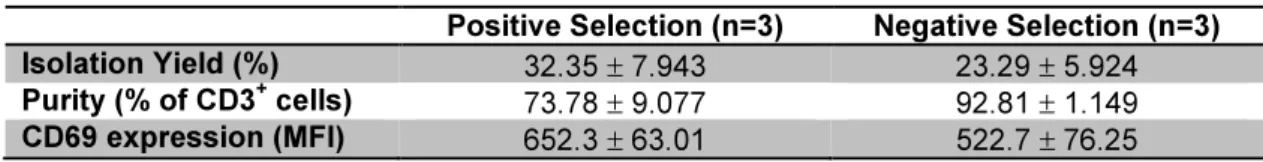
Different protocols for T cells isolation were compared: positive versus negative immunomagnetic selection and isolation using nylon wool fiber columns. Positive selection provided the highest isolation yield (32.35%), while negatively selected cells had the highest purity (92.81%). Although nylon wool fiber column was the fastest and cheapest method, unlike the immunomagnetic methods, it did not allow complete separation of T from B lymphocytes.
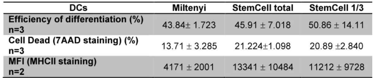
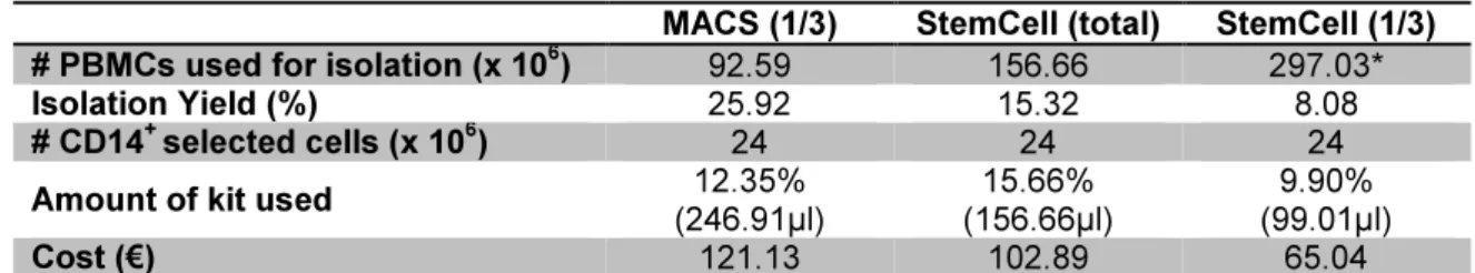
Positive selection of monocytes was compared using two widely used commercial kits. Miltenyi’s kit provided the highest isolation yield (25.92%), recovery rate (86.70%) and purity (95.01%). Monocytes isolated with StemCell kit presented a higher cell complexity, and when differentiated into dendritic cells (DCs), showed a more mature phenotype. Differences between both kits are probably caused by the nature of the magnetic beads, suggesting caution when choosing one or other kit, as it may have an impact on DCs’ function.
Overall, although dealing with apparently straight forward methodologies, our results show that testing commercial products and optimizing protocols is very important and contribute for a better quality of products and services.
Key words: sera, cell isolation, cryopreservation, cell culture, PBMCs (peripheral blood mononuclear
IX
RESUMO
O uso de células humanas primárias fornece a investigadores de diferentes áreas, dados irrefutavelmente mais relevantes do que usando linhas celulares ou células de sangue de animais. Este trabalho foi realizado no âmbito dos Serviços de Biologia Celular @ CEDOC, com o objectivo de fornecer células primárias e produtos humanos viáveis e de confiança. Tínhamos três objectivos principais: optimização de protocolos para o isolamento cultura e criopreservação de células do sangue; estimativas de custo e divulgação dos serviços.
Revimos protocolos padrão e comparámos diferentes estratégias para o isolamento de células do sangue. O impacto dessas metodologias foi avaliado tendo em conta o rendimento e pureza celular, características funcionais celulares e o custo. Também desenvolvemos um método para o isolamento de soro a partir de plasma em buffy coats de sangue. O soro resultante era estéril e adequado para uso em culturas leucocitárias.
Foram comparados diferentes protocolos para o isolamento de células T: selecção imunomagnética positiva versus negativa e isolamento usando colunas de fibra de lã de nylon. A selecção positiva forneceu o rendimento celular mais alto (32,35%), enquanto as células seleccionadas negativamente tinham a maior pureza (92,81%). Apesar de a fibra de lã de nylon ser o método mais rápido e barato, ao contrário dos métodos imunomagnéticos, não permitiu uma separação completa de linfócitos T e B.
A selecção positiva de monócitos foi comparada usando kits comerciais. O kit da Miltenyi forneceu o maior rendimento de isolamento (25,92%), taxa de recuperação (86,70%) e pureza (95,01%). Os monócitos isolados com o kit da StemCell apresentavam uma maior complexidade celular e quando diferenciados em células dendríticas, mostravam um fenótipo mais maturo. As diferenças entre os dois kits são provavelmente causadas pela natureza das esferas magnéticas, sugerindo cautela aquando da escolha entre um kit ou outro, pois este pode ter impacto na função das células dendríticas.
No geral, apesar de lidarmos com metodologias aparentemente lineares, os nossos resultados mostram que testar produtos comerciais e optimizar protocolos é muito importante e contribui para uma melhor qualidade de produtos e serviços.
Palavras-chave: soro, isolamento de células, criopreservação, cultura de células, PBMCs (células
XI
The work developed until the present date has originated:
• One poster:
Graça S. Marques; Inês Iria; Zélia Silva; Paula A. Videira. 2013. Cell Biology Services at CEDOC/FCM. XXXVIII Jornadas Portuguesas de Genética. Porto, Portugal.
• One oral presentation:
XIII
INDEX
I. INTRODUCTION ... 1
I.1. The immune system ... 1
I.2. The cells of the immune system ... 1
I.2.1. Lymphocytes ... 2
I.2.2. Monocytes, macrophages and dendritic cells ... 3
I.2.3. Granulocytes ... 4
I.3. Cell separation ... 5
I.3.1. Cell density ... 5
I.3.2. Cell size ... 6
I.3.3. Cell adherence ... 7
I.3.4. Antibody-based techniques ... 8
I.3.4.1. Magnetic-activated cell sorting ... 8
I.3.4.2. Fluorescence-activated cell sorting ...13
I.4. Cell culture ...14
I.4.1. Requirements of culture media ...14
I.4.2. Serum ...16
I.4.2.1. Heat inactivation ...17
I.5. Cell cryopreservation ...17
I.5.1. Principals of cryopreservation ...18
I.5.2. The cryoprotective agent ...18
I.5.3. Sample preparation and equilibration ...19
I.5.4. Cooling rate ...19
I.5.5. Storage ...20
I.5.6. Cell thawing ...20
I.6. Cell viability studies ...20
I.7. Context and objectives of the work developed for this thesis ...21
II. MATERIALS AND METHODS ...23
II.1. Human peripheral blood ...23
II.2. Isolation of plasma and defibrinated plasma ...23
II.3. Isolation of peripheral blood mononuclear cells ...24
II.3.1. Lyse solution ...25
II.4. Isolation of CD3+ T cells using MACS technology from Miltenyi ...25
II.4.1. Positive selection ...26
II.4.2. Negative selection ...26
II.5. Isolation of CD3+ T cells by nylon wool fiber column ...27
II.6. Isolation of CD14+ monocytes by positive selection ...27
XIV
II.6.2. Isolation of CD14+ monocytes using EasySep platform from StemCell ...28
II.7. Generation of dendritic cells ...29
II.8. Testing of serum samples ...29
II.8.1. Protein electrophoresis ...30
II.8.2. Performance when used in culture media ...30
II.9. Cell count using the Neubauer chamber ...31
II.10. Isolation yield ...32
II.11. Cell preservation ...32
II.11.1. Cell freezing and thawing ...32
II.11.2. DMSO Removal ...33
II.12. Flow cytometry ...34
II.12.1. The flow cytometer: basic principals ...34
II.12.2. Data analysis ...35
II.13. Isolation purity ...35
II.14. Cell recovery rate ...35
II.15. Economic viability evaluation of different types of cell isolations ...36
II.16. Statistical analysis ...36
II.17. Divulgation of the Cell Biology services ...36
III. RESULTS ...39
III.1. Optimization of a protocol to obtain human serum from buffy coat plasma ...39
III.1.1. Electrophoretic profile of serum samples ...39
III.1.2. Influence of using sera isolated in our laboratory in culture media ...41
III.1.2.1. Influence of different sera in the viability of the PBMCs and in the proportion of different cell types ...42
III.1.2.2. Response of PBMCs to a LPS stimulus ...44
III.2. Isolation Yield ...46
III.3. Influence of using a lyse solution when isolating PBMCs ...48
III.4. MACS CD3+ T cell separation: positive selection versus negative selection ...50
III.5. T Cell separation using nylon wool fiber columns ...51
III.6. CD14+ monocytes positive selection: Miltenyi vs. StemCell ...53
III.7. Cell preservation ...56
III.7.1. Shelf life of PBMCS at -80ºC ...56
III.7.2. DMSO immediate removal vs. after 24 hours ...57
III.7.3. Effect of centrifugation speed: 110 x g vs. 250 x g ...58
III.8. Economic viability evaluation ...59
III.8.1. Isolation of CD3+ T cells ...59
III.8.2. Isolation of CD14+ monocytes ...60
XV
IV. DISCUSSION ...65
IV.1. Protocol optimizations ...65
IV.1.1. Serum isolation from buffy coats of human peripheral blood and its performance in cell culture ...65
IV.1.2. Comparison between different protocols for the isolation of lymphocytes and monocytes from PBMCs ...67
IV.1.3. Cell preservation: shelf time and cryoprotectant removal ...70
IV.2. Cost estimations ...70
IV.3. Cell Biology services divulgation ...71
IV.4. Concluding remarks and future work ...71
V. REFERENCES ...73
XVII
INDEX OF FIGURES
Fig.I.1 Scheme portraying the different types of leucocytes (white blood cells) ... 1
Fig.I.2 Blood sample after incubation with the RosetteSepTM Cocktail from StemCell and prior to perform a density gradient centrifugation ... 6
Fig.I.3 Steps of MACS separation using technology from Miltenyi: magnetic labeling, magnetic separation and elution of the labeled cells . ... 9
Fig.I.4 Cell separation by positive selection using MACS technology ...10
Fig.I.5 Cell separation by depletion strategy using MACS technology ...10
Fig.I.6 Cell separation by untouched isolation using MACS technology ...11
Fig.I.7 Cell separation by depletion followed by positive selection using MACS technology ...12
Fig.I.8 Cell separation by two subsequent positive selections using MACS technology ...12
Fig.I.9 Magnetic labeling using StemCell products ...13
Fig.II.1 Buffy coat processing workflow applied in Cell Biology Services @ CEDOC ...23
Fig.III.1 Electrophoretic profile of plasma and serum samples ...40
Fig.III.2 Culture media supplemented with different types of sera ...41
Fig.III.3 Flow cytometry analysis of PBMCs kept in culture for a 7-day period ...42
Fig.III.4 Percentage of debris and dead cells in cultures of PBMCs after 7 days ...43
Fig.III.5 PBMCs in culture using media supplemented with different types of sera ...44
Fig.III.6 Analysis of the MFI values obtained with HLA-DR staining in samples of PBMCs kept in culture using media supplemented with different types of sera ...45
Fig.III.7 Variation of the average number of PBMCs obtained per isolation, according to the age group and the blood type ...46
Fig.III.8 Isolation yield for CD3+ selection using protocols for positive and negative selection ...47
Fig.III.9 Isolation yield for CD14+ monocytes selection using protocols from different brands (positive selection) ...48
Fig.III.10 Comparative analysis by flow cytometry of PBMCs isolated using lyse solution and without using lyse solution ...49
Fig.III.11 Percentage of lymphocytes and monocytes upon isolation of PBMCs, depending on the utilization, or not, of a lyse solution ...49
Fig.III.12 Flow cytometry analysis of the CD3+ population isolated using positive selection and negative selection ...51
Fig.III.13 Flow cytometry analysis of cells isolated using a nylon wool fiber column and stained with CD3-APC and CD14-FITC ...52
Fig.III.14 Relative proportion of different cell populations among PBMCs separated using a nylon wool fiber column ...52
Fig.III.15 Flow cytometry analysis of FSC and SSC profiles of the CD14+ singlet population isolated by positive selection ...54
XVIII
Fig.III.17 Evolution of cell viability and cell recovery rate throughout a 6 month period ...57
Fig.III.18 Comparison between immediate DMSO removal and after 24 hours ...58
Fig.III.19 Cell recovery rate and cell viability using different centrifugations parameters ...59
Fig.III.20 Homepage of the Cell Biology Services @ CEDOC ...62
XIX
INDEX OF TABLES
Table I.1 Main cells of the peripheral blood ... 2
Table III.1 Comparison between positive and negative selection ...50
Table III.2 Comparison between CD14+ monocytes isolated using positive selection
kits from different vendors: Miltenyi and StemCell ...53 Table III.3 Comparison between mo-DCs differentiated from CD14+ monocytes isolated
using positive selection kits from different vendors: Milyenyi and StemCell ...55 Table III.4 Comparison between prices and limitations of CD3+ T cells using different
types of selection ...60 Table III.5 Comparison between prices and limitations of CD14+ monocytes positive
selection kits from Miltenyi and StemCell ...61 Table. III.6 Comparison between costs associated with CD14+ monocytes positive
XXI
INDEX OF APPENDIXES
APPENDIX I Blood Collection and Handling Process by the Portuguese
Blood Institute (Instituto Portugês do Sangue - IPS) ...81
APPENDIX II Constitution of the buffer solutions and media used in this work ...83
XXIII
ABBREVIATIONS
7-AAD 7-amino-actinomycin D
APC Aloficocianine
BSA Bovine Serum Albumin
BVDV Bovine Viral Diarrhea Virus
CMC Carboximethylcellulose
CPD Citrate Phosphate dextrose
DCs Dendritic Cells
DMSO Dimethyl sulfoxide
FAQs Frequently Asked Questions
FBS Fetal Bovine Serum
FITC Fluorescein isothiocyanate
FSC Forward Scatter
GM-CSF Granulocyte Macrophage Colony-Stimulating Factor
HES Hydroxyethyl starch
IL-4 Interleucin-4
IPS “Instituto Português do Sangue” (Portuguese Blood Institute)
Tc T cytotoxic (lymphocytes)
Th T helper (lymphocytes)
Treg/s T regulatory/suppressor (lymphocytes)
LPS Lipopolysaccharide
MFI Mean Fluorescence Intensity
MHC-II Major Histocompatibility Complex Class II
Mo-DCs Monocyte-derived dendritic cells
MRBC Monkey Red Blood Cells
NCS Newborn Calf Serum
NK Natural Killer (cells)
PBMC Peripheral Blood Mononuclear Cells
PBS Phosphate Buffered Saline
PEG Polyethylene glycol
PMN Polimorphonuclear (cells)
PE Phycoerythrin
PVP Polyvinylpyrrolidone
SRBS Sheep Red Blood Cells
1
I.
INTRODUCTION
I.1. The immune system
The human body possesses an important and highly discriminatory immune system that is
essential for survival. This system is composed by specific organs, lymphoid tissue, and various types
of cells and soluble factors, all of which are specifically adapted for their respective role. The lymphatic
organs are responsible to trigger the majority of immunological responses that will efficiently protect
the host (Arosa et al., 2007).
The role of the immune system concerns much more than regulating the homeostatic
equilibrium of the organism (Arosa et al., 2007). Not only keeps invaders from taking advantage of the rich source of nutrients provided by the host but also protects the organism from altered internal cells.
Thus, the immunological system has to be able to differentiate cells that belong to the host and the
commensal flora that populate the skin, gut and other tissues from the pathogenic invading organisms.
It is also important that the host is sophisticated enough not to reject tissue that is demonstrably
foreign, as the case of the fetus (Male et al., 2006).
I.2. The cells of the immune system
As already mentioned, the immune system is composed by various types of cells with specific
characteristics and purposes. The white blood cells or leucocytes are responsible for regulating the
immune response and are composed by polimorphonuclear and mononuclear cells. Fig.I.1
schematically represents the different types of leucocytes, as well as the respective molecules
expressed in their membranes that allow their identification.
Fig.I.1 – Scheme portraying the different types of leucocytes (white blood cells). For each cell type is
presented a representative image, as well as a suggestion of the molecules expressed on cell surface
that can be used for their identification. Adapted from:
http://cnx.org/content/m46701/latest/; and
2
The polimorphonuclear (PMN) cells, also known as granulocytes, have cytoplasmic granules and a nucleus with variable shape, as indicated by the name. This type of cells can be distributed into 3 groups, differentiated by the type of the respective granules, namely: neutrophils, eosinophils and basophiles.
In the case of mononuclear cells, the name once more reflects one of the most general characteristics of this class: the existence of a single nucleus. The mononuclear cells are divided in lymphocytes and monocytes. Both these cell classes can present granules, although, they are more visible in some types of lymphocytes (Arosa et al., 2007).
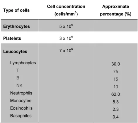
In Table I.1 it is possible to compare the percentages in which the previously mentioned cells can be found in the peripheral blood.
Table I.1 – Main cells of the peripheral blood (Arosa et al., 2007).
Type of cells Cell concentration
(cells/mm3)
Approximate
percentage (%)
Erythrocytes 5 x 10⁶
Platelets 3 x 10⁶
Leucocytes Lymphocytes T B NK Neutrophils Monocytes Eosinophils Basophiles
7 x 10⁶
30.0 75 15 10 62.0 5.3 2.3 0.4
I.2.1. Lymphocytes
Lymphocytes result from differentiation of stem cells produced on the bone marrow and are divided in essentially two types that differ among themselves regarding the place where they are developed. Thus, B lymphocytes or B cells are maturated in the bone marrow of adult mammals, while T lymphocytes or T cells are maturated in the thymus (Male et al., 2006).
These cells are programmed to recognize pathogens and to initiate the adequate immune response; however each type of cell has its specialized roles.
3
particular antibody that upon the eventuality of finding its specific antigen, will bind to it, multiply and differentiate into a plasma cell. These cells will then produce great quantities of the antibody in a soluble form. Soluble antibodies are large glycoproteins that can be found in blood as well as tissue fluids. Being a soluble form of the original antibody, it will still bind the same antigen as the B cell that was initially activated (Male et al., 2006).
The T lymphocytes can be divided in 3 different classes: T helpers (Th), T cytotoxic (Tc) and T regulatory/suppressors (Treg/Ts). Each type of cells is identified by the expression of a different molecule: Th cells express CD4, Tc cells express CD8 (Arosa et al., 2007), and finally Treg/Ts cells can express one or the other (Arosa et al., 2007; Tsai et al., 2011).
Th cells, as indicated by their name, act by aiding other cells performing their respective jobs, namely the antibody production by B cells and the microbicide function of macrophages. Tc cells act for example on the destruction of cells that are neoplastic or infected by viruses, thus having a more direct effective role. Finally, Treg/s cells can suppress specific immune responses. On the contrary to what happens with B cells, T cells do not directly recognize antigens; instead, they have to be processed and presented on the surface of cells associated with the major histocompatibility complex (Arosa et al., 2007).
Both types of lymphocytes are small, with little cytoplasm and a round nucleus where the chromatin is highly condensate. However, after cell activation, the amount of cytoplasm increases and the chromatin becomes less condensate (Arosa et al., 2007).
Finally, there is a third type of lymphocytes, designated as Natural Killer (NK cells); this term results from the ability of this type of cells to recognize and destroy aberrant cells, not needing any previous activation. NK cells also produce chemokines and cytokines, which are soluble factors that can either promptly act as microbicides or activate other cells of the immune system. Similarly to B and T cells, the nucleus of NK cells is round, however in some occasions it presents a small indentation in one of the sides. Also, NK cells have many granules in their cytoplasm, resembling Tc cells. Regarding their size, NK cells are bigger than non-activated T cells (Arosa et al., 2007).
I.2.2. Monocytes, macrophages and dendritic cells
Usually, monocytes are the biggest cells circulating in the peripheral blood; their nucleus presents a shape similar to a horseshoe due to its lobed morphology. Like the nucleus, the limits of these cells are also irregular and the cytoplasm can present vacuoles. Monocytes circulate temporarily in the peripheral blood; eventually they can enter in the tissues and differentiate into macrophages or dendritic cells (DCs) (Arosa et al., 2007).
4
be activated when stimulated by specific soluble factors, resulting in further increase in their phagocytic capacity and secretion of many immunoregulatory soluble factors (Arosa et al., 2007)
DCs are distinguished by their starry, dendritic shape, and when in an immature state, are responsible for collecting antigens. The posterior migration of these cells into the T areas of the lymphoid organs is very important for antigen presentation to T cells, and is concomitant with phenotypic and functional alterations in the dendritic cells. These alterations are caused by a variety of stimulus and result in the maturation of DCs, which at this point are responsible for T cell stimulation. There are two different lineages of DCs, lymphoid and myeloid, depending on their origin or phenotypic characteristics. According to different types of regulatory signals, DCs can direct T cells to different types of immune responses. It is also possible to identify different types of DCs according to their location in the human body: when in the epidermis of the skin, DCs are called Langerhans cells; immature DCs located in the interstitial spaces drained by lymphatic vessels from organs like the kidney, liver, heart, lung, pancreas and intestine are called interstitial DCs; DCs present in T zones of secondary lymphoid organs are called interdigitating; immature or maturating DCs, not yet presenting a ramified morphology and located in afferent lymph or blood are designated by veiled cells (or circulating DCs); DCs located in B zones of secondary lymphoid organs are called follicular DCs (Arosa et al., 2007).
I.2.3. Granulocytes
There are 3 main types of granulocytes: neutrophils, eosinophils and basophiles.
Neutrophils have a nucleus that is divided into 2 to 5 lobules linked by thick heterochromatin covered by nuclear membrane, forming a circle that rounds up the centrosome. Neutrophils have a short life and are one of the first peripheral blood cells to reach an inflammation site. There, they phagocyte and eliminate the pathogens using many mechanisms. One of the main processes consists in using peptides or proteins, with specific microbicide properties that are accumulated in the granules; a selection of the type of granules that are used allows to destroy intracellular or extracellular microorganisms (Arosa et al., 2007).
Eosinophils have a bilobed or eventually trilobed nucleus and act essentially against parasites. They are characterized by a low phagocytic capacity; instead they act by releasing the content of their granules into the extracellular region (Arosa et al., 2007).
5
I.3. Cell separation
Whenever we need to perform functional studies on a particular cell type we have to isolate the cell population from others present in the sample. Therefore it may be necessary to use physical or immunological separation methods (Freshney, 2010a).
Regardless the technique used, the separation principles fall in one or in a combination of parameters related to the specific characteristics of each cell type. Cell adherence (Tomlinson et al., 2012), cell density and the affinity of antibodies to specific molecules in the cell surface are 3 relatively simple and cheap methods where no high technology is required. Methods relying on cell size and light scatter or differences in the emission of fluorescence, detected by flow cytometry require more advanced and expensive material (Freshney, 2010a).
Thus, some methods involving these principles will be further developed in the following sections.
I.3.1. Cell density
Cell separation according to different density profiles can be performed on a density gradient solution by centrifugation (Al-Mufti et al., 2004). Basically, in order to efficiently accomplish this purpose, a cell sample has to be centrifuged using enough centrifugal force, during a period of time that is long enough to allow cells to arrive the point in the gradient that equals their own density, i.e., their isopycnic density, reason why this process is referred as isopycnic centrifugation (Pretlow and Pretlow, 1989).
The density gradient medium needs to be precisely defined regarding its osmolarity, temperature, pH and composition in order to still allow reproducible results regardless eventual alterations that cell properties may suffer, caused by external conditions (Shortman, 1984). Thus, it is important that at high densities (1,10 g/ml), the media is not toxic nor viscous; also it should apply little osmotic pressure onto the cell solution (Pretlow and Pretlow, 1989; Freshney, 2010a). Probably, the most known density gradient media used to perform cell separation are Ficoll and Percoll. The first is a sucrose polymer, thought to cause less osmotic trauma to the cells than sucrose solutions themselves (Sykes et al., 1970). Percoll consists in colloidal silica spheres coated with polyvinylpyrrolidone (PVP) that allow the formation of density gradients at high-speed centrifugation (Pertoft et al., 1978). Compounds as bovine serum albumin (BSA) (Turner et al., 1966), dextran (Schulman, 1967) and metrizamide (Munthe-Kaas and Seglen, 1974) have also been used. Obviously, each compound has determined particularities in what concerns the formation of the density gradient.
6
used for this process is, as already mentioned, SRBC; dog and pig red blood cells also showed some
adherence, however in a smaller extent. All these red blood cells have the ability to form rosette
structures with T cells; monkey red blood cells (MRBC) also formed rosettes, but to lymphocytes
identified as B cells (Pellegrino et al., 1975). Eventually, this method can be combined with antibody binding technology as happens with the RosetteSepTM kit from StemCell. In this kind of separation,
cells are incubated with an antibody cocktail solution against specific epitopes of the unwanted cells;
consequently, these will crosslink the unwanted cells to erythrocytes, forming immunorosettes as
represented in Fig.I.2.
Fig.I.2 – Blood sample after incubation with the RosetteSepTM Cocktail from StemCell, and prior to
perform a density gradient centrifugation. Erythrocytes (red blood cells) are linked to an unwanted cell,
forming an immunorosette (StemCell).
Regardless the fact that antibody binding is or not involved, the isolation principle is the same:
erythrocytes form complexes with the unwanted cells, thus creating immunorosettes. These
complexes are considerably denser than the mononuclear cells of interest; therefore, after
centrifugation it is possible to remove the wanted cell fraction in the mononuclear cell phase, while the
immunorosettes will constitute the pellet (Strelkauskas et al., 1975; Tomlinson et al., 2012).
I.3.2. Cell size
Cell separation can also be performed based on cell size, which is a determinant factor for
sedimentation velocity.
The majority of cell separations using this principle use a cell sorter (see section I.3.4.2) or
centrifugal elutriation. Elutriation refers to when separation is performed by washing; therefore, in
centrifugal elutriation the centrifugal force is opposed to a counter flow of medium. This process
occurs in a separation chamber and it is the balance between these two forces that allows separation.
With elutriation it is possible to process a large number of cells in a fast way; moreover, despite the
shear forces applied to the cells, their viability is generally maintained (Sanders and Soll, 1989).
However, there is also a simpler process based on cell size that does not involve centrifugal
methods and it is called unit gravity. This method only requires letting cells settle over a gradient
7
relation between their radius and their sedimentation velocity. This method is more useful when cells present major differences regarding cell size, or when separating aggregates; on the other side, it is generally not suitable to separate large number of cells, except if the mean size of the cells is different and each cell population is homogeneous in size (Sanders and Soll, 1989; Freshney, 2010a).
I.3.3. Cell adherence
The utilization of techniques based on cell adherence allows performing cell separation in a simple and not expensive way. However, these types of techniques depend on the affinity of the cells of interest to adhere to a certain material. Eventually, there may be more than one type of adherent cells, in these cases the target cells have also to be able to outcompete the other cell types for adherence. Moreover, the results are not specific, resulting in lower cell purity when compared to other separation methods (Tomlinson et al., 2012).
One of the oldest methods used to separate T and B cells (besides the previously mentioned rosetting – see section I.3.1), rely precisely on specific adherence of B cells to determined materials. It has been proven that these cells have a preference to stick to glass beads (Rabinowitz, 1965), cotton wool (Hogg and Greaves, 1972) or nylon wool (Eisen et al., 1972; Julius et al., 1973; Wong and Mittal, 1981).
Regardless the fact that the theoretical basis for cell adherence to nylon wool separation is still unclear, this method has been widely used due to its simplicity and because it requires only standard laboratory equipment. Moreover, lymphocytes handled by this process are not exposed to antigens, complement, enzymes or electric fields. As a result, with this method it is expected to obtain a nylon adherent fraction, which is enriched with B cells and a non-adherent fraction, enriched with the T cells. Each fraction is expected to be depleted from the cells of the opposite type, resulting in high purity values (Trizio and Cudkowicz, 1974). On the downside, there is the fact that some antigen presenting cells also have adherence to nylon wool, thus being retained together with B cells. Moreover, it has also been described that T cells purified using a nylon wool column presented alterations regarding proliferation, activation response and production of cytokines (Wohler and Barnum, 2009). It has also been confirmed that is possible to use nylon wool as a solid support to which specific monoclonal antibodies may be associated, in order to separate T cells subpopulations (Kokkinopoulos et al., 1992).
8
I.3.4. Antibody-based techniques
As portrayed by the name, antibody-based techniques relay on the specificity of a selected antibody to bind to the correct epitope on the surface of a specific cell type. This principle can be applied to many techniques in order to separate a large variety of cell types (Freshney, 2010a). Among the cell isolation techniques that have as basic principle antibody binding, the most commonly used are magnetic-activated cell sorting (MACS) and fluorescence-activated cell sorting (FACS).
In one hand, both techniques have in common the use of specific antibodies against cell surface antigens. On the other hand, MACS technology uses antibodies linked to iron oxide microbeads that will link to target cells, and consequently requires a magnetic field to separate those cells from the remaining ones; whereas FACS technology fluorescently labels its antibodies, and afterwards, the excitation of determined fluorophores above a specific threshold will signal the associated cell to be separated (Tomlinson et al., 2012). Both methods will be approached in the following sections, with special highlight to MACS separation (section I.3.4.1).
I.3.4.1. Magnetic-activated cell sorting
MACS technology by Miltenyi (Klein et al., 1994) is probably one of the most used for this type of separation. Nonetheless, there are other companies that also have their own magnetic sorting technologies as the EasySepTM platform from StemCell (Shin et al., 2012). Both technologies have
been used in our laboratory in order to separate different types of cells from human peripheral blood. Therefore, their main characteristics will be presented below.
MACS technology combines the use of high gradient magnetic fields with small superparamagnetic microparticles, as first suggested by Molday and Molday (Molday and Molday, 1984) and also with fluorescent labeling (Miltenyi et al., 1990).
Separation by MACS (Miltenyi) is effective for small or large scale isolations and requires 3 different components: MACS MicroBeads, MACS Columns and MACS Separators (Miltenyi Biotec, 2013). MACS MicroBeads are biodegradable, non-toxic superparamagnetic particles conjugated to highly specific antibodies against a cell surface antigen. These particles are as small as 50 nm, reason why they do not saturate cell surface epitopes neither activate cells themselves; also they do not have to be removed from the cellular suspension prior to subsequent assays. Moreover, MACS MicroBeads are supplied in a colloidal suspension; this fact associated with the small dimensions of the beads enables short labeling steps, fast binding kinetics, and consistency among different lots and at the same time avoids cell clumping. Furthermore, after this process, cells are only minimally labeled in order to assure that concurrent antibody staining will still have enough free epitopes to bind to.
9
column, they never get to bind to the column itself; instead, the cells stay in suspension, thus
minimizing the stress caused to them. The remaining cells will simply be allowed to pass through the
column as there is enough space between the spheres for that to happen.
Finally, MACS Separators can be found in different formats, which are specifically designed
for the appropriated type of column, considering the isolation at hands and the volume that is being
applied.
The basic overall principle of this method is described in 3 simple steps that are represented in
Fig.I.3: magnetic labeling, magnetic separation and elution of the labeled cells (Miltenyi Biotec, 2013).
First of all, a single cell suspension is prepared, magnetically labeled with the intended MACS
MicroBeads and applied to an adequate MACS Column, previously set in a MACS separator for the
magnetic separation. The unlabeled cells will be eluted and the labeled cells will be retained within the
column. After washing the column, it can be removed from the separator in order to also elute the
labeled cells. Consequently, this method allows both cell fractions to be isolated with high purity.
Fig.I.3 – Steps of MACS separation using technology from Miltenyi: magnetic labeling, magnetic
separation and elution of the labeled cells. When applied to a magnetic field, the magnetically labeled
cells will be retained in the column; those cells can be eluted after removing the column from the
magnetic field (Miltenyi Biotec, 2013).
Concerning the labeling methods, they can be performed in a direct or indirect way. In the first
case, the MicroBeads will specifically bind to the respective epitopes on the surface of the cells. This
case represents the fastest way to label cells as it only requires one incubation step and a minimal
number of washing steps, what also minimizes cell loss. On the case of indirect magnetic labeling, two
steps are required. Firstly, the cells are labeled with an antibody against a specific cell marker. This
primary antibody can be unconjugated, or conjugated with biotin or a fluorochrome. The second step
corresponds to the magnetically labeling itself and consists on the binding of the MACS MicroBeads to
the primary antibody (Anti-Immunoglobulin MicroBeads) or to the respective conjugated molecule
(Anti-Biotin MicroBeads or Anti-Fluorochrome MicroBeads). Indirect labeling is useful to isolate
untouched cells, as it allows the use of a cocktail of primary antibodies that will concurrently label the
10
MACS technology uses essentially 4 types of separation strategies: positive selection,
depletion, untouched isolation (also referred as negative selection) and sequential sorting (Miltenyi
Biotec, 2013).
In positive selection (see Fig.I.4), a specific cell type is magnetically labeled (direct or
indirectly), thus being the one retained during separation, while the remaining cell types flow through.
Then, after the column had been washed and removed from the magnetic field, the target cells can be
eluted. To use this strategy is the fastest way to isolate a specific cell type.
Fig.I.4 – Cell separation by positive selection using MACS technology. Target cells are magnetically
labeled; then the cell suspension is applied in a magnetic field: unlabeled cells flow through the
column while the target cells are retained, thus needing to be eluted after removing the column from
the magnetic field (Miltenyi Biotec, 2013).
In a depletion strategy (see Fig.I.5), an unwanted cell type is magnetically labeled in order to
be removed from a cell mixture. This cell type will be retained in the column, while all the remaining
cells that represent the target population are collected in the flow-through. Nonetheless, the unwanted
cell fraction can still be eluted after the column had been removed from the magnetic field.
Fig.I.5 – Cell separation by depletion strategy using MACS technology. Unwanted cells are
magnetically labeled. When the suspension is applied to the magnetic field, the target cells will be
acquired in the flow through while the unwanted ones will be retained in the column and eventually
11
Untouched isolation (Fig.I.6) is the most adequate strategy to obtain a target cell type without
labeling it (untouched form). In this case, the unwanted cell types are the ones being magnetically
labeled and consequently retained in the column and depleted. Being unlabeled, the target cells will
be collected in the flow-through. If desired, it is also possible to elute the labeled fraction, after it had
been removed from the magnetic field. In this type of isolation strategy it is used indirect labeling,
therefore, the respective kits are supplied with a cocktail of titrated antibodies and MACS MicroBeads.
Fig.I.6 – Cell separation by untouched isolation using MACS technology. Unwanted cells are
magnetically labeled and consequently retained in the magnetic field. Target cells are collected in the
flow through (Miltenyi Biotec, 2013).
Sequential sorting combine two successive separations using more than one marker, in order
to obtain a specific cell subset; even in situations where there is no marker defined for that type of cell.
In these cases, the isolation strategies may be of different types (depletion followed by positive
selection) or of the same type (two subsequent positive selections); both situations will be briefly
described next.
When unwanted and target cells, both have the same marker, first of all it is necessary to
deplete the undesired cells that also express that marker by magnetically labeling them via antigens
different from the common ones. Then, the unlabeled cells obtained in the flow-through must be
magnetically labeled with beads for the common marker in order to proceed for their positive selection.
12
Fig.I.7 – Cell separation by depletion followed by positive selection using MACS technology.
Unwanted cells that express a common marker with the target cells are labeled and depleted. Then
the target cells obtained in the flow through are labeled for the common marker and obtained by
positive selection (Miltenyi Biotec, 2013).
The other possible situation requires a multiparameter sorting using two different markers
sequentially. In this case, the first step would be to use the appropriate MACS MultiSort Microbeads to
the first marker and proceed to the first positive selection. Before labeling the cells against the second
marker it is necessary to enzymatically remove the first beads by using the MultiSort Release
Reagent. Only then it is possible to continue for the second positive selection. This process is
represented in Fig.I.8.
Fig.I.8 – Cell separation by two subsequent positive selections using MACS technology. First of all,
cells are labeled for the first maker and collected by positive selection. Then the labeling is
enzymatically removed in order to allow a second labeling with a second marker for the target cells,
which will also be collected by positive selection (Miltenyi Biotec, 2013).
The EasySepTM platform (StemCell) uses the same magnetic separation principle, however
has the advantage of not requiring columns or washes, resulting in a simpler and faster process that
can be completed in about 25 minutes. In this case, instead of columns, standard polystyrene tubes
can be used; those are directly placed into an EasySepTM Magnet, which is capable of generating a
high-gradient field. There are different magnets available, suited for processing different sample
13
samples that do not require the removal of the magnetic particles prior to eventual downstream
applications (StemCell, 2013).
In order to isolate cells using EasySepTM, the target cells are linked to dextran-coated
magnetic particles by antibody complexes(see Fig.I.9). The next step consists in placing the tube
containing the cell suspension into the EasySepTM Magnet: the labeled cells (positive fraction) are
attracted to the sides, thus remaining in the tube; however the unlabeled cells (negative fraction) can
be poured off into another tube.
Fig.I.9 – Magnetic labeling using StemCell products. Antibody complexes link the target cells to the
dextran-coated magnetic beads (StemCell, 2013).
This technology allows cell isolation through positive selection, negative selection and
depletion (StemCell, 2013), much alike to what happens with the technology form Miltenyi, however in
less time. The number of washes required by the protocols from Miltenyi, together with the variable
time that the cell suspension and subsequent washes take to flow through the column, makes them
more time consuming.
I.3.4.2. Fluorescence-activated cell sorting
As previously stated, fluorescence-activated cell sorting is an antibody-binding method that
uses an instrument capable of separating cells according to different fluorescent markers (Bonner et al., 1972).
The fluorescence-activated cell sorter acquires cells one-by-one by projecting them in a
stream through a laser that detects the emission signals from each single cell. Then, the instrument
processes the information, presenting it in a dot plot format, for example. Thus, it is possible to gate
specific populations in the display, targeting them for separation. Consequently, the cell sorter will
separate each cell that has properties matching the selected coordinates, regarding their light scatter
and/or fluorescence (Freshney, 2010a).
This technology has been used to separate several types of cells; however hematopoietic cell
separation seems to be the most common application (Yeung and So, 2009).
Comparing FACS technology to the MACS one, FACS has the advantage of allowing a cell to
14
the MicroBeads in order to relabel the same cell. Moreover, the fact that FACS technology considers each cell at a time results in more specific results; on the downside this makes the separation much slower than with MACS technology (Tomlinson et al., 2012).
I.4. Cell culture
I.4.1. Requirements of culture media
There are many types of culture media, adapted to better suit the needs of different cell types, however there are some properties that are overall indispensable, some of which will be briefly mentioned (Freshney, 2010b).
Thus, one of the first points is the pH level. The majority of cells require a pH of approximately 7.2–7.4 and its maintenance between these values is crucial for optimum cell culture conditions (Sigma Life Tecnhologies and ECACC, 2010). Regular assessment of the pH level of culture media is very important, reason why, commercial culture media, such as the RPMI-1640 from Sigma that we use in the laboratory, already include phenol red as a pH indicator. That facilitates a constant control of the pH status of the media: at pH 7.4 phenol red presents a red coloration, an increase in pH causes it to turn pink at pH 7.4 and purple at pH 7.6; on the other side, a decrease to pH 7.0 will turn the media orange, at pH 6.5 it will be yellow and bellow that value it will acquire a lemon yellow coloration (Freshney, 2010b). There are essentially two main forms by which the pH can be buffered, one natural and one chemical. In the natural option, the CO3/HCO3 content of the culture media is balanced with a CO2 incubator, allowing that a 5-10% CO2 to be maintained in this bicarbonate/CO2 buffering system. The chemical option requires the use of HEPES, a zwitterion with a high buffering capacity in the range 7.2-7.4 that does not require a controlled gaseous atmosphere. Comparing this two methods, the first one has the advantage of being low-cost and non-toxic, what not always happens with the chemical system (Sigma Life Tecnhologies and ECACC, 2010).
Inorganic salts are also important because they provide sodium, potassium and calcium ions. They are necessary for cell matrix attachment as enzyme cofactors, to retain the osmotic balance of the cells and to regulate the membrane potential (Sigma Life Tecnhologies and ECACC, 2010). The osmolality of the cultured cells is also very important and should be kept between 260 mOsm/kg and 320 mOsm/kg (human plasma osmolality is 290 mOsm/kg so this is inferred as an adequate value for cells in culture). When incubation is performed using a Petri dish or open plates, eventual evaporations can be compensated by a faintly hypotonic medium. Changes in the osmolality may be necessary, for example to the addition of HEPES, and can be achieved by altering the sodium chloride concentration (Freshney, 2010b).
15
It is also critical to add amino acids (glutamine in particular) to the culture media as their concentration will determine cell proliferation: amino acids are the constituents of proteins, though they cannot be produced by cells themselves, therefore, from the time they are no longer available to be used by the cells, cell density will no longer increase. Non-essential amino acids can also be added in order to prolong cell viability in culture and stimulate growth (Sigma Life Tecnhologies and ECACC, 2010).
Regarding vitamins, those are usually obtained as part of serum (serum will be further developed in section I.4.2), but can also be added as supplements. Riboflavin, thiamine and biotin are examples of vitamins that are frequently used in culture media, although some media also require B group vitamins or increased levels of vitamins A and E. By supplementing culture media with vitamins, it becomes suitable for a larger variety of cell lines, as vitamins act as precursors for many co-factors (Sigma Life Tecnhologies and ECACC, 2010).
Proteins, peptides, fatty acids and lipids are usually present in serum, making them particularly relevant in culture media. Albumin, transferrin, fibronectin and fetuin are the proteins and peptides added to the medium as replacement. Cholesterol (fatty acid) and steroids (lipids) may also be added for specialized cells (Sigma Life Tecnhologies and ECACC, 2010).
Eventually, more specialized types of cells may also require to supplement the culture media with trace elements as copper, zinc, selenium and tricarboxylic acid intermediates (Sigma Life Tecnhologies and ECACC, 2010).
Factors as temperature and viscosity may also influence the cell culture. The optimal temperature may depended for example on the body temperature of the animal that “provided” the cells as well as on mechanical variations, however most warm-blooded animal cell lines can be kept at 37 ºC; usually, the temperature is set to a slightly inferior value as to prevent damage caused by eventual oscillations (overheating can be more dangerous than underheating). Viscosity takes particular importance when the cells are agitated or dissociated after tripsinization, mainly if there are low or inexistent concentrations of serum. Increasing the viscosity of the medium may be useful to decrease eventual damages upon these procedures and may be accomplished for example by adding carboxymethylcellulose (CMC) (Freshney, 2010b).
16
I.4.2. Serum
As already mentioned in the previous section, serum is composed by several constituents, such as albumins, growth factors, growth inhibitors and hormones. Besides providing nutrients, the main roles performed by serum in culture media are: increase the buffering capacity, protect against any mechanical damage caused by handling the cells, and bind and neutralize toxins. Serum has the advantage of suiting a large variety of cell types, regardless their distinct growth factors requirements. On the downside, standardization of serum production is difficult as it varies a lot from batch-to-batch; moreover, the presence of serum in the culture media may difficult downstream processes, as protein purification, for example (a 10% supplement of Fetal Bovine Serum (FBS) adds an extra 4.8 mg of protein per milliliter of the cell culture fluid). (Sigma Life Tecnhologies and ECACC, 2010).
It is also very important to access the quality of each serum batch, reason why several tests are performed in order to detect the presence or level of the following: virus contamination (e.g. bovine viral diarrhea virus – BVDV), Mycoplasma contamination, endotoxin, haemoglobin, total protein, immunoglobulin, osmolality, hormone testing, pH at room temperature and sterility (Sigma Life Tecnhologies and ECACC, 2010).
There are many types of serum commercially available, which may vary according to their range of applications. FBS, bovine calf serum, newborn calf serum (NCS) and human serum are probably the most commonly used.
FBS is one of the most popular types of serum due to its wide range of applications. Its contribution involves providing growth promoting factors as well as improving cell survival.
NCS is also widely used, not only for cell culture, but also for organ preservation. Its cell culture applications include oocyte maturation, tubular epithelial cell functional studies and primary cell culture (e.g. visceral adipocytes). Bovine calf serum is also available, although it has more restrict applications, such as propagation of viruses, Helicobacter pylori and Plasmodium falciparum.
Regarding human serum, type AB is probably the most used one, being associated with a great variety of applications: tissue engineering, transplantation and some kinds of cell therapy.
The most common way to isolate human sera is by allowing blood to clot. The resulting clear portion will be free of blood cells, fibrinogen and some coagulant factors, being designated as serum (Lewis and Tatsumi, 2006; MeSH-NCBI: Serum). However, if an anti-coagulant agent is added to the blood, it will not be allowed to coagulate; in such cases, the clear phase originated upon centrifugation will be plasma, not serum. Nonetheless, there are also protocols to isolated serum from plasma. As an example we have the method used by Sigma: calcium is added to the plasma in order to activate the clotting cascade; then the preparation is centrifuged and the resultant liquid phase will be serum (Sigma-Aldrich – FAQs).
17
I.4.2.1. Heat inactivation
The heat inactivation procedure is performed in order to inactivate the complement cascade which if active, could lead to the activation of lymphocytes and macrophages (Sigma Life Tecnhologies and ECACC, 2010). The functions of the complement include opsonization (increase susceptibility to phagocytosis of antigens), chemotaxis (cell attraction based on concentration gradients) and cell lysis (Pacheco and Cardoso, 2007).
Heat inactivation also reduces the cytotoxic action of immunoglobulins without being harmful to polypeptide growth factors (Freshney, 2010b). Moreover, heat inactivation can also protect the cell culture from being contaminated by viruses, as some can be inactivated by this process (Sigma Life Tecnhologies and ECACC, 2010). On the down side, heat inactivation may cause the removal of labile constituents, and this can be prejudicial to the cell culture (Freshney, 2010b). Thus, heat inactivation of serum is not an obligatory requirement for cell culture (Sigma Life Tecnhologies and ECACC, 2010). By principle, in the case of cultures of PBMCs, the fact that the complement is or not inactivated should not be a problem as normal CD4+ T cells do not express complement receptor in a
large enough level to be of influence (Montefiori Laboratory, 2011).
The heat inactivation process is very simple, though it may present some variations. The most common method requires serum to be heated to 56 ºC for 30 minutes (Sigma-Aldrich, 2013c). However, there are protocols that suggest an incubation period of 1h if samples have not been previously heat-inactivated; when they have already been previously heat-inactivated for 30 minutes or less, a new incubation step at 56 ºC should be performed during 30-45 minutes (Montefiori Laboratory, 2011).
I.5. Cell cryopreservation
Cell cryopreservation, is an almost self-explaining term that refers to the process of conserving and protecting the structure, form and chemical composition of cells by means of freezing (MeSH-NCBI, 1990).
Frequently, in most laboratories, it is necessary to stabilize materials in order to preserve their sub-cellular components. Mainly, when working with cell lines it is vital to protect them from several mishaps such as genotypic drift, phenotypic instability, transformation and acquisition of non-desired characteristics, senescence and eventual extinction (Freshney, 2010c). Also, it is important to avoid contamination by other microorganisms or even by other cell lines (cross-contamination) (Capes-Davis et al., 2010). So, before proceeding to the cryopreservation process itself it is important to check for
contamination and register the cell line characteristics including eventual genetic modifications or infectiousness (Simione, 1992; Stacey and Dowall, 2007).
18
treatments, such as DC-based vaccinations, it is useful to store some aliquots of PBMCs to differentiate into DCs later on (Westermann et al., 2003; Heo et al., 2009).
For all the referred reasons, cryopreservation is a practical solution; it is also a helpful solution when distributing samples to other investigators or even as a way to save time, by avoiding the maintenance of the cell lines that are not being used at that moment (Freshney, 2010c).
I.5.1. Principals of cryopreservation
The objective of any cryopreservation process is not only to preserve cells but also to allow the maximum recovery rate possible. In order to accomplish such goals it is important that certain principles are followed. First of all, it is essential that an adequate cryoprotectant is used, and then samples have to be frozen with an appropriate cooling rate and stored under restrict conditions. Finally, the thawing process also has to be controlled in order to maximize cell viability and recovery (Freshney, 2010c).
I.5.2. The cryoprotective agent
The utilization of a cryoprotective agent in a freezing process is fundamental; its main role in the process is to minimize damage caused by the formation of ice crystals and the subsequent osmotic imbalance.
Dimethylsulfoxide (DMSO - Me2SO) and glycerol are clearly the ones most commonly used (Simione, 1992); however PVP (Suzuki et al., 1995), polyethylene glycol (PEG) (Monroy et al., 1997) and hydroxyethyl starch (HES) (Pasch et al., 2000) are examples of compounds already suggested and tested as cryoprotectants.
Handling DMSO requires some caution as it is a penetrating agent that when in direct contact with skin is rapidly absorbed and may transport other potentially harmful substances along with it (Pope and Oliver, 1966). On the other side, this facility in penetrating cells comes as an advantage in its role as a cryoprotective agent, especially in larger and more complex types of cells, because it encourages the dehydration of the cell before ice crystals start to form in its interior. Glycerol is not as penetrating as DMSO, nonetheless is often preferred due to its inferior toxicity. Moreover, these 2 cryoprotective agents are not used simultaneously, in the same freezing media, unless we are handling plant cells (Simione and Thermo Fisher Scientific, 2009).
19
I.5.3. Sample preparation and equilibration
Usually, samples are frozen at a high cell concentration in order to compensate eventual low cell recovery after thawing (Freshney, 2010c); the values are highly variable, but 5 x 10⁶-107 cells/ml
are commonly used concentrations when freezing PBMCs (Stacey and Dowall, 2007). Also after thawing, the cryoprotective agent needs to be removed by centrifugation or diluted in the cell suspension. In the case of dilution, the initial cell concentration should allow a dilution factor of 10 or 20 fold. When centrifugation is not the preferred option, a higher cell concentration prior freezing will allow a higher sample dilution (Freshney, 2010c).
Another variable that is important to consider when freezing cells is the equilibration process. This refers to the period of time that starts when the cryoprotective agent is added to the cell suspension, and goes up until the beginning of the cooling process. This period of time should take place at room temperature and may be used to distribute the cell suspension through the intended cryovials. Equilibration should occur in a period of approximately 15-45 minutes, in order to allow the cryoprotective agent to enter the cells. However it may never exceed 60 minutes as the cryoprotective agent may be toxic to the cells after longer periods (Simione and Thermo Fisher Scientific, 2009).
I.5.4. Cooling rate
When cells are not able to use water for their usual chemical processes their metabolism stops. That is what occurs upon freezing, as all water is converted to ice (Simione and Thermo Fisher Scientific, 2009).
Cell freezing causes ice crystals to form inside and outside the cells. The rate at which cells are frozen has great influence on the overall success of the process. If the cooling rate is too slow, ice crystals will start forming outside the cell first, diminishing the amount of water in a liquid state and increasing solute concentration. To compensate this imbalance, intracellular water will start coming to the extracellular environment, thus increasing the concentration of solute inside the cell too. Therefore, the overall result of this osmotic imbalance will be less intracellular ice crystals being formed and higher solute concentrations. On the other side, if the cooling rate is too fast, ice crystals will form uniformly both inside and outside the cell, not causing a big alteration in solute concentration. However, as water did not have much time to migrate to the extracellular environment, there will be more ice forming inside the cell. The formation of too much ice crystals inside the cell may damage its components, and this, together with eventual damage caused by recrystallization during thawing can be very prejudicial to the cells (Simione, 1992; Simione and Thermo Fisher Scientific, 2009).
20
Different cells may require different rates of cooling but usually, a compromise is found at 1 ºC/min (Leibo and Mazur, 1971).
The easiest way of achieving a controlled freezing rate consists in using a programmable-rate cell freezing apparatus that according to its sophistication may allow the selection of one or more cooling rates throughout all the freezing period. These units have the disadvantage of being expensive, reason why cheaper and simpler options are available for similar purposes. For instance, Thermo Scientific Nalgene Mr. Frosty simulates a controlled-rate cooling process by allowing a decrease in temperature close to 1 ºC per minute. Nonetheless, this container still needs to be placed in a mechanical freezer at -60 ºC to -80 ºC (Simione and Thermo Fisher Scientific, 2009).
I.5.5. Storage
Cell recovery and survival rates are highly dependent of the temperature of storage. Usually, longer viable storage periods are associated with colder freezing temperatures, nonetheless it is important that the freezers are functioning properly and have no leaks (Simione and Thermo Fisher Scientific, 2009).
Short-term storage of cells is possible with temperatures of -80 ºC (Simione, 1992), however for long-term storage the best results are obtained with freezing temperatures below -130 ºC, because that temperature marks the limit of biological mechanisms (Mazur, 1984).
I.5.6. Cell thawing
Cell thawing is actually a very simple process, only requiring cells to be warmed as fast as possible, usually in a water bath at 37 ºC (Simione and Thermo Fisher Scientific, 2009). Delaying this step will increase the formation of ice crystals inside the cell, intensifying the risk of cell damage or even death. Afterwards, depending on the sensibility of the cells being handled, the cryoprotective agent might need to be removed by centrifugation; this is usually the case of cells capable of growing in suspension. On the other side, there are cells that only require to be gradually diluted or to have their media changed the following day (Freshney, 2010c).
I.6. Cell viability studies