Thickened Pituitary Stalk Associated with a Mass
in the Sphenoidal Sinus: An Alarm to Suspect
Hypophysitis by Immunoglobulin G4?
Rafael Loch Batista
1Luciano Silva Ramos
1Valter Angelo Cescato
2Nina Rosa Castro Musolino
2Clarissa Groberio Borba
2Gilberto Ochman Silva
2Lilian Hupfeld Moreno
1Malebranche Bernardo Carneiro Cunha Neto
21Neurosurgical Unit, Universidade de São Paulo, São Paulo, SP, Brazil 2Funcional Neurosurgery Unit, Universidade de São Paulo, São Paulo,
SP, Brazil
Int Arch Otorhinolaryngol 2015;19:273–276.
Address for correspondence Rafael Loch Batista, MD, Neurosurgical Unit, USP, Alameda Joauime Eugênio de Lima, 1058/102, Sao Paulo, Sao Paulo 01403002, Brazil
(e-mail: [email protected]; [email protected]).
Introduction
Hypophysitis is a chronic inflammation of the pituitary gland of complex and still incompletely defined pathogenesis. It belongs to the group of non-hormone-secreting sellar masses, sharing with them comparable clinical presentation and radiographic appearance. These similarities often make it difficult to establish a diagnosis with certainty before pitui-tary surgery and pathologic examination of the resected pituitary tissue.1Immunoglobulin G4 (IgG4)-related diseases, including IgG4-related hypophysitis, are recently character-ized entities marked by elevated serum IgG4 levels and tissue
infiltration by IgG4-positive plasma cells.2,3It wasfirst diag-nosed on clinical grounds in 20044and then by pathology in 2007.5We reported a patient with IgG4-related hypophysitis and summarize the current relevant literature.
Review of Literature
Hypophysitis has been classified in several ways based on anatomic location of the pituitary involvement, cause, and histopathologic appearance (►Table 1).6
When surgery of the pituitary gland is performed, the pituitary pathology reveals two more common forms Keywords
►
pituitary diseases
►
hypopituitarism
►
sphenoid sinus
Abstract
Introduction
Hypophysitis is a chronic in
fl
ammation of the pituitary gland of complex
and still incompletely de
fi
ned pathogenesis. It belongs to the group of
non-hormone-secreting sellar masses, sharing with them comparable clinical presentation and
radiographic appearance.
Objectives
Describe the case of immunoglobulin G4 (IgG4)-related hypophysitis
presenting as a mass in the sphenoid sinus.
Resumed Report
A 40-year-old Brazilian man had a diagnosis of central diabetes
insipidus since 2001 associated with pituitary insuf
fi
ciency. Pituitary magnetic
reso-nance imaging revealed a centered pituitary stalk with focal nodular thickening and the
presence of heterogeneous materials inside the sphenoid sinus. The patient was treated
with testosterone replacement therapy. Laboratory results revealed increased IgG4
serum.
Conclusion
IgG4-related hypophysitis should be considered in patients with pituitary
insuf
fi
ciency associated with sellar mass and/or thickened pituitary stalk. IgG4 serum
measurement for early diagnosis of IgG4-related hypophysitis should be performed.
received
October 3, 2014
accepted
November 24, 2014
published online
March 15, 2015
DOI http://dx.doi.org/ 10.1055/s-0034-1397333.
ISSN 1809-9777.
Copyright © 2015 by Thieme Publicações Ltda, Rio de Janeiro, Brazil
THIEME
(lymphocytic and granulomatous) and three rarer variants (xanthomatous, necrotizing, and IgG4-producing plasma cells).6 IgG4-related hypophysitis wasfirst reported in 2004 in a 66-year-old woman with multiple pseudotumors in the salivary glands, pancreas, and retroperitoneum,4and then the entity was more extensively described in 2006 in a 70-year-old man with swelling of the salivary glands caused by a marked infiltration with lymphocytes and IgG4-positive plasma cells.7Since thefirst IgG4-related hypophysitis case was described in 2004, more than 20 histogenetically proven cases have been reported, mostly from Japan.8–11All were accompanied by complications of pituitary insufficiency, but Hattori et al described thefirst case of IgG4-related hypophysitis without pituitary insufficiency.12 Our patient had pituitary insufficiency.
This disease is typically part of a multifocal systemic disease recently called“IgG4-related autoimmune disease,”13 which emphasizes the contribution of IgG4 in establishing the diagnosis.6 IgG4, the least abundant of the four IgG antibodies, has long been associated with autoimmune and allergic diseases.14IgG4 antibodies were ignored for diagnos-tic purposes until 2001, when Hamano et al linked them to autoimmune pancreatitis and made it possible to recognize that many diseases associated with autoimmune pancreatitis share similar pathologic features, thus defining the existence of a multifocal systemic disease.15At the present time, the pathogenetic mechanism and underlying immunologic abnormalities remain unclear.16 A recent report identified autoimmune antibodies against GH (growth hormone) and
adrenocorticotropic hormone in a patient with IgG4-related hypophysitis.17Nonetheless, collecting further evident cases and analysis is required to characterize the pathophysiology of IgG4-related hypophysitis.12
Leporati et al suggestedfive criteria to diagnose IgG4-related hypophysitis: (1) pituitary histopathology, (2) magnetic reso-nance imaging (MRI) of the pituitary, (3) biopsy-proven involve-ment in other organs, (4) serology with increased serum IgG4, and (5) response to glucocorticoids.6Leporati et al also proposed that the diagnosis of IgG4-related hypophysitis is established when any of the following is fulfilled: criterion 1 alone or criteria 2þ3 or criteria 2þ4þ5 (►Table 2).6 According to the criteria, a pituitary biopsy is not essential; however, there have been eight cases diagnosed by pituitary biopsy. Our patient fulfilled two diagnostic criteria suggested by Leporati et al to be associated with mass in the sphenoidal sinus and complication of pituitary insufficiency.
Case Report
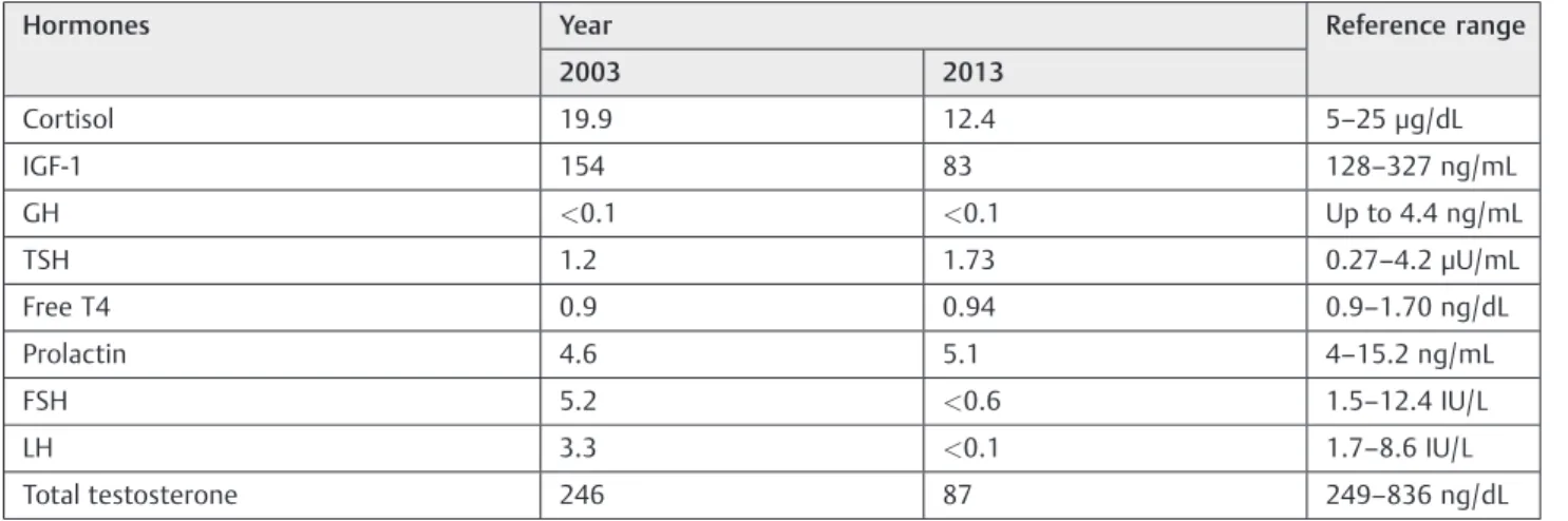
A 40-year-old man with diagnosis of central diabetes insipidus since 2001, using oral desmopressin 0.3 mg/d, complained of frontal headache, sexual impotence, and decrease in libido. Endocrine assessment in 2003 revealed low levels of testoster-one and gonadotropins and decreased insulin-like growth factor 1 (IGF-1) serum (►Table 3). The patient was treated with
replacement therapy of testosterone decanoate 250 mg injec-tions every 21 days and sexual impotence and libido improved. In 2005, MRI revealed a centered pituitary stalk with focal nodular thickening measuring 6 mm at the lower portion characterized by isointensity on T1-weighted images, hypoin-tensity on T2-weighted images, and a heterogeneous intense Table 1 Current classifications of hypophysitis
Based on the anatomic location of pituitary involvement
Adenohypophysitis
Infundibuloneurohypophysitis
Panhypophysitis
Based on the histologic appearance
Lymphocytic
Granulomatous
Xanthomatous
Necrotizing
Immunoglobulin G4 plasmacytic
Mixed forms (lymphogranulomatous, xanthogranulomatous)
Based on the cause
Primary (isolated or as part of a multiorgan systemic disease)
Secondary to:
Sellar diseases (germinoma, Rathke cleft cyst, craniopharyngioma, pituitary adenoma)
Systemic diseases (Wegener’s granulomatosis, tuberculosis, sarcoidosis, syphilis)
Injection of immunomodulatory drugs (CTLA-4 blocking antibody, interferon-alpha)
Abbreviation: CTLA-4, T-lymphocyte-associated protein-4. Note: Adapted of Leporati et al.6
Table 2 Diagnostic criteria for IgG4-related hypophysitis
Criterion 1: Pituitary histopathology
Mononuclear infiltration of the pituitary gland, rich in lymphocytes and plasma cells, with more than 10 IgG4-positive cells per high-powerfield
Criterion 2: Pituitary MRI
Sellar mass and/or thickened pituitary stalk
Criterion 3: Biopsy-proven involvement in other organs
Association with IgG4-positive lesions in other organs
Criterion 4: Serology
Increased serum IgG4 (>140 mg/dL)
Criterion 5: Response to glucocorticoids
Shrinkage of the pituitary mass and symptom improvement with steroids
Diagnosis of IgG4-related hypophysitis is established when any of the following is fulfilled:
Criterion 1 OR
Criteria 2 and 3 OR
Criteria 2, 4, and 5
Abbreviations: IgG4, immunoglobulin G4; MRI, magnetic resonance imaging. Note: Adapted from Leporati et al.6
International Archives of Otorhinolaryngology Vol. 19 No. 3/2015
Sphenoidal Sinus and Thickened Pituitary Stalk Batista et al.
enhancement after gadolinium administration. Heterogeneous materials inside the sphenoidal sinus were also present (►Fig. 1).
MRIs from 2008, 2010, and 2011 showed no changes on the images.
In 2012, based on the clinical and radiologic findings, a diagnosis of autoimmune hypophysitis with associated sinusi-tis was suspected. We observed unreactive levels of autoim-mune antibodies, including anti-Sjögren syndrome antigen A (anti SS-A/Ro) and antigen B (anti SS-B/La) antibodies, ANA (antinuclear antibody) anti-Sm, rheumatoid factor, thyroglob-ulin, thyroperoxidase, and parietal cell. Adenosine deaminase, angiotensin I-converting enzyme, and tumor markers were negative. In 2013, there was elevation of gonadotropins, IGF-1, and total testosterone levels (►Table 3). Laboratory results
revealed the following: serum IgG, 1,644 mg/dL (reference range 952 to 1,538 mg/dL), and serum IgG4, 2,040 mg/dL (reference range 84 to 888 mg/dL). IgG4 was titled in two different analyses. The patient’s test results strongly favored a diagnosis of IgG4-related hypophysitis.
Discussion
IgG4 serum levels are typically elevated in IgG4-related hypophysitis, similar to our patient, but they can decrease
after glucocorticoid therapy initiation and in later disease stages. The involvement of the sphenoidal sinus is yet another manifestation of the multifocal IgG4-related autoimmune disease.6Our patient showed a concomitant involvement of both pituitary and sphenoidal tissues, showing that hypophysitis by IgG4 should be in the differential diagnosis of masses in the sphenoid sinus.
The typical therapy for IgG4-related hypophysitis is
unde-fined; however, glucocorticoids are recommended as afi rst-line therapy against IgG4-related disease.10On the basis of treatment of autoimmune pancreatitis, an initial oral pred-nisolone dose of 0.6 mg/kg for 2 to 4 weeks is suggested, tapered by 5 mg every 1 to 2 weeks for 2 to 3 months to determine a maintenance dose (2.5 to 5 mg/d), which should be discontinued within 3 years.10,13
Final Comments
IgG4-related hypophysitis should be considered in patients with pituitary insufficiency associated with sellar mass and/or thickened pituitary stalk. The presence of a mass in the sphenoidal sinus together with the thickening of the pituitary stalk could also suggest IgG4 hypophysitis. Fur-thermore, IgG4 serum measurement for early diagnosis of IgG4-related hypophysitis should be performed. Finally, a Table 3 Endocrine assessment
Hormones Year Reference range
2003 2013
Cortisol 19.9 12.4 5–25μg/dL
IGF-1 154 83 128–327 ng/mL
GH <0.1 <0.1 Up to 4.4 ng/mL
TSH 1.2 1.73 0.27–4.2μU/mL
Free T4 0.9 0.94 0.9–1.70 ng/dL
Prolactin 4.6 5.1 4–15.2 ng/mL
FSH 5.2 <0.6 1.5–12.4 IU/L
LH 3.3 <0.1 1.7–8.6 IU/L
Total testosterone 246 87 249–836 ng/dL
Abbreviations: FSH, follicle-stimulating hormone; GH, growth hormone; IGF-1, insulin-like growth factor-1; LH, luteinizing hormone; TSH, thyroid-stimulating hormone.
Fig. 1 Magnetic resonance imaging showing thickening of the stalk and presence of heterogeneous materials inside sphenoid sinus. (A) T1-weighted coronal image without gadolinium. (B) T1-T1-weighted coronal image with gadolinium.
International Archives of Otorhinolaryngology Vol. 19 No. 3/2015
correct diagnosis is critical because it can spare the patient from a major surgery for a disease that responds well to glucocorticoids.
Acknowledgments
We thank Stefan Menon, director of ONE-I agency, for help with the images.
Declaration of Interest
Authors declare that there is no conflict of interest that could be perceived as prejudicing the impartiality of the research reported.
References
1 Howlett TA, Levy MJ, Robertson IJ. How reliably can autoimmune hypophysitis be diagnosed without pituitary biopsy. Clin Endo-crinol (Oxf) 2010;73(1):18–21
2 Stone JH, Zen Y, Deshpande V. IgG4-related disease. N Engl J Med 2012;366(6):539–551
3 Okazaki K, Umehara H. Are classification criteria for IgG4-RD now possible? The concept of IgG4-related disease and proposal of comprehensive diagnostic criteria in Japan. Int J Rheumatol 2012; 2012:357071
4 van der Vliet HJ, Perenboom RM. Multiple pseudotumors in IgG4-associated multifocal systemicfibrosis. Ann Intern Med 2004; 141(11):896–897
5 Wong S, Lam WY, Wong WK, Lee KC. Hypophysitis presented as inflammatory pseudotumor in immunoglobulin G4-related sys-temic disease. Hum Pathol 2007;38(11):1720–1723
6 Leporati P, Landek-Salgado MA, Lupi I, Chiovato L, Caturegli P. IgG4-related hypophysitis: a new addition to the hypophysitis spectrum. J Clin Endocrinol Metab 2011;96(7):1971–1980
7 Yamamoto M, Takahashi H, Ohara M, et al. A case of Mikulicz’s disease (IgG4-related plasmacytic disease) complicated by auto-immune hypophysitis. Scand J Rheumatol 2006;35(5):410–411 8 Tanabe T, Tsushima K, Yasuo M, et al. IgG4-associated multifocal
systemicfibrosis complicating sclerosing sialadenitis, hypophysi-tis, and retroperitonealfibrosis, but lacking pancreatic involve-ment. Intern Med 2006;45(21):1243–1247
9 Isaka Y, Yoshioka K, Nishio M, et al. A case of IgG4-related multifocalfibrosclerosis complicated by central diabetes insipidus. Endocr J 2008;55(4):723–728
10 Shimatsu A, Oki Y, Fujisawa I, Sano T. Pituitary and stalk lesions (infundibulo-hypophysitis) associated with immunoglobulin G4-re-lated systemic disease: an emerging clinical entity. Endocr J 2009; 56(9):1033–1041
11 Kotera N, Isogawa A, Uchida L, et al. [Case report: IgG4-related hypophysitis presenting with secondary adrenal insufficiency and central diabetes insipidus in a type 1 diabetes patient]. Nippon Naika Gakkai Zasshi 2011;100(4):1044–1047
12 Hattori Y, Tahara S, Ishii Y, et al. A case of IgG4-related hypophysitis without pituitary insufficiency. J Clin Endocrinol Metab 2013; 98(5):1808–1811
13 Kamisawa T, Funata N, Hayashi Y, et al. A new clinicopathological entity of IgG4-related autoimmune disease. J Gastroenterol 2003; 38(10):982–984
14 Kotani T, Kato E, Hirai K, Kuma K, Ohtaki S. Immunoglobulin G subclasses of anti-thyroid peroxidase autoantibodies in human autoimmune thyroid diseases. Endocrinol Jpn 1986;33(4):505–510 15 Hamano H, Kawa S, Horiuchi A, et al. High serum IgG4 concen-trations in patients with sclerosing pancreatitis. N Engl J Med 2001;344(10):732–738
16 Umehara H, Okazaki K, Masaki Y, et al; Research Program for Intractable Disease by Ministry of Health, Labor and Welfare (MHLW) Japan G4 team. A novel clinical entity, IgG4-related disease (IgG4RD): general concept and details. Mod Rheumatol 2012;22(1): 1–14
17 Landek-Salgado MA, Leporati P, Lupi I, Geis A, Caturegli P. Growth hormone and proopiomelanocortin are targeted by autoantibod-ies in a patient with biopsy-proven IgG4-related hypophysitis. Pituitary 2012;15(3):412–419
International Archives of Otorhinolaryngology Vol. 19 No. 3/2015
Sphenoidal Sinus and Thickened Pituitary Stalk Batista et al.