Strana 606 VOJNOSANITETSKI PREGLED Vojnosanit Pregl 2013; 70(6): 606–608.
Correspondence to: Slavica Stojnev, Faculty of Medicine, University of Niš, Zorana Djindjiýa 81, 18 000 Niš, Serbia. Phone.: +381 64 32 333 90. E-mail: [email protected]; [email protected]
C A S E R E P O R T UDC: 616-002.2::616.62-07/-08
DOI: 10.2298/VSP1306606R
Malakoplakia mimics urinary bladder cancer: A case report
Malakoplakija imitira karcinom mokra
ü
ne bešike
Ana Ristiü-Petroviü*, Slavica Stojnev*, Ljubinka Jankoviü-Veliþkoviü*, Goran Marjanoviü†
*Institute of Pathology, †Institute of Hematology and Immunology, Faculty of Medicine, University of Niš, Niš, Serbia
Abstract
Introduction. Malakoplakia is an unusual and very rare chronic inflammatory disease. In bladder especially it can mimic malignancy and lead to serious misdiagnosis. Case report. We presented a case of a middle-aged woman with persistent macrohematuria and cystoscopically polypoid bladder mass that resembled a neoplastic process. The final diagnosis was based on cystoscopic biopsy and microscopic findings of acidophilic, foamy histiocytes with the presence of Michaelis-Gutmann inclusions which are characteristic for diagnosis of malakoplakia. Immunohistochemistry con-firmed diagnosis by demonstrating CD68-positive macro-phages. Conclusion. Urinary bladder malakoplakia should be considered in patients with persistent urinary tract infec-tions and tumor mass at cystoscopy. Early identification with prompt antibiotic treatment can be helpful in avoiding unnecessary surgical interventions and in preventing devel-opment of possible complications.
Key words:
malakoplakia; urinary bladder; diagnosis, differential; urinary bladder neoplasms; immunohistochemistry.
Apstrakt
Uvod. Malakoplakija je neobiÿna i veoma retka hroniÿna za-paljenska bolest koja može imitirati maligni tumor u mokraý -noj bešici i dovesti do pogrešne dijagnoze. Prikaz bolesnika.
Predstavljena je bolesnica srednjih godina sa perzistentnom makrohematurijom i cistoskopskim nalazom polipoidne mase nalik na neoplastiÿni proces. Krajnja dijagnoza je postavljena na osnovu cistoskopske biopsije i mikroskopskim nalazom acidofilnih, penastih histiocita u kojima su uoÿene Michaelis-Gutmann-ove inkluzije, karakteristiÿne za malakoplakiju. Imunohistohemijska analiza potvrdila je dijagnozu,
prikazuju-ýi prisustvo CD68 pozitivnih makrofaga. Zakljuÿak. Mala-koplakiju mokraýne bešike trebalo bi uzeti u obzir kod boles-nika sa upornim infekcijama urinarnog trakta i prisutnom tu-morskom masom na cistoskopiji. Rano postavljanje dijagnoze i blagovremena primena antibiotika mogu spreÿiti nepotrebne hirurške intervencije i razvoj moguýih komplikacija.
Kljuÿne reÿi:
malakoplakija; mokraýna bešika; dijagnoza, diferencijalna; mokraýna bešika, neoplazme; imunohistohemija.
Introduction
Malakoplakia is an unusual and very rare chronic, in-flammatory disease which may be presented as a plaque or a nodule. The clinical presentation of malakoplakia varies and depends on the affected organ. In bladder, especially, it can lead to misdiagnosis of a malignant condition 1–3. Even though it usually occurs in the genitourinary tract, it has been described in almost all body organs. It should be pointed out that various organs can be affected simultaneously 4–6. The disease is more frequent in immunocompromised patients 2.
In the past twenty years a single case of urinary bladder malakoplakia has not been seen at the Institute of Pathology in the town of Niš. According to these clinical data, we pre-sented middle-aged woman diagnosed with malakoplakia of
the urinary bladder highlighting pathological aspects, since histopathology was the most important in establishing the correct diagnosis.
Case report
ex-Volumen 70, Broj 6 VOJNOSANITETSKI PREGLED Strana 607
Ristiý-Petroviý A, et al. Vojnosanit Pregl 2013; 70(6): 606–608. amination due to constant changes in urine color, urgency and difficulty in micturition over the past few months.
Urine analysis showed albuminuria, macrohematuria, pyuria and significant bacteriuria. Cystoscopy revealed mul-tiple foci of thickened mucosa of the bladder that resembled a neoplastic mass. Yellowish polypoidal lesions of the blad-der, 2 to 3 cm in size, were removed from trigonal area, left ureteric orifice, posterior wall and bladder roof.
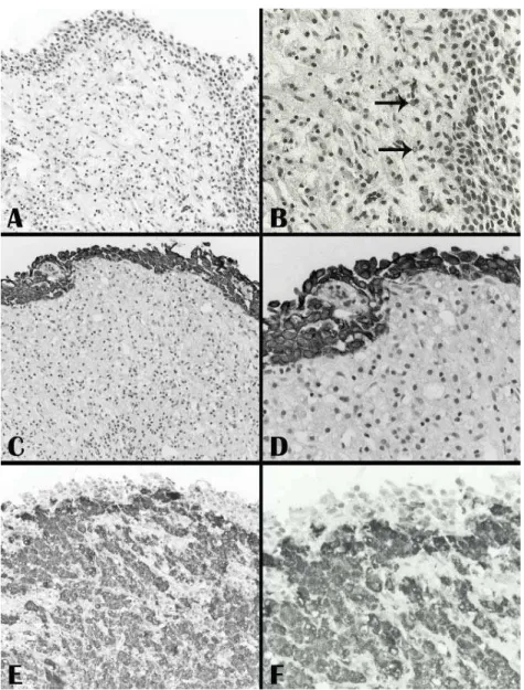
Histologically, on hematoxylin and eosin (HE) staining, aggregates of large macrophages with fine eosinophilic granular cytoplasm (von Hansemann cells) (Figure 1A) ad-mixed with basophilic inclusions (Michaelis-Gutmann bod-ies) (Figure 1B) and infiltrated by dense collections of lym-phocytes, and plasma cells were seen in lamina propria of urinary bladder. The macrophages were immunohistochemi-cally negative for citokeratin (Figures 1C, 1D) and positive for CD68 (Figures 1E, 1F). Immunohistochemical examina-tion of proliferative activity measured by Ki-67 in malako-plakia was negative. Pearls staining demonstrated deposition of calcium in Michaelis-Gutmann inclusions (Figure 2).
Fig. 2 – Malakoplakia,Michaelis Gutmann inclusion (black arrow) (Pearls staining, u 400).
Fig. 1 – A) Malakoplakia. Normal urothelium and von Hansemann’s cells (HE, u 200); B) Black arrows show Michaelis-Gutmann bodies (HE, u 400); C) Normal urothelium positive and von Hansemann’s cells negative for citokeratin
Strana 608 VOJNOSANITETSKI PREGLED Volumen 70, Broj 6
Ristiý-Petroviý A, et al. Vojnosanit Pregl 2013; 70(6): 606–608. Discussion
The first case of malakoplakia was described by von Hansemann who introduced the term “malakoplakia” 7. Malakoplakia is a chronic inflammatory disorder that occurs mostly in the genitourinary tract with a special affinity for bladder. Although malakoplakia in genitourinary tract is four times more common in women, in general, men above the age of 50 years are more frequently affected 5.
The symptoms of bladder malakoplakia are hematuria and irritative voiding symptoms such as frequency, hesitancy and dysuria 1, 2, 5. Macroscopically, as clinically, malakopla-kia can simulate tumors or abscesses, like we presented in this case.
The accurate pathogenesis has not been fully clarified, but it is thought to be a result of chronic infections by coli-forms in patients with chronic weariness or immunosupres-sion 1. The etiopathogenesis of malakoplakia appearance mainly include damaged host defenses and deficient phago-cytosis. Inadequate killing of bacteria, most commonly Es-cherichia coli, as a consequence of a defect in monocytes and macrophages phagolysosomal activity, results in an ac-cumulation of bacterial degradation products and a granu-lomatous reaction 8. However, partially digested bacteria ac-cumulate in macrophages, eventually become mineralized, forming the pathognomonic calcified intracellular inclusions called Michaelis-Gutmann bodies. Nevertheless, infectious etiology often remains only a suspicion as patients some-times have scarce symptoms and Gram staining does not al-ways succeed in revealing any bacteria 1, 2. Malakoplakia can be associated with inflammatory bowel disease which
sup-ports the theory that the malakoplakia is a consequence of chronic inflammation and altered regulation of the immune response 9.
Abundant accumulation of macrophages in lamina pro-pria of urinary bladder causes the intraluminal protrusion of bladder mucosa, like we presented in this particularl case. Since this clinical entity is very rare and nearly always oc-curs with dramatic hematuria it can easily be misdiagnosed.
According to the aforementioned we emphasize that the final diagnosis of malakoplakia is based only on cystoscopic biopsy and microscopic findings of characteristic acidofilic, foamy histiocytes with the presence of Michaelis-Gutmann in-clusions. Immunohistochemistry demonstrates CD68-positive macrophages.
Conclusion
Urinary bladder malakoplakia should be considered in immunocompromised or patients with neglected, persistent urinary tract infections and tumor mass at the cystoscopy. Early identification with prompt antibiotic treatment can be helpful in avoiding unnecessary surgical interventions and in preventing development of possible complications.
Declaration of interest
The authors declare no conflict of interest.
Acknowledgement
This research was supported by the Ministry of Educa-tion, Science and Technological Development of the Repub-lic of Serbia (grant No 175092).
R E F E R E N C E S
1. Shaktawat SS, Sissons MC. Malakoplakia of the appendix, an
uncommon entity at an unusual site: a case report. J Med Case Reports 2008; 2:181.
2. Patnayak R, Reddy MK, Subramanian S, Jena A, Ravisankar G,
Dandu RS. An unusual case of bilateral hydroureterone-phrosis caused by uretero-vesico malakoplakia in a young male: a case report and review of the literature. Cases J 2009; 2: 7527.
3. Velásquez-López JG, Vélez-Hoyos A, Uribe-Arcila JF.
Malakopla-kia in urology: a report of six cases and review of the literature. Actas Urol Esp 2006; 30(6):610î8. (Spanish)
4. Bylund J, Pais VM Jr. A case of acute renal failure caused by
bilateral, multifocal malacoplakia lesions of the bladder and ureters. Nat Clin Pract Urol 2008; 5(9): 516î9.
5. Chen YC, Kuo HC. Malakoplakia of ipsilateral kidney, ureter
and bladder. Tzu Chi Med J 2010; 22(2): 103î5.
6. Schwartz DA, Ogden PO, Blumberg HM, Honig E. Pulmonary
malakoplakia in a patient with the acquired immunodeficiency syndrome. Arch Pathol Lab Med 1990; 114(12): 1267î71.
7. Dasgupta P, Womack C, Turner AG, Blackford HN. Malacoplakia:
von Hansemann’s disease. BJU Int 1999; 84(4): 464î9.
8. Shah V, Rupani A, Pathak HR. Malacoplakia of urinary bladder.
Bom Hosp J 2010; 52(1):144î5.
9. Chung DE, Carr LK, Sugar L, Hladunewich M, Deane LA.
Xan-thogranulomatous cystitis associated with inflammatory bowel disease. Can Urol Assoc J 2010; 4(4): E9