Controversies in the diagnosis and treatment of deep vein
thrombosis for vascular ultrasound
Controvérsias no diagnóstico e tratamento da trombose venosa profunda pela ecografia vascular
Marcio Vinicius Lins Barros1, Virgínia Soares Rodrigues Pereira2, Daniel Mendes Pinto3
Introduction
Deep vein thrombosis (DVT) is a severe clinical entity characterized by the formation of thrombi in deep veins, no-tably in the lower limbs (80-95% of cases). DVT is the third leading cause of cardiovascular disease in the US, with appro-ximately 200 thousand new cases per year1. In Brazil, the
in-cidence is around 0.6 per 1,000 inhabitants/year. hree main factors are directly related to the genesis of thrombi: blood stasis, endothelial injury and hypercoagulability. Among the major complications, we can mention chronic venous insuiciency (post-phlebitic syndrome) due to injury of the venous valves leading to venous relux, and pulmonary embolism, which presents a high mortality rate, most ca-ses occurring among hospitalized patients, even though it
could be avoided with prophylactic measures such as the use of anticoagulants2.
Since Talbot3, in 1983, irst diagnosed thrombi in the
subclavian vein of a patient complaining of sudden pain and swelling in the arm using high-resolution ultrasono-graphy imaging, vascular echoultrasono-graphy became the method of choice for the diagnosis and follow-up of patients with DVT. he sensitivity and speciicity of this method compa-red to investigations with lebography is about 96%4.
However, several issues regarding the use of vascular ultrasound in the diagnosis of DVT remain controversial, such as the protocol to be used, the time for exam perfor-mance, and calf plexus thrombosis. he aim of this litera-ture review is to bring about a discussion on these issues based on current knowledge.
Abstract
Deep vein thrombosis is a potentially serious clinical entity, responsible for high morbidity and mortality. he vascular ultrasound is the diagnostic methods of choice in the diagnosis and monitoring of patients with this disease. However, several issues remain controversial, such as the initial approach of patients with suspected deep vein thrombosis, protocols to be used, the time for the exam and thrombosis in the calf plexus. he objective of this review is to discuss these issues in light of current knowledge.
Keywords: venous thrombosis; ultrasonography, Doppler, color; diagnostic techniques, cardiovascular.
Resumo
A trombose venosa profunda é uma entidade clínica potencialmente grave, responsável por elevada morbimortalidade. A ecograia vascular representa o método propedêutico de escolha no diagnóstico e acompanhamento dos pacientes com essa doença. Entretanto, várias questões permanecem controversas, tais como a abordagem inicial do paciente com suspeita de trombose venosa profunda, os tipos de protocolo a serem usados, o tempo para a realização do exame e a trombose no plexo de panturrilha. O objetivo dessa revisão é discutir esses assuntos à luz dos conhecimentos atuais.
Palavras-chave: trombose venosa; ultrassonograia Doppler em cores; técnicas de diagnóstico cardiovascular.
Study carried out at the Hospital Mater Dei – Belo Horizonte (MG), Brazil.
1 PhD in Medicine from Universidade Federal de Minas Gerais (UFMG); Coordinator of the Service of Echocardiography and Vascular Ultrasound at Hospital Mater Dei – Belo Horizonte (MG), Brazil; 2 Resident in Vascular Ultrasound at Hospital Mater Dei – Belo Horizonte (MG), Brazil.
3 Full member of the Brazilian Society of Angiology and Vascular Surgery (SBACV); Coordinator of the Service of Angiology and Vascular Surgery at Hospital Mater Dei – Belo Horizonte (MG), Brazil.
Financial support: none.
Deep vein thrombosis diagnosis at emergency
Facing a patient with suspected DVT, many questions are raised: what is the best diagnostic strategy? What is the most appropriate time to perform diagnosis? Should I start treatment right away?
Although DVT cause few specific symptoms, well-directed anamnesis and physical examination are essential in the initial management of patients with fin-dings that may suggest DVT. Knowing the main factors related to the thrombotic process genesis, such as pre-vious surgery, immobilization for more than three days, neoplasms and hormone therapy with estrogen associa-ted with pain and unilateral limb edema are strongly re-lated to DVT and can be classified according to clinical prediction models5. Wells et al. developed a model of
patient classification based on signs and symptoms, risk factors and alternative diagnoses, thus estimating a pre-test probability as low, medium and high-risk for DVT (Table 1). This classification has been proved useful in the initial management of patients6,7.
Once clinical indings not always correlate with patho-logical changes (clinical diagnosis is correct only in 50% of cases) and, as undiagnosed DVT can lead to fatal pulmona-ry embolism, an totally preventable condition when the ap-propriate treatment is established in time, complementary exams and speciic vascular propedeutics is recommended to conirm or exclude this diagnosis8,9.
Dosage of D-dimer is one of the tests used for initial evaluation of patients with suspected DVT and is used in any situation with ibrin formation and degradation; there-fore, it is not a speciic marker. D-dimer negative predictive value is 94-95%10, which indicates an incidence of DVT of
5-6% ater the test, which is not sensitive enough to exclude the hypothesis of deep venous thrombosis11. Studies have
established two main features in the diagnosis of venous thromboembolism: the need to combine D-dimer determi-nation with pretest clinical probability score before procee-ding with the diagnostic investigation.
hese tests combined will reduce the incidence of DVT ater the test to less than 0.5% and the need for ultrasono-graphy to 40-50%12.
Several studies have suggested that consecutive asses-sments of clinical score, D-dimer test and vascular ultra-sound bring better results when it comes to the cost-efec-tiveness of DVT diagnosis, and are related to a signiicant reduction of ultrasound requests and increase in time for the patient and the physician
In patients with low risk for DVT, negative D-dimer score is related to a negative predictive value to reducing the need for other imaging examinations, and the use of clinical criteria associated with D-dimer score has a good cost-efectiveness value.
But how long can we wait to perform vascular ultra-sound examination? he clinical suspicion of thrombosis represents a major impact for the patient and demands immediate investigation. In Brazil, only physicians can perform ultrasonography and the 24-hour shits of health professionals represent a challenge for hospital costs mana-gement. Some studies, however, have shown protocols that enable proper diagnosis that do not demand the professio-nal to work out of their shit13,14.
Based on these studies and diagnostic guidelines15-17,
we have been using a protocol that enables an eicient diag-nostic method without therapeutic loss (Figures 1-4):
• outpatients with low risk for DVT and negative
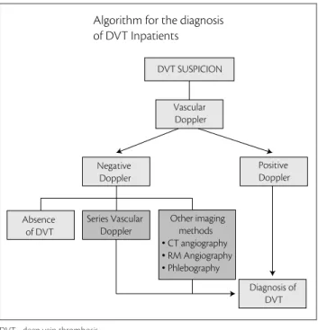
D-dimer. hese factors have a high negative pre-dictive value when associated. hus, there is no need for imaging tests to exclude DVT (Figure 1); patients with moderate to high probability require imaging studies (such as vascular ultrasound, in cases of suspected cavo-iliac thrombosis, CT angiography or magnetic resonance angiography) (Figures 2 and 3); hospitalized patients require imaging studies (such as vascular ultrasound in cases of suspected cavo-iliac thrombosis, CT angiography or magnetic resonance angiography) for diagnosis (Figure 4).
Protocols
Diferent protocols have been proposed for the ultra-sonographic evaluation in DVT diagnosis: assessing all
Ongoing neoplasms (under treatment in the last 6 months): 1 point
Palsy, paresia or recent imobilization of lower limbs: 1 point
Recent need for prescribed rest for mor than 3 days OR major surgery that required general raquidian anesthesia or in the last 12 weeks: 1 point
Pain at palpation of the deep venous path of lower limbs: 1 point
Whole limb swelling: 1 point
Bigger Cacifo sign on the afected limb: 1 point
Swelling of the afected calf, with 3 cm of diference compared to the contralateral limb (measure 10 cm below tibial tuberosity): 1 point
Superitial collateral veins (non varicose): 1 point
More likely diferential diagnosis:<2 points
Source: modiied from Rollo et al.17.
Risk interpretation score: 0 points – low; 1 to 2 points – moderate; >2 points – high.
venous segments of the lower limb, as well as the entire pro-ximal (femoropopliteal) segment, and even the two-point evaluation (common femoral and popliteal veins).
Ultrasonography with two-points compression for DVT investigation on the lower limbs, performed by physicians in the emergency room were proven accurate for the identification thrombosis18,19. A randomized
stu-dy published in 2008 showed that both diagnostic stra-tegies (conventional and two-point protocol) were equi-valent when used for the management of symptomatic outpatients with suspicion of DVT of the lower limbs in relation to the incidence of venous thromboembolism (VTE) after three months of follow-up20.
Although several protocols which address the pro-ximal segment only have shown excellent short-term prognosis, we believe that evaluating the whole venous system is essential to the adequate approach, for, al-though an infrapopliteal DVT cannot determine unfa-vorable short-term outcomes, proper diagnosis is extre-mely important for the patient, as a matter of secondary prevention approach facing a recurrence. Furthermore, examining the infrapopliteal segment allows diagnosis of other pathologies such as Baker’s cyst, hematomas, and muscle ruptures.
Should deep vein thrombosis investigation be bilateral?
he evaluation of bilateral DVT in patients with symp-toms in only one of the lower limbs is a controversial issue. Garcia et al. found no signs of DVT in the asympto-matic limbs of outpatients with unilateral symptoms at vascular ultrasound, so the investigation of the symp-tomatic limb was enough to diagnosis. However, inpa-tients with unilateral symptoms were diagnosed with thrombosis on the symptomatic side in 24% of cases, on the asymptomatic limb in only 5%, and on both limbs in 5% of cases21. In another study, Lemech et al. found
about 10% of bilateral DVT in patients with unilateral symptoms, thus suggesting that inpatients should have both limbs investigated22.
Pennell et al. showed that inpatients have a high incidence of clinically silent contralateral thrombosis (34%) and usually must undergo bilateral examination, as well as patients with malignant disease, whose inci-dence of asymptomatic blood clots is 38%. Outpatients with unilateral symptoms and without risk factors for thrombosis should undergo unilateral examination and be treated properly according to the results. Algorithms to select patients for unilateral studies should include
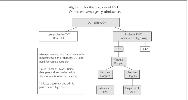
Less probable DVT (low risk)
Probable DVT (moderate to high risk) DVT SUSPICION
Vascular Doppler
DD+
Absence of DVT
DD
-Negative Doppler
Positive Doppler
Absence of DVT
Diagnosis of DVT
Management options for patients with low risk, DD+ and referred to vascular Doppler:
* Give 1 dose of LMWH (initial therapeutic dose) and schedule the examination for the next day
* Initiate treatment and admit patients with high risk.
Algorithm for the diagnosis of DVT Outpatients/emergency admittances
DVT - deep vein thrombosis; DD - negative D-dimer; DD+ - positive D-dimer; LMWH - low-molecular-weight heparin.
Less probable DVT (low risk)
Probable DVT (moderate to high risk)
Positive Doppler
Diagnosis of DVT DVT SUSPICION
Vascular Doppler
DD - DD+
Negative Doppler
Absence of DVT Management options for patients with
moderate to high probability, DD- and need for vascular Doppler:
* Give 1 dose of LMWH (initial therapeutic dose) and schedule the examination for the next day
* Initiate treatment and admit patients with high risk.
Algorithm for the diagnosis of DVT Outpatients/emergency admittances
DVT - deep vein thrombosis; DD - negative D-dimer; DD+ - positive D-dimer.
Figure 2. Algorithm for the diagnosis of deep vein thrombosis for outpatients or emergency admittances – moderate to high risk of DVT and DD-.
Series Vascular Doppler. Repeat examination in 24-48h
Positive Doppler Negativ
Doppler
Another imaging method
Diagnosis of DVT DVT SUSPICION
Vascular Doppler
DD- DD+
Initiate treatment with LMWH (therapeutic dose) Probable DVT
(moderate to high risk)
Absence of DVT Management options for patients with moderate to high probability, DD+ and need for vascular Doppler:
* Initiate treatment and admit patients with high risk.
* Give 1 dose of LMWH (initial therapeutic dose) and schedule the examination for the next day
*If Doppler is negative for thrombosis, consider series Doppler or another imaging method.
Algorithm for the diagnosis of DVT Outpatients/emergency admittances
DVT - deep vein thrombosis; DD - negative D-dimer; DD+ - positive D-dimer.
data from active malignant diseases, recent trauma or surgery, pregnancy, hormone therapy or history of thrombophilia23.
DVT in calf muscle veins
he distal DVT or calf veins occurs in infrapoplíte-al veins, i.e., posterior tibiinfrapoplíte-al veins, peroneinfrapoplíte-al veins and cinfrapoplíte-alf muscle veins (gastrocnemius and soleus plexus). While sensitivity and speciicity of compression ultrasonography in proximal DVT are high and the treatment with anticoa-gulants is well established, distal DVT is less severe (50-75% sensitivity and 90-95% speciicity). Unlike proximal DVT, the distal DVT diagnosis and treatment approach remain controversial24-26.
Lagerstedt et al. (1985) showed that the use of anticoa-gulant for three months in patients with venous thrombosis in the calves signiicantly reduced recurrences and compli-cations in symptomatic patients, compared to patients trea-ted by other agents27.
Philbrick et al., in a literature review of 20 studies, sho-wed that calf thrombosis may spread proximally in about 20% of cases, and that anticoagulation in symptomatic patients may prevent the spread, embolization, and early recurrence. Follow-up for 1 week to assess the thrombus propagation is an alternative to anticoagulation28. A study
by Lohr et al. showed that about 32% of patients presented signs of progression, and 75 patients (5%) presented signs suggestive of pulmonary embolism29.
he CALTHRO study, conducted with 431 patients wi-thout proximal DVT, which evaluated the distal segment, showed that 15.3% of the sample had distal DVT. here was a signiicant diference in the onset of new events in three months among patients with distal DVT (5/64, 7.8% ver-sus 3/351, 0.8%, p=0.003). he study then led the medical
community to the conclusion that a negative outcome in patients with distal DVT who received no treatment may be relevant30.
However, recent studies have shown that the importan-ce of infrapopliteal DVT diagnosis and treatment can be at least questioned due to the absence of improvement as to recanalization, progression and complications, besides the fact that an increase in the number of diagnoses of distal DVT using vascular ultrasound may lead to an increase in the number of patients receiving oral anticoagulant therapy, thus resulting in excess treatment25.
Cliford et al. performed a retrospective study and found no signiicant diference as to disease progression with anticoagulant treatment in patients with distal DVT31.
In a randomized trial with 107 patients, 54 using low-molecular-weight heparin for a short period and 53 patients with venous compression, Schwarz et al. found no differences between groups as to pulmonary embo-lism, death occurrences, hemorrhage, and degree of re-canalization32. Sule et al., on the other hand, showed no
significant differences between the group receiving anti-coagulation and patients who did not received it when it comes to the progression of disease, recanalization, pul-monary embolism and death occurrences33.
Further randomized clinical trials evaluating the true efectiveness of anticoagulation in the treatment of distal DVT are thus needed. Righini et al. have been developing the CACTUS study, which was initiated in 2008 and is expected to be concluded in 2013. he authors expect to allocate about 600 patients in a randomized, double-blind study aimed to determine the efectiveness of nadroparin treatment (low-molecular-weight heparin) compared to placebo in patients with the irst episode distal DVT.
he current recommendation of the American College of Chest Physicians34 is to treat distal DVT with
anticoagu-lants for three months. Given the conlicting results of stu-dies presented here, the management of patients with distal DVT remains controversial in clinical practice. Recently, a systematic review by Masuda et al. analyzing over 1,500 ar-ticles on the subject, although there were no data that could clarify the controversy surrounding the best treatment for
Positive Doppler
Diagnosis of DVT DVT SUSPICION
Other imaging methods
• CT angiography
• RM Angiography
• Phlebography
Vascular Doppler
Negative Doppler
Absence of DVT
Series Vascular Doppler
Algorithm for the diagnosis of DVT Inpatients
DVT - deep vein thrombosis.
infrapopliteal DVT, showed that due to the risk of propa-gation, pulmonary embolism, and recurrence, not taking any approach facing distal DVT should be unacceptable. In the absence of strong evidence, both anticoagulation and follow-up with imaging methods and selective anticoagu-lation remain as the acceptable treatment methods35. De
Martino et al., in a recent meta-analysis aimed to evaluate the efectiveness and safety of anticoagulation in patients with calf DVT, showed that episodes of pulmonary em-bolism and propagation of thrombosis were less frequent among patients who received anticoagulants36.
Vascular ultrasound has revolutionized the diagno-sis and management of DVT, enabling a non-invasive and high-accurate management of several anatomical and functional features determined by the thrombus for-mation and sequelae. The clinical practice over the last 30 years have enabled a better understanding of many controversial issues, such as those presented throughout this literature review. However, there are still gaps that may only be filled by further studies conducted with ade-quate methodology.
References
1. White RH. he epidemiology of venous thromboembolism. Circulation. 2003;107:I4-8
2. Mafei FHA, Rollo HA. Trombose venosa profunda dos mem-bros inferiores: incidência, patologia, patogenia, isiopatologia e diagnóstico. In: Mafei FHA, Lastória S, Yoshida WB, Rollo HA. Doenças vasculares periférica. 3ª ed. Rio de Janeiro, MEDSI, 2002. p. 1363-86.
3. Talbot SR. Use of real-time imaging in identifying deep venous obstruction: a preliminary report. Bruit. 1982;6:41.
4. Goodacre S, Sampson F, homas S, van Beek E, Sutton A. Systematic review and meta-analysis of the diagnostic accuracy of ultrasonography for deep vein thrombosis. BMC Medical Imaging. 2005;5:6.
5. Mafei FHA, Caiafa JS, Ramacciotti E, Castro AA para o Grupo de Elaboração de Normas de Orientação Clínica em Trombose Venosa Profunda da SBACV. Normas de orientação clínica para prevenção, diagnóstico e tratamento da trombose venosa profun-da (revisão 2005) Salvador: SBACV; 2005. J Vas Bras. 2005;4(Suppl 3):S205-20.
6. Wells PS, Hirsh J, Anderson DR, et al. Accuracy of clinical assess-ment of deep-vein thrombosis. Lancet .1995;345(8961):1326-30.
7. Anderson DR, Wells PS, Stiell I, et al. hrombosis in the emer-gency department: use of a clinical diagnosis model to safely avoid the needfor urgent radiological investigation. Arch Intern Med. 1999;159(5):477-82.
8. Seidel AC, Silva JCCB, Miranda Jr F. Diagnóstico clínico e exames subsidiários da trombose venosa profunda. Rev Bras Clin Med. 2003;I(3):74-82.
9. Seidel AC, Miranda Jr F, Cavalheri Jr G. he role of duplex ultraso-nography in the diagnosis of lower-extremity deep vein thrombo-sis in non-hospitilized patients. Int Angiol. 2008;27(5):377-84.
10. Heim SW, Schectman JM, Siadaty MS, Philbrick JT. D-dimer tes-ting for deep venous thrombosis: a metaanalisys. Clin Chem. 2004;50(7):1136-47.
11. Júnior JEA, Jardim C, Souza R. D-Dímero para exclusão de trom-bose venosa profunda e tromboembolismo pulmonar. Rev Assoc Med Bras. 2004;50:232-3.
12. Michiels JJ, Freyburger G, Van der Graaf F, Janssen M, Oortwijn W, Van Beek EJ. Strategies for the safe and efective exclusion and diagnosis of deep vein thrombosis by the sequential use of clinical score, D-dimer testing, and compression ultrasonography. Semin hromb Hemost. 2000;26(6):657-67.
13. Arnaoutakis GJ, Pirrucello J, Brooke BS, Reifsnyder T. Venous duplex scanning for suspected deep vein thrombosis: results before and after elimination of after-hours studies. Vasc Endovascular Surg. 2010;44(5):329-33.
14. Rathbun SW, Whitsett TL, Raskob GE. Exclusion of irst-episode deep-vein thrombosis after-hours using D-dimer. Blood Coagul Fibrinolysis. 2007;18(8):795-800.
15. Qaseem A, Snow V, Barry P, et al. Current diagnosis of venous thromboembolism in primary care: a clinical practice guideli-ne from the American Academy of Family Physicians and the American College of Physicians. Ann Intern Med. 2007;146(6):454-8
16. Bharadia V. D-dimer for the exclusion of acute venous thrombosis and pulmonary embolism. A systematic reveiw. Ann Intern Med. 2004;140:589-602.
17. Rollo HA, Fortes VB, Junior ATF, Yoshida WB, Lastória S, Mafei FHA. Abordagem diagnóstica dos pacientes com suspeita de trombose ve-nosa profunda dos membros inferiores. J Vasc Bras. 2005;4(1):79-92.
18. Cogo A, Lensing AW, Prandoni P, Hirsh J. Distribution of thrombosis in patients with symptomatic deep vein thrombosis. Implications for simplifying the diagnostic process with compression ultra-sound. Arch Intern Med. 1993;153(24):2777-80.
19. Crisp, Jonathan G, Lovato LM, Jang TB. Compression phy of the lower extremity with portable vascular ultrasonogra-phy can accurately detect deep venous thrombosis in the emer-gency department. An Emerg Med. 2010;56:611-3.
20. Bernardi E, Camporese G, Büller HR, et al. Serial 2-point graphy plus D-dimer vs whole-leg color-coded Doppler ultrasono-graphy for diagnosing suspected symptomatic deep vein throm-bosis: a randomized controlled trial. JAMA. 2008;300:1653-9.
21. Garcia ND, Morasch MD, Ebaugh JL, et al. Is bilateral ultrasound scanning of the legs necessary for patients with unilateral symp-toms of deep vein thrombosis? J Vasc Surg. 2001;34(5):792-7.
22. Lemech LD, Sandroussi C, Makeham V, Burnett A, Harris JP. Is bi-lateral duplex scanning necessary in patients with symptoms of deep venous thrombosis? ANZ J Surg. 2004;74(10):847-51.
23. Pennell RC, Mantese VA, Westfall SG. Duplex scan for deep vein thrombosis-deining who needs an examination of the contralate-ral asymptomatic leg. J Vasc Surg. 2008;48:413-6.
25. Righini M. Is it worth diagnosing and treating distal deep vein thrombosis? No. J hromb Haemost 2007;5 (Suppl. 1):55-9.
26. Lautz TB, Abbas F, Walsh SJ, et al. Isolated gastrocnemius and soleal vein thrombosis: should these patients receive therapeutic antico-agulation? Ann Surg. 2010;251(4):735-42.
27. Lagerstedt CI, Olsson CG, Fagher BO, Oqvist BW, Albrechtsson U. Need for long-term anticoagulant treatment in symptomatic calf--vein thrombosis. Lancet. 1985;2(8454):515-8.
28. Philbrick JT, Becker DM. Calf deep venous thrombosis. A wolf in sheep’s clothing? Arch Intern Med. 1988;148:2131-8.
29. Lohr JM, Kerr TM, Lutter KS, Cranley RD, Spirtof K, Cranley JJ. Lower extremity calf thrombosis: to treat or not to treat? J Vasc Surg. 1991;14(5):618-23.
30. Palareti G, Cosmi B, Lessiani G, Rodorigo G, Guazzaloca G, Brusi C,. Evolution of untreated calf deep-vein thrombosis in high risk symptomatic outpatients: he blind, prospective CALTHRO study. hromb Haemost. 2010:104(5):1063-70.
31. Cliford MS, Faheem H, Rami B, Frances S. Management of iso-lated soleal and gastrocnemius vein thrombosis. J Vasc Surg. 2010;52(5):1251-4.
32. Schwarz T, Buschmann L, Beyer J, Halbritter K, Rastan A, Schellong S. herapy of isolated calf muscle vein thrombosis: A randomized, controlled study. J Vasc Surg 2010;52(5):1246-50.
33. Sule AA, Chin TJ, Handa P, Earnest A. Should symptomatic, isola-ted distal deep vein thrombosis be treaisola-ted with anticoagulation? Int J Angiol 2009;18:83-87.
34. Kearon C, Kahn SR, Agnelli G, Goldhaber S, Raskob GE, Comerota AJ. Antithrombotic therapy for venous thromboem-bolic disease: American College of Chest Physicians Evidence-Based Clinical Practice Guidelines (8th Edition). Chest. 2008;133(6 Suppl):454S-545S.
35. Masuda EM, Kistner RL, Musikasinthorn C, Liquido F, Geling O, He Q. The controversy of managing calf vein thrombosis: A systematic review. J Vasc Surg. 2012;55(2):550-61. Epub 2011 Oct 26.
36. De Martino RR, Wallaert JB, Rossi AP, Zbehlik AJ, Suckow B, Walsh DB. A meta-analysis of anticoagulation for calf deep venous thrombosis. J Vasc Surg. 2011 Dec 29.
Correspondence Márcio Vinícius Lins Barros Rua Carangola, 57, apto. 1.201 – Santo Antonio
CEP 30330-240 – Belo Horizonte (MG), Brazil E-mail: [email protected]