INNOVATION
Technical note: ultrasonography evaluation of aortic
aneurysms treated with endoprosthesis
Nota técnica: avaliação ultrassonográfica de aneurismas da aorta tratados com endopróteses
Sergio Xavier Salles Cunha1
Introduction
Aortic aneurysms treated with endoprosthesis may de-velop endoleaks1,2. An endoleak is a leak of blood low, or
prosthesis, or a branch of the aorta into the aneurysm. hey have been classiied according to their anatomical location3.
his technical note emphasizes the hemodynamics of en-doleaks’ low, describes the concepts of endoseepage and intermittent endoleak, and summarizes the technique of intravascular ultrasound virtual histology (IVUS-VH) for the detection of blood or luids, acute, subacute or ibrotic thrombi inside the aneurysm.
Endoleak flow types
Endoleaks have been identiied with at least three types of low:
1. similar to that of a pseudoaneurysm; 2. channel low through the aneurysm; 3. direct low into the aneurysm.
Pseudoaneurysm flow
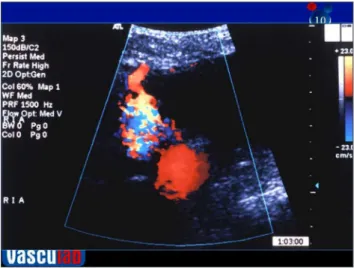
Figure 1 shows the flow of an endoleak into a pseu-doaneurysm inside an aortic aneurysm treated with
Abstract
Aortic aneurysms treated with endovascular techniques may develop endoleaks. In addition to anatomic location, these endoleaks should be classiied based on the type of blood low. We describe the types of blood low into the endoleaks as detected by Doppler ultrasonography: in-and-out low as a “pseudoaneurysm”, channel low through the aneurysm, and direct low into the aneurysm. Endoleaks may be intermittent and actually be endoseepage, detected by aneurysmal growth without an apparent inlow. Ultrasound virtual histology is suggested to detect endoseepage. Relative quantity and location of blood/liquid in the aneurysm can be detected by evaluating pixel brightness of the ultrasonographic B-mode image. Such anatomofunctional information allows for quantitative follow-up and emphasizes the risk of an endoleak with direct low into the aneurysm.
Keywords: aortic aneurysm; endovascular procedures; endoleak; ultrasonography.
Resumo
Aneurismas da aorta tratados com endopróteses podem desenvolver endoleaks. Esses endoleaks devem ser classiicados com base nos tipos de luxo, além da localização anatômica. Enfatizamos tipos de luxo dos endoleaks detectados pela ultrassonograia com Doppler: luxo entra-e-sai como “pseudoaneurisma”, luxo de canal e luxo direto. Ao se notar a possibilidade de endoleaks intermitentes, salientamos o conceito de endoseepage detectáveis por crescimento de dimensões sem luxo entrante aparente. Sugerimos o uso da histologia virtual ultrassonográica para detecção de iniltrações. Quantidade relativa e localização de sangue ou líquido dentro do saco aneurismático pode ser detectado pela avaliação dos níveis de brilho da ultrassonograia modo B. Essa informação permite um seguimento compreensivo do aneurisma tratado com endoprótese e salienta o risco maior deum endoleak de luxo direto entrante no aneurisma.
Palavras-chave: aneurisma aórtico; procedimentos endovasculares; endoleak; ultrassonograia.
Study carried out at the Angiolab Curitiba – Curitiba (PR), Brazil.
1 Consultant of non-invasive techniques for vascular diagnosis at Angiolab Curitiba – Curitiba (PR), Brazil; e no Angiloab Vitória – Vitória (ES), Brazil.
Financial support: none.
Ultrasound of aortic aneurysm with endoprosthesis - Cunha SXS J Vasc Bras 2012, Vol. 11, Nº 2 151
endoprosthesis. The flow into the pseudoaneurysm is characterized as in- and outflow. This type of endoleak does not seem to contribute with increase in the diam-eter or volume of the treated aneurysm. Occasionally, the pseudoaneurysm may thrombose and occlude. However, thrombosis of the pseudoaneurysm does not guarantee the closure of the endoleak. The flow can change direction, creating a new pseudoaneurysm in other area of the aneurysm. Periodic monitoring of this type of endoleak is recommended, even if the first pseudoaneurysm has been closed. This type of endole-ak may be intermittent.
Channel flow
Figure 2 shows a blood low channel inside the aortic aneurysm treated with endoprosthesis. his channel may have varied sources of blood low, such as the endoprosthe-sis itself, or a branch of the aorta or the iliac arteries. he drainage of this channel also varies, but it is generally found at a branch of the aneurysm. his channel is apparently restricted by a chronic thrombosed tissue, that is, a blood thrombus in various stages of ibrosis. he blood leakage from the channel into the aneurysm depends on the type of tissue around this false lumen. Apparently, this type of
Figure 2. Endoleak of aortic aneurysm treated with endoprosthesis. Channel low inside the aneurysm.
Figure 3. Endoleak of aortic aneurysm treated with endoprosthesis. Direct blood low inside the aneurysm. he high speed shows aliasing – light color at the end of the prosthesis.
Ultrasound of aortic aneurysm with endoprosthesis - Cunha SXS J Vasc Bras 2012, Vol. 11, Nº 2
152
endoleak does not dramatically afect the diameter or vol-ume of the treated aneurysm. However, it is this type of en-doleak is thought to be more dangerous than the enen-doleak similar to that of a pseudoaneurysm. A closer follow-up is recommended in such cases.
Direct flow
Figure 3 shows a direct low of endoleak into the an-eurysm sac. his type of endoleak is classiied as high-risk. he volume of blood drained into the aneurysm may quickly rupture the aneurysm wall. his aneurysm ruptured two hours ater the ultrasound and, as a conse-quence, the 93-year-old patient, who was not candidate for revascularization, died. he vascular surgeon should be in-formed immediately in case of endoleak direct low shown at ultrasonography.
Endoseepage
Diameter measurements illustrate cases of endo-seepage. he continuing growth of 2 cm in diameter per year of an aneurysm is equivalent to 0.05 mm per day or 0.002 mm per hour. It is not well established whether en-doseepage can be identiied by any modern low measure-ment technique. he concept of endoseepage was then created for aneurysms that grow in size without an appar-ent source of blood low.
Intermittent endoleak
Some studies suggest that Doppler low ultrasound is more accurate than radiographic techniques used to de-tect endoleaks, while other studies show the other way
around4-6. In general, ultrasonography has detected more
endoleaks than computed tomography scan; the low posi-tive predicposi-tive value of ultrasonography can be analyzed as CT scan false-negative result.
Another explanation for these discrepancies is that these techniques were not performed simultaneously, and that the endoleak could be intermittent. An intermit-tent endoleak could explain the contradictory results, the change in direction of the pseudoaneurysm-like endoleaks in follow-up, or the growth of the aneurysm dimensions without an apparent source of endoleak. Monitoring of the dimensions, diameter and even of the volume parallel to the low is recommended.
Ultrasound-virtual histology
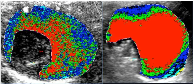
Figure 4 shows two Ultrasound-virtual histology im-ages of aneurysms treated with endoprosthesis. his tech-nique has artiicial color brightness levels of the B-mode
ul-trasound imaging7-10. he image shows regions with echoes
similar to blood, lipids, muscle, iber and calcium. In the case of aortic aneurysms, it would be interpreted as blood or luid, recent thrombus or thrombi in various stages of
Ultrasound of aortic aneurysm with endoprosthesis - Cunha SXS J Vasc Bras 2012, Vol. 11, Nº 2 153
ibrosis. Based on intravenous studies, the acute throm-bus would generate images similar to lipids (green stain in Figure 4); the subacute thrombus would tend to appear in bright blue8,9.
he virtual histology technique creates a range of brightness in the B-mode ultrasound imaging. Usually, these images have 256 levels of brightness. In these appli-cations, the blood (or what appears in black) is zero in the scale. he adventitia (or what is in white at the prosthesis) is 200 in the scale. Blood, lipids, muscle, iber, and calcium have echoes in the intervals 0-4, 8-26, 41-76, 112-196 e 211-2557.
Evaluation of endoleaks, endoseepage and dimensions is recommended in periodic follow-up of aneurysms with hypoechoic regions deined by B-mode virtual histology imaging.
Conclusions
Simple analysis of ultrasound B-mode imaging or the complex processing of the image using the technique of Ultrasound-virtual histology can identify hypoechoic re-gions inside the aneurysm treated with endoprosthesis. Duplex and Doppler ultrasound imaging can show the lo-cation and type of endoleak low. he results of these imag-ing techniques can be used to determine the follow-up of the aneurysm or the need for emergency care of an aneu-rysm with direct-low endoleak.
References
1. Silvestre JMS, Motta F, Sardinha WE, et al. Tratamento endovas-cular do aneurisma da aorta abdominal infrarrenal em pacien-tes com anatomia favorável para o procedimento – experiência inicial em um serviço universitário. J Vas Bras. 2011;10(1):31-9
2. Mendonça CT, Moreira RCR, Timi JRR, et al. Comparação entre os tratamentos aberto e endovascular dos aneurismas da aor-ta abdominal em pacientes de alto risco cirúrgico. J Vasc Bras. 2005;4(3):232-42.
3. Cao P, De Rango P, Verzini F, et al. Endoleak after endovascular aortic repair: classiication, diagnosis and management following
endovascular thoracic and abdominal aortic repair. J Cardiovasc Surg (Torino). 2010;51(1):53-69.
4. Schmieder GC, Stout CL, Stokes GK, et al. Endoleak after endovas-cular aneurysm repair: duplex ultrasound imaging is better than computed tomography at determining the need for intervention. J Vasc Surg. 2009;50(5):1012-7.
5. Raman KG, Missig-Carroll N, Richardson T, et al. Color-low du-plex ultrasound scan versus computed tomographic scan in the surveillance of endovascular aneurysm repair. J Vasc Surg. 2003;38(4):645-51.
6. Manning BJ, O’Neill SM, Haider SN, et al. Duplexultrasound in aneurysm surveillance following endovascular aneurysm repair: acomparison with computed tomography aortography. J Vasc Surg. 2009;49(1):60-5.
7. Lal BK, Hobson RW 2nd, Pappas PJ, et al. Pixel distribution analysis of B-modeultrasound scan images predicts histologic features of atherosclerotic carotidplaques. J Vasc Surg. 2002;35(6):1210-7.
8. Cassou-Birckholz MF, Engelhorn CA, Salles-Cunha SX, et al. Assessment of deep venous thrombosis by grayscale median anal-ysis of ultrasound images. Ultrasound Q. 2011;27(1):55-61.
9. Menezes FH, Silveira SAF, Salles-Cunha SX. Pixel characterization for development of ultrasound-based virtual histology of deep venous thrombosis. Apresentado no Congresso Anual da Society of Vascular Ultrasound (SVU), Chicago, IL, 15-18 de Junho, 2011, e-published na svunet.org (members only), Presentation Abstract 109, p. 3
10. Engelhorn AL, Engelhorn CA, Salles-Cunha SX. Initial evaluation of virtual histology ultrasonographic techniques applied to a case of renal transplant. Apresentado no Congresso Anual da Society of Vascular Ultrasound (SVU), Chicago, IL, USA, 15-18 de Junho, 2011, e-published na svunet.org (members only) Poster Abstract 412, p.20
Correspondence
Sergio Xavier Salles Cunha Rua Ernesto Zwarg, 215 – Satélite CEP 11740-000 – Itanhaém (SP), Brasil E-mail: [email protected]
Authors’ contributions