ABSTRACT Alexandre Wada
Marcus Castro Ferreira
Paulo Tuma Júnior
Gino Arrunátegui
ORIGINAL AR
TICLE
Experience with local negative
pressure (vacuum method) in the
treatment of complex wounds
Division of Plastic Surgery, Hospital das Clínicas, Faculdade de
Medicina da Universidade de São Paulo, São Paulo, Brazil
CONTEXT AND OBJECTIVE:Use of topical nega-tive pressure over diffi cult-to-heal wounds has been studied. The objective of this study was to analyze the effects from negative pressure in the treatment of complex wounds.
DESIGN AND SETTING: Case series developed at Hospital das Clínicas, Faculdade de Medicina da Universidade de São Paulo.
METHODS:Twenty-nine patients with complex wounds were treated with the vacuum system and then underwent a surgical procedure to close the wound.
RESULTS: 85% of the skin grafts took well, and 87.5% of the local fl aps were successful, thus demonstrating adequate wound preparation. The wounds were closed within shorter times than observed using other conventional treatments In two cases, the vacuum system was also used to stabilize the skin grafts over the wounds. CONCLUSIONS: Use of the vacuum method is safe and effi cient for preparing wounds for surgi-cal closure. It allows for an improvement of losurgi-cal wound conditions, and healthy granulation tissue develops with control over local infection. KEY WORDS: Wound healing. Vacuum. Plastic surgery. Leg ulcer. Decubitus ulcer.
INTRODUCTION Treatment of wounds is one of the most challenging situations that a plastic surgeon can face. Wound healing consists of a series of events that differ one from the other, leading to uncertainty that frequently makes the surgeon choose incorrect or inadequate methods for treating the problem. Also, the wide range of dressings and drugs that are available for treat-ing chronic wounds means that it is not always clear which of these is most indicated. The surgeon should decide which surgical technique is best indicated for the specifi c case by consid-ering its effectiveness, safety and cost.
Nowadays, because of greater severity of patients’ illnesses, chronic wounds have become prevalent. In a study conducted in our hospital, about 8% of the patients had chronic wounds, and this situation may be more critical in geriatric hospitals or intensive care units.* The wounds included pressure sores, wounds on diabetic feet, and wounds of vascular origin. The morbidity rate and treatment costs are high and social integration after hospital discharge may be compromised. Recent studies have shown the importance of faster treatment of wounds among in-hospital patients in order to reduce such costs.
The treatment of complex wounds has changed greatly in wound centers over the last decade. It has included aggressive surgical debridement for removal of necrotic tissue, thus allowing better control over local infec-tion. As a second stage, the wound bed should be prepared so as to lead to the formation of healthy granulation tissue and thus reach the third stage: surgical closure using skin grafting, local fl aps or microsurgical fl ap transplanta-tion. Wound contraction and spontaneous epithelization should be kept to a minimum, thus avoiding prolonged hospital stay and recurrence of infection.
New dressings that help in wound prepa-ration have been described, as well as physical methods such as electric current and others. Local negative pressure may be included in this group of agents that improve bed preparation, since this stimulates granulation tissue and provides mechanical drainage of the wound.
The use of negative pressure over the wound surface was fi rst described by Morikwas and Argenta in 1997.1,2 It was demonstrated
that a vacuum device removing fl uid from the wound but not drying it completely produced a better environment for epithelial prolifera-tion and fi broblast migraprolifera-tion. Because of this capacity for removing fl uids, vacuum condi-tions also remove bacteria from the wound, thus reducing the bacterial population. Nega-tive pressure applied over the current surface accelerates the formation of granulation tissue, and promotes increased capillary circulation. Moreover, the mechanical effect of the vacuum attracts the borders of the wound towards the center, thus reducing the wound volume.
This method has been approved by the United States Food and Drug Administration (FDA). Because there was no previous experi-ence with this method in Brazil, we started to use it in Hospital das Clínicas, Sao Paulo, in 2002. Preliminary results from three patients were published in 20033 and we now present
our more extensive experience, with results from 29 patients.
OBJECTIVE The objective of this study was to evaluate the use of negative pressure on the treatment of chronic wounds.
METHODS Twenty-nine patients with complex wounds were treated in a tertiary hospi-tal (Hospihospi-tal das Clínicas, Faculdade de
*incidence and prevalence of chronic wounds in Hospital das Clínicas, FMUSP.
151
Medicina da Universidade de São Paulo), between March 2002 and February 2003. Their mean age was 59.8 years (range: 29-78). Eleven patients were female, and 18 were male. The wound locations were grouped according to topography: 19 in the lower extremities, seven sacral ulcers and three in other locations (one case of dehiscence following abdominoplasty, one dehiscence following breast reconstruction and one traumatic skin avulsion). Among the 19 patients with wounds in the lower extremities, eleven had diabetes (Figure 1), two had vasculitis, and three had wound caused by saphenectomy dehiscence.
The scheme adopted for vacuum use was that it should start forty-eight hours after the necrotic tissue had been surgically debrided.
The vacuum device (VAC, Kinetic Con-cepts Inc, USA) consisted of a sponge that was applied directly to the wound, connected to a tube and covered by a transparent adhesive plastic fi lm (Figure 2). The system was linked to a machine that produced negative pressure in a controlled manner (Figure 3). Fluids were collected in a disposable receptacle.
The machine was installed by the plastic surgery staff, and regulated to a 125 mmHg negative pressure, in continuous mode. The sponges were changed every 48 hours, and the reservoir cleared only when completely
full. The patients were allowed to stay for a maximum of one hour without negative pres-sure, during bath time. The device was used for eight days.
After an eight-day period with negative pressure (Figure 4), the patients underwent surgical closure of the wound, either using a skin graft or a local fl ap. In 20 cases, nineteen in the lower extremities and one case follow-ing breast reconstruction dehiscence, the use of negative pressure enabled surgical closure using split-thickness skin graft.
In two cases (breast wound and diabetic foot wound), the vacuum method was also used to stabilize the skin graft. The skin graft was meshed 1:1.5, and a gauze was fi tted over it. A sponge was used to cover the skin graft and gauze, and fi lm was applied over the system. The vacuum device was installed with 100 mmHg negative pres-sure, for fi ve days. After this period, it was removed and the skin graft was evaluated (Figures 5 and 6).
In eight patients, all with sacral pressure ulcers, local fl ap rotation was performed after the eight-day vacuum period.
The results were assessed according to the percentage take of the skin graft, by visual evaluation, or when a local fl ap was used, according to the percentage survival and wound recurrence.
RESULTS One patient with a traumatic skin avulsion from thigh to anterior thorax underwent the vacuum method in order to improve the local and systemic conditions for a skin graft. This patient died on day 6, due to sepsis that was unrelated to the use of the vacuum method, and was the only death during this study.
Among the 20 patients who received skin grafts, there were three partial skin losses (15%) that required new skin grafts in order to achieve complete closure. Minor skin graft losses occurred in fi ve patients (25%), but these patients did not require additional surgical procedures (Table 1). In no case was there wound recurrence. It took an average of 15 days from when surgical debridement was carried out for the wounds to close.
The eight patients with sacral ulcers achieved wound closure by means of a local fasciocutaneous or myocutaneous fl ap. There was one case of fl ap necrosis that required another surgical operation in order to per-form fl ap rotation again. Minor fl ap losses occurred in two patients, and these only required simple dressing changes in order to achieve total wound closure. The average time required for achieving closure among the patients with fl ap rotation was 15 days, which was similar to the time required by the skin graft patients.
Figure 3. Negative pressure applied to sponge.
Figure 2. Sponge fi tted onto a diabetic foot wound.
Figure 1. Ulcer on diabetic foot.
Figure 4. After eight days of negative pressure on diabetic foot.
Figure 5. Skin loss after breast recon-struction.
Figure 6.Wound in the breast area after eight days of negative pressure.
152
DISCUSSION Local negative pressure is a relative new procedure, but it has already been used in a wide variety of situations, including chronic exudative wounds and some acute wounds. The applications and principles have been studied both experimentally and in the clini-cal setting. However, there was no previous clinical experience of its use in Brazil.
Hospital das Clínicas is a tertiary medical center and a reference center for the treat-ment of complex wounds in Brazil. Because of this profi le, and the particular character-istics of the public health system in Brazil, complicated wound cases have been seen at
an increasing frequency, especially among clinically impaired patients.
Our experience has shown that the vacuum system is an appropriate method for wound management. Complicated cases should not be expected to heal spontane-ously by secondary intention. Surgical treat-ment removes necrotic tissue and the sub-sequent use of vacuum conditions enables adequate formation of granulation tissue. Thus, the negative pressure system improves local conditions, which is paramount for surgical closure of a wound, and maintains such conditions while the patient’s clinical state is stabilized. This allows the surgery
to be safer and more predictable, which is critical for a successful outcome.
It is still not completely clear how negative pressure promotes faster and more effi cient wound preparation. Continuous drainage of the wound should have a role, since this removes secretions and helps to control infec-tion. Granulation tissue is then able to grow faster, but there may be an additional effect from stimulation of growth factors, induced by controlled ischemia.
In this study, we did not compare the results from the vacuum method with any results from other wound dressings, since the variety of wounds did not allow us to group
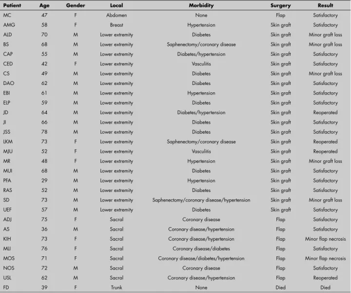
Table 1. Patients treated for wound healing with a vacuum device, and surgical outcomes
Patient Age Gender Local Morbidity Surgery Result
MC 47 F Abdomen None Flap Satisfactory
AMG 58 F Breast Hypertension Skin graft Satisfactory
ALD 70 M Lower extremity Diabetes Skin graft Minor graft loss
BS 68 M Lower extremity Saphenectomy/coronary disease Skin graft Minor graft loss
CAP 55 M Lower extremity Diabetes/hypertension Skin graft Satisfactory
CED 42 F Lower extremity Vasculitis Skin graft Satisfactory
CS 49 M Lower extremity Diabetes Skin graft Minor graft loss
DAO 62 M Lower extremity Diabetes Skin graft Satisfactory
EBI 61 M Lower extremity Hypertension Skin graft Satisfactory
ELP 59 M Lower extremity Diabetes Skin graft Satisfactory
JD 64 M Lower extremity Diabetes/hypertension Skin graft Reoperated
JI 66 M Lower extremity Diabetes Skin graft Satisfactory
JSS 78 M Lower extremity Diabetes Skin graft Satisfactory
LKM 73 F Lower extremity Saphenectomy/coronary disease Skin graft Reoperated
MJU 52 F Lower extremity Vasculitis Skin graft Reoperated
MR 48 F Lower extremity Hypertension Skin graft Minor graft loss
MUI 68 M Lower extremity Diabetes Skin graft Satisfactory
PFA 29 M Lower extremity Hypertension Skin graft Satisfactory
RAS 52 M Lower extremity Diabetes Skin graft Satisfactory
SD 73 M Lower extremity Saphenectomy/coronary disease/hypertension Skin graft Minor graft loss
UEF 57 M Lower extremity Diabetes Skin graft Satisfactory
ADJ 75 F Sacral Coronary disease Flap Satisfactory
AS 36 M Sacral Coronary disease/hypertension Flap Satisfactory
KIH 73 F Sacral Coronary disease/hypertension Flap Minor fl ap necrosis
MLI 76 F Sacral Coronary disease/diabetes Flap Satisfactory
MOS 71 F Sacral Coronary disease/diabetes/hypertension Flap Minor fl ap necrosis
NOS 72 M Sacral Coronary disease Flap Satisfactory
USL 62 M Sacral Coronary disease/hypertension Flap Reoperated
FD 39 F Trunk None Died Died
153
them as a single category of wounds. However, when these results were compared with our previous experience, it could be seen that the vacuum method allowed the surgical staff to perform the surgical procedures more safely after its introduction. Thus, adequate wound preparation could be achieved, even in clini-cally compromised patients.
It is important to emphasize the role of the vacuum method in preparing the wound for defi nitive surgical closure. It can be used to completely close wounds that are not very large. However, the time needed to establish
total closure in larger wounds is frequently excessive. Therefore, we accept negative pres-sure as an auxiliary method for improving the condition of the wound prior to surgical closure, rather than as a defi nitive method for wound closure.4 In our experience, it
has helped to reduce hospital stays, thereby reducing the morbidity that is associated with immobilized patients.
By enabling earlier closure, the device might also reduce the total cost of treatment. Its use over skin grafts is another possible advantage of the system.5,6 As demonstrated
in two cases, the vacuum promoted safe at-tachment of the skin graft to the wound. In some cases, it may even allow patients to walk while negative pressure is applied.
CONCLUSIONS This study demonstrated that this new method was safe, with minimal complica-tions. It was effi cient for adequately preparing the wound bed for defi nite surgical closure, which is an obligatory stage in most cases of complex wounds.
1. Morykwas MJ, Argenta LC, Shelton-Brown EI, McGuirt W. Vacuum-assisted closure: a new method for wound control and treatment: animal studies and basic foundation. Ann Plast Surg. 1997;38(6):553-62.
2. Argenta LC, Morykwas MJ. Vacuum-assisted closure: a new method for wound control and treatment: clinical experience. Ann Plast Surg. 1997;38(6):563-76; discussion 577. 3. Ferreira MC, Wada A, Tuma Júnior P. The vacuum assisted
closure of complex wounds: report of 3 cases. Rev Hosp Clin
Fac Med Univ São Paulo. 2003;58(4):227-30.
4. Sposato G, Molea G, Di Caprio G, Scioli M, La Rusca I, Ziccardi P. Ambulant vacuum-assisted closure of skin-graft dressing in the lower limbs using a portable mini-VAC device. Br J Plast Surg. 2001;54(3):235-7.
5. Scherer LA, Shiver S, Chang M, Meredith JW, Owings JT. The vacuum assisted closure device: a method of securing skin grafts and improving graft survival. Arch Surg. 2002;137(8):930-3; discussion 933-4.
6. Moisidis E, Heath T, Boorer C, Ho K, Deva AK. A prospec-tive, blinded, randomized, controlled clinical trial of topical negative pressure use in skin grafting. Plast Reconstr Surg. 2004;114(4):917-22.
Sources of funding: None
Confl ict of interest: None
Date of fi rst submission: June 20, 2005
Last received: May 22, 2006
Accepted: May 30, 2006
REFERENCES
AUTHOR INFORMATION Alexandre Wada, MD. Attending physician in the
Division of Plastic Surgery, Hospital das Clínicas, Facul-dade de Medicina da UniversiFacul-dade de São Paulo, São Paulo, Brazil.
Marcus Castro Ferreira, MD. Full professor and chair-man, Division of Plastic Surgery, Hospital das Clínicas, Faculdade de Medicina da Universidade de São Paulo, São Paulo, Brazil.
Paulo Tuma Júnior, MD. Attending physician in the Division of Plastic Surgery, Hospital das Clínicas, Facul-dade de Medicina da UniversiFacul-dade de São Paulo, São Paulo, Brazil.
Gino Arrunátegui, MD. Attending physician in the Division of Plastic Surgery, Hospital das Clínicas, Facul-dade de Medicina da UniversiFacul-dade de São Paulo, São Paulo, Brazil.
Address for correspondence:
Alexandre Wada
Alameda Joaquim Eugênio de Lima, 1360 Apto. 29 — Jardim Paulista
São Paulo (SP) — Brasil – CEP 01403-002 Tel. (+55 11) 3559-8671
E-mail: [email protected]
Copyright © 2006, Associação Paulista de Medicina
RESUMO Experiência com pressão negativa local (método a vácuo) no tratamento de feridas complexas
CONTEXTO E OBJETIVO: Foi estudado o uso tópico de pressão negativa em feridas de difícil resolução. O objetivo deste estudo foi analisar os efeitos da pressão negativa como tratamento de feridas complexas. TIPO DE ESTUDO E LOCAL:Série de casos atendidos em um hospital terciário (Hospital das Clínicas – Fac-uldade de Medicina da Universidade de São Paulo).
MÉTODOS:Vinte e nove pacientes com feridas complexas foram tratados com o sistema a vácuo e, então, submetidos a um procedimento cirúrgico para o fechamento da ferida.
RESULTADOS: 85% dos enxertos de pele tiveram boa “pega” e 87,5% dos retalhos locais foram mobilizados com êxito, o que aponta para um preparo adequado do leito da ferida. As feridas foram fechadas em tempo mais curto que o que nós observamos em outros tratamentos convencionais. Em dois casos, o vácuo também foi usado para a estabilização dos enxertos de pele em cima das feridas.
CONCLUSÕES: O vácuo é um método seguro e efi ciente no preparo de feridas para o fechamento cirúrgico. Permite melhora das condições locais da ferida, com desenvolvimento de tecido de granulação saudável e controle da infecção local.
PALAVRAS-CHAVE: Cicatrização de feridas. Vácuo. Cirurgia plástica. Úlcera da perna. Úlcera de decúbito.