Case series
preparation among diabetic patients: case series
Terapia por pressão negativa (vácuo) no preparo do leito da ferida
em pacientes diabéticos: série de casos
Marcus Castro Ferreira
1, Viviane Fernandes de Carvalho
2, Fábio Kamamoto
3, Paulo Tuma Junior
4, André Oliveira Paggiaro
5Plastic Surgery Division, Faculdade de Medicina da Universidade de São Paulo (FMUSP), São Paulo, Brazil
1MD, PhD. Full professor and chairman, Plastic Surgery Division, Faculdade de Medicina da Universidade de São Paulo (FMUSP), São Paulo, Brazil. 2PhD. Attending physician at Hospital das Clínicas, Faculdade de Medicina da Universidade de São Paulo (FMUSP), São Paulo, Brazil. 3MD, MSc. Attending physician at Hospital Universitário, Faculdade de Medicina da Universidade de São Paulo (HC/FMUSP), São Paulo, Brazil. 4MD, PhD. Attending plastic surgeon at Hospital das Clínicas, Faculdade de Medicina da Universidade de São Paulo (HC/FMUSP), São Paulo, Brazil. 5MD. Medical preceptor, Plastic Surgery Division, Faculdade de Medicina da Universidade de São Paulo (FMUSP), São Paulo, Brazil.
ABSTRACT
CONTEXT: Complications from diabetes mellitus affecting the lower limbs occur in 40 to 70% of such patients. Neuropathy is the main cause of ulceration and may be associated with vascular impairment. The wound evolves with necrosis and infection, and if not properly treated, amputation may be the end
result. Surgical treatment is preferred in complex wounds without spontaneous healing. After debridement of the necrotic tissue, the wound bed needs to be prepared to receive a transplant of either a graft or a lap. Dressings can be used to prepare the wound bed, but this usually leads to longer duration
of hospitalization. Negative pressure using a vacuum system has been proposed for speeding up the treatment. This paper had the objective of analyzing the effects of this therapy on wound bed preparation among diabetic patients.
CASE SERIES: Eighty-four diabetic patients with wounds in their lower limbs were studied. A commercially available vacuum system was used for all patients after adequate debridement of necrotic tissues. For 65 patients, skin grafts completed the treatment and for the other 19, skin laps were used. Wound bed preparation was achieved over an average time of 7.51 days for 65 patients and 10 days for 12 patients, and in only one case was not
achieved.
CONCLUSIONS: This experience suggests that negative pressure therapy may have an important role in wound bed preparation and as part of the treatment for wounds in the lower limbs of diabetic patients.
RESUMO
CONTEXTO: Complicações do diabetes mellitus que afetam os membros inferiores ocorrem em 40 a 70% dos pacientes. A neuropatia é a principal causa de ulceração e pode estar associada com problemas vasculares. A ferida evolui com necrose e infecção, e se não for corretamente tratada
poderá terminar em amputação. O tratamento cirúrgico é preferido em feridas complexas, quando não há cicatrização espontânea. Após desbridamento cirúrgico do tecido necrótico do leito da ferida este precisa ser preparado para receber um transplante, seja um enxerto ou um retalho. Curativos podem
ser usados para o preparo do leito da ferida, mas frequentemente levam a um longo tempo de hospitalização. A pressão negativa usada através de um sistema vácuo foi proposta para acelerar o tempo de tratamento. O presente trabalho teve como inalidade analisar os efeitos desta terapia no preparo
do leito de feridas em pacientes diabéticos.
SÉRIE DE CASOS: Oitenta e quatro pacientes diabéticos com feridas em membros inferiores foram estudados. Um sistema vácuo de uso comercial foi utilizado em todos os pacientes após adequado desbridamento de tecidos necróticos. Em 65 pacientes enxertos de pele completaram o tratamento e em outros 19 retalhos cutâneos. O preparo do leito da ferida foi conseguido, em média, em 7,51 dias em 65 pacientes, em 10 dias para 12 pacientes
e em somente um caso não foi efetivo.
CONCLUSÃO: A experiência sugere que a terapia por pressão negativa possa ter um papel importante no preparo do leito e como parte do tratamento de feridas nos membros inferiores de pacientes diabéticos.
KEY WORDS: Diabetic foot. Skin transplantation
Surgical laps.
Negative-pressure wound therapy.
Wound healing.
PALAVRAS-CHAVE: Pé diabético.
Transplante de pele Retalhos cirúrgicos.
Tratamento de ferimentos com pressão negativa.
INTRODUCTION
Diabetes mellitus is a chronic multifactorial disease. he worldwide prevalence was 120 million cases in 1996 but the prediction for 2025 is for 250 million cases, because of longer life expectancy, greater obesity and greater sedentarism.1 In Brazil, it has been estimated that around
10 million people are diabetic.2
he complications from this disease are serious and those afecting the lower limbs represent 40% to 70% of these patients.3 Such patients
usually present lesions on their lower limbs that do not heal primarily. Although simply controlling blood glucose levels is important, this is not necessarily followed by healing of these ulcers.4 he wounds may
evolve with extensive necrosis and infection, which may lead to ampu-tation of parts of or even the whole limb.5,6
Obstruction of major blood vessels is responsible for less than 20% of these wounds.7 he main cause recognized today is neuropathy,
con-sisting of progressive degeneration of sensitive nerves in the foot, in-duced by microangiopathy of the small vessels to the nerve fascicles. his is sometimes associated with external compression at anatomical sites, such as the tarsal tunnel.8-10 Small traumas sufered by feet that
have abnormal (decreased) sensation can cause ulcerations that do not heal primarily.11
Such wounds are considered to be complex and are best treated sur-gically, including debridement of necrotic tissues. his provides prepa-ration for the wound bed, for subsequent skin replacement by means of skin grafts or laps.12
he concept of wound bed preparation was introduced in 2002 by Schultz et al.13 he aim was to create favorable conditions that would
speed up the endogenous cure of the wound. More recently, wound bed preparation has been used to improve the acceptance of skin grafts.12
he preparation includes controlling the microorganisms, reducing the exudate volume and stimulating the granulation tissue.14
Nonetheless, in many complex wounds, the conventional dress-ings that are used to prepare the wound bed still take a long time to achieve ideal conditions. Some alternatives have been tried out over the last 10 years.15
he use of negative pressure was proposed by Argenta et al.16 and
Morykwas et al.17 in 1997. his consisted of developing a mechanical
system (vacuum-assisted closure) to help the healing process. Negative pressure is created by a machine that is connected by a plastic tube to a sponge placed over the wound bed. he pressure is adjusted to between -50 and -125 mmHg, continuously or intermittently. he wound bed should be completely covered by the sponge, thus creating an environ-ment under vacuum when the machine is switched on.
We introduced this method to Hospital das Clínicas, Faculdade de Medicina da Universidade de São Paulo (HCFMUSP) in 2002, to treat complex wounds. An initial paper reported three cases of wounds for which the vacuum system was used.18 A more extensive report on our
clinical experience with the vacuum system, mostly on pressure sores, was presented by our group in 2006.19
he purpose of the present paper is to present a series of 84 cases of negative pressure therapy on lower-limb wounds among diabetic patients.
CASE SERIES
Eight-four diabetic patients with a single complex wound on one lower limb were treated using negative pressure (vacuum system) at the Plastic Surgery Division of HCFMUSP, between 2004 and 2006.
All the patients were informed about the proposed treatment and consented to the use of this treatment. his study was approved by the Ethics Committee of HCFMUSP.
he patients were irst seen by the vascular surgery service or were referred by their primary physician for a consultation with our complex wound group of the Plastic Surgery Division. All the patients underwent Doppler examination regarding the vascular status of their legs and were considered satisfactory. here was no indication for revascularization or amputation of the limb in any case. he patients’ diabetes status was controlled by their primary physician and was not part of this study.
In most cases, the ulcer had been present for a time of between two and three months. It had been treated by means of conventional dress-ings alone, while awaiting spontaneous closure.
Surgical debridement was usually indicated, if needed, and any ne-crotic tissue was removed as soon as possible, as dictated by the pa-tient’s general condition. After removal of the necrosis, a less-than-ideal wound bed was almost always revealed. he quality of the wound bed was assessed in terms of the poor vascularity, gross inlammatory reac-tion or signs of infecreac-tion that left doubts regarding whether the “take” of the skin graft would be assured.
he vacuum system (V.A.C.® Vacuum Assisted Closure, KCI Inc., San Antonio, Texas, USA) was then installed. he sponge was placed di-rectly on the wound, and the connections with the machine enabled a pressure of minus 125 mmHg. Sponge shrinkage within the sealed en-vironment indicated that the machine was efective. he sponge was changed every 72 hours, and the condition of the wound bed was reas-sessed at that time.
he wound bed was considered prepared, i.e. ready for surgical clo-sure (mostly by means of a meshed skin graft, but sometimes by laps), when the volume of exudate was less than 20 ml/day, no signs of infec-tious process were seen around the wound and healthy granulation tis-sue could be observed over at least 75% of the wound surface. he time elapsed between installation of the vacuum machine at the irst consul-tation and the reconstruction (wound preparation) was recorded, along with the outcome after the plastic surgery.
Skin grafts were indicated as the irst choice for diabetic ulcers. Lo-cal laps were used on smaller wounds or when specialized structures like tendons or bones were exposed and needed a thicker composite tis-sue. In some special indications, a microvascular muscle-free lap had to be transferred.
For 65 (77.3%) of the patients, the mean time taken to prepare the wound was 7.51 ± 1.87 days. For another 12 patients (14.2%), it was 10.87 ± 1.48 days and for one patient (1.3%), the negative pressure ther-apy was not eicient for improving the quality of the wound bed. For six patients (7.14%), the wound closed in nine days without the need for surgical reconstruction: only the vacuum system was used (Figure 1).
1 Non Response
49 Skin Grafts
28 Flaps
17 Local Flaps
11 Free Flaps 6 Spontaneously
closed
84 Patients
Figure 1. Distribution of the patients according to the treatment chosen.
of the grafts were meshed (using a mesh graft machine) (Zimmer® Inc., Dover, Ohio, USA) and the vacuum system was applied over the graft, in order to enhance the integration of the skin grafting (Figure 2).
For 28 patients (33.4%), skin or muscle-skin laps were the method chosen for closing the wound, usually because there was some exposed structure (bone or tendon) that would be better treated by lap coverage.
Nineteen (22.6%) cases presented a satisfactory outcome, with no necrosis or dehiscence of the lap, and nine (10.7%) presented small ne-crotic areas that healed after some short delay (Figure 3). No complica-tions relating to the use of the vacuum device were observed.
DISCUSSION
For proper resolution of any chronic wound, a logical sequence of events should be followed in order to transform the wound bed into a
healthier condition and then achieve healing. his may take place us-ing intrinsic mechanisms alone, or it may be helped by autogenous tis-sue transplantation, skin grafts or laps. hese sequential events con-sist of debridement (removal of necrotic tissue), edema control, reduc-tion of exudate, reducreduc-tion of the bacteria count and increasing the num-bers of blood vessels, with correspondingly better granulation tissue.13
he negative pressure action elicited by the vacuum system has been associated with controlling the exudate, reducing the bacterial population and stimulating the formation of granulation tissue since the irst patient with a complex wound was treated by Argenta et al.,16
16 years ago.
hese efects were clearly demonstrated in the present series. hey led to improvements in the treatment of the diabetic ulcer, over a short-er time. Although we did not conduct any comparisons with controls, it is well known from practical experience of any surgery procedure that, with conventional dressings, much more time is needed to prepare a wound bed. Furthermore, although we did not investigate the costs, the reduction in the time taken to close the wound will have had a positive impact regarding reduction of the cost of the treatment.
he mechanism through which the negative pressure achieved the increase in the blood vessel count, i.e. an angiogenic efect, is not com-pletely clear yet. he vacuum system promotes the removal of exces-sive luids in the wound, thus reducing the bacterial population and the edema. here is consequently an improvement in the blood low to the area, thereby leading to better-quality granulation tissue.20,21 he
mechanical force applied to the wound bed by the vacuum may ex-plain the cell proliferation, since this would act as a physical substitute for the normal extracellular matrix. his is important for restarting the proliferation phase of wound healing.22,23 Another possible explanation
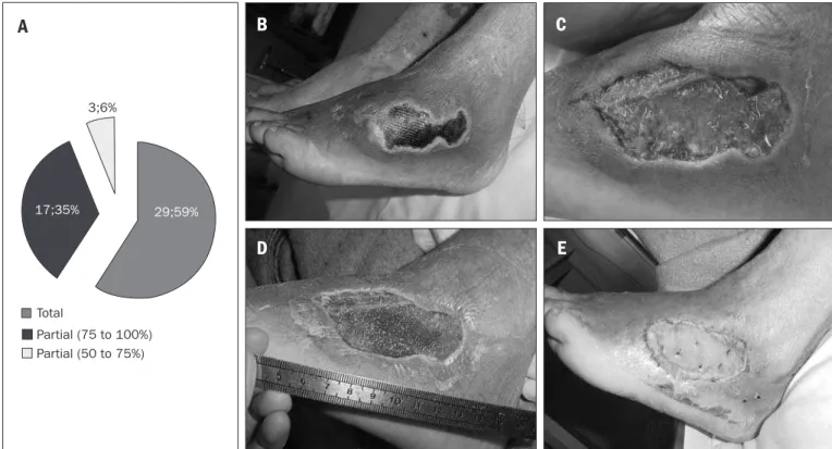
Figure 2. Results from skin grafts. A – Results from patients undergoing skin graft surgery. B - Wound on dorsum of foot before surgical debridement. C - Wound after surgical debridement. D - Wound after seven days of negative pressure therapy. E - Wound after skin grafting.
17;35%
3;6%
29;59%
Total
Partial (75 to 100%) Partial (50 to 75%)
A
B
D
C
grafts. In cases of diabetic foot ulcer, as observed in the present series, this efect will correspond to a reduction in the duration of hospitaliza-tion and the need for antibiotics.
he decrease in the bacterial population, down to under the level of 105 colonies in the tissue cultures, will have been important not only because it reduced the expenditure on antibiotics, but also because it en-hanced the oxygen and nutrient levels. hese are important as healing mechanisms and would have been diverted for the needs of the bacte-ria. So far, no consensus has been reached regarding the role of negative pressure therapy on bacteria clearance from the wound, although Mo-rykwas et al.17 showed that there was a decrease in bacterial counts in the
vacuum group, compared with controls.
In our hospital, quantitative counting of tissue bacteria is not done routinely and for this reason, we cannot airm that the vacuum therapy applied to the wound had the efect of decreasing the bacterial count. Nonetheless, the clinical appearance of the wound, the quality of the exudate and even the smell of the wound pointed towards diminished colonization among most of the diabetic patients that we treated.
he low percentage of skin graft losses also points towards this efect of infection control in the wound. It is well known that skin grafts do not incorporate when the wound is massively infected.26 In most cases
here, antibiotics were not prescribed during the vacuum system treat-ment, which usually lasted for one to two weeks.
Contraction of the wound bed was observed. It is well known that this is one the natural mechanisms of reduction of the size of the wound and promotion of primary healing. In our series, contraction was not important because we performed operations on our patients and cov-ered the wounds with grafts or laps, with the exception of six cases. for the action of the vacuum system could be that it removes
prote-olytic enzymes, especially the metalloproteinases and cytokines in the exudate. hese enzymes degrade the extracellular matrix and prevent wound resolution.24
In an experimental study on pigs, Morykwas et al.17 showed that the
blood low around the wound increased gradually with each elevation of 25 mmHg of vacuum applied to the wound. he improved blood low was optimized at -125 mmHg. On the other hand, when the vac-uum was greater than -400 mmHg, the blood low fell back to below the baseline.
he vacuum system has also been shown to enhance the quality of the granulation tissue in the wound. Granulation tissue is a mixture of blood vessels and connective tissue and it plays an essential role in cell growth in the wound and thus in its closure. It is considered, clini-cally, to have a good appearance when it presents a bright red color, a “beefy” appearance.
In the same experimental study,17 two groups of wounds were
com-pared: one using the vacuum system and the other covered by saline dressings alone. It was observed that there was a signiicantly higher growth rate for the granulation tissue in the vacuum group. Moreover, intermittent negative pressure at -125 mmHg seemed to be more ei-cient than continuous application was. Chen et al.25 showed
experimen-tally that negative pressure caused an increase in the number of capil-lary blood vessels that sprouted, as measured by biopsies and compared with controls.
his angiogenic efect from negative pressure therapy seems to be a very important factor in reducing the time needed for wound bed prep-aration and thus for the wound to be able to receive and integrate skin
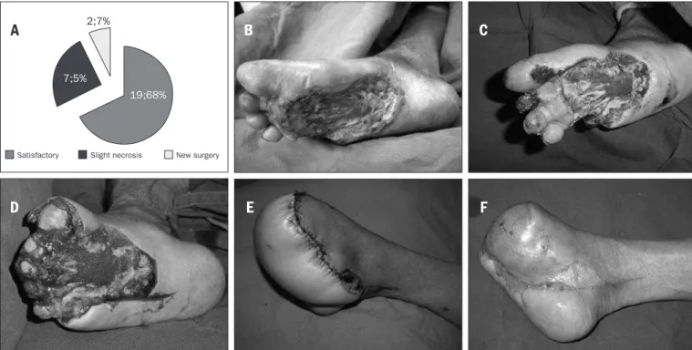
Figure 3. Results from treatment with laps. A - Distribution of patients undergoing skin lap surgery. B - Wound on foot before surgical debridement. C – Appearance of the wound after irst debridement and seven days of negative pressure therapy. Amputation of the necrotic toes was indicated. D - Wound located on the sole of the same diabetic patient after second surgical debridement and amputation of the toes. The heads of the metatarsal bones can be seen. E – Anterolateral microsurgical lap from thigh. F – Result after six months of defatting of the lap.
New surgery
7;5%
19;68%
2;7%
Slight necrosis Satisfactory
A
B
C
he negative efect of wound contraction was prevented by the tissue transfer.
Our previous clinical experience with the treatment of diabet-ic ulcers has shown that, if clindiabet-ical dressings alone are used, it has to be expected that a long time will be required (usually more than two months) for the wound bed to reaches a good condition for accepting skin grafts.
CONCLUSIONS
In this series of patients, we observed that negative pressure ther-apy had a positive efect on the treatment of complex wounds among diabetics. It enabled wound bed preparation in only a short time, and allowed us to successfully close the wound using skin transplants. It is possible that the duration of hospitalization and the overall treatment time might be shorter than with conventional dressing, but further ran-domized clinical trials are still needed to conirm this hypothesis.
REFERENCES
1. Wild S, Roglic G, Green A, Sicree R, King H. Global prevalence of diabetes: estimates for the year 2000 and projections for 2030. Diabetes Care. 2004;27(5):1047-53.
2. Malerbi DA, Franco LJ. Multicenter study of the prevalence of diabetes mellitus and impaired glucose tolerance in the urban Brazilian population aged 30-69 yr. The Brazilian Cooperative Group on the Study of Diabetes Prevalence. Diabetes Care. 1992; 15(11):1509-16. 3. Singh N, Armstrong DG, Lipsky BA. Preventing foot ulcers in patients with diabetes. JAMA.
2005;293(2):217-28.
4. Jeffcoate WJ, Price P, Harding KG; International Working Group on Wound Healing and Treat-ments for People with Diabetic Foot Ulcers. Wound healing and treatTreat-ments for people with diabetic foot ulcers. Diabetes Metab Res Rev. 2004;20(Suppl 1):S78-89.
5. Armstrong DG, Lipsky BA. Diabetic foot infections: stepwise medical and surgical manage-ment. Int Wound J. 2004;1(2):123-32.
6. Miyajima S, Shirai A, Yamamoto S, Okada N, Matsushita T. Risk factors for major limb ampu-tations in diabetic foot gangrene patients. Diabetes Res Clin Pract. 2006;71(3):272-9. 7. Abbott CA, Carrington AL, Ashe H, et al. The North-West Diabetes Foot Care Study: incidence
of, and risk factors for, new diabetic foot ulceration in a community-based patient cohort. Diabet Med. 2002;19(5):377-84.
8. Dellon AL. From there to here: a personal viewpoint after three decades of neuropathy research. Clin Podiatr Med Surg. 2006;23(3):497-508.
9. Valdivia JM, Dellon AL, Weinand ME, Maloney CT Jr. Surgical treatment of peripheral neuropathy: outcomes from 100 consecutive decompressions. J Am Podiatr Med Assoc. 2005;95(5):451-4.
10. Aszmann O, Tassler PL, Dellon AL. Changing the natural history of diabetic neuropathy: in-cidence of ulcer/amputation in the contralateral limb of patients with a unilateral nerve decompression procedure. Ann Plast Surg. 2004;53(6):517-22.
11. Mayield JA, Reiber GE, Sanders LJ, Janisse D, Pogach LM; American Diabetes Association.
Preventive foot care in diabetes. Diabetes Care. 2004;27(Suppl 1):S63-4.
12. Ferreira MC, Tuma P Jr, Carvalho VF, Kamamoto F. Complex wounds. Clinics (Sao Paulo). 2006;61(6):571-8.
13. Schultz GS, Sibbald RG, Falanga V, et al. Wound bed preparation: a systematic approach to wound management. Wound Repair Regen. 2003;11(Suppl 1):S1-28.
14. Sibbald RG, Orsted H, Schultz GS, Coutts P, Keast D; International Wound Bed Preparation Advisory Board; Canadian Chronic Wound Advisory Board. Preparing the wound bed 2003: focus on infection and inlammation. Ostomy Wound Manage. 2003;49(11):23-51. 15. Falanga V. Wound healing and its impairment in the diabetic foot. Lancet. 2005;
366(9498):1736-43.
16. Argenta LC, Morykwas MJ. Vacuum-assisted closure: a new method for wound control and treatment: clinical experience. Ann Plast Surg. 1997;38(6):563-76; discussion 577. 17. Morykwas MJ, Argenta LC, Shelton-Brown EI, McGuirt W. Vacuum-assisted closure: a new
method for wound control and treatment: animal studies and basic foundation. Ann Plast Surg. 1997;38(6):553-62.
18. Ferreira MC, Wada A, Tuma Júnior P. The vacuum assisted closure of complex wounds: report of 3 cases. Rev Hosp Clin Fac Med Univ São Paulo. 2003;58(4):227-30.
19. Wada A, Ferreira MC, Tuma Júnior P, Arrunátegui G. Experience with local negative pressure (vacuum method) in the treatment of complex wounds. Sao Paulo Med J. 2006;124(3):150-3.
20. DeFranzo AJ, Marks MW, Argenta LC, Genecov DG. Vacuum-assisted closure for the treatment of degloving injuries. Plast reconstr surg. 1999;104(7):2145-8.
21. Lambert KV, Hayes P, McCarthy M. Vacuum assisted closure: a review of development and current applications. Eur J Vasc Endovasc Surg. 2005;29(3):219-26.
22. Saxena V, Hwang CW, Huang S, Eichbaum Q, Ingber D, Orgill DP. Vacuum-assisted closure: mi-crodeformations of wounds and cell proliferation. Plast Reconstr Surg. 2004;114(5):1086-96; discussion 1097-8.
23. Chen KD, Li YS, Kim M, et al. Mechanotransduction in response to shear stress. Roles of receptor tyrosine kinases, integrins, and Shc. J Biol Chem. 1999;274(26):18393-400. 24. Yager DR, Nwomeh BC. The proteolytic environment of chronic wounds. Wound Repair Regen.
1999;7(6):433-41.
25. Chen SZ, Li J, Li XY, Xu LS. Effects of vacuum-assisted closure on wound microcirculation: an experimental study. Asian J Surg. 2005;28(3):211-7.
26. Krizek TJ, Robson MC. Evolution of quantitative bacteriology in wound management. Am J Surg. 1975;130(5):579-84.
Sources of funding: None
Conlict of interest: None
Date of irst submission: June 11, 2007
Last received: July 2, 2009
Accepted: July 13, 2009
Address for correspondence: Marcus Castro Ferreira
Rua Barata Ribeiro, 483 — Conj. 161 e 162 Bela Vista — São Paulo (SP) — Brasil CEP 01308-000