Journal of Feline Medicine and Surgery Open Reports
1 –7
© The Author(s) 2016 Reprints and permissions:
sagepub.co.uk/journalsPermissions.nav DOI: 10.1177/2055116916646584 jfmsopenreports.com
This paper was handled and processed by the American Editorial Office (AAFP) for publication in JFMS Open Reports
Creative Commons Non Commercial CC-BY-NC: This article is distributed under the terms of the Creative Commons Attribution-NonCommercial 3.0 License (http://www.creativecommons.org/licenses/by-nc/3.0/) which permits non-commercial use, reproduction and distribution of the work without further permission provided the original work is attributed as specified on the SAGE and Open Access pages (https://us.sagepub.com/en-us/nam/open-access-at-sage).
Case description
A 9-year-old, male neutered domestic shorthair cat with outdoor access was evaluated for a 3 week history of leth-argy and coughing. One week prior to presentation, the owner reported that the cat had an episode of self-resolv-ing dyspnea. A complete blood count (CBC [IDEXX Procyte Dx Hematology Analyzer; IDEXX Laboratories]) performed by the referring veterinarian was normal. Referral veterinarian imaging included thoracic radio-graphs, which demonstrated a severe, diffuse, mixed, predominantly bronchial pulmonary pattern with struc-tured interstitial (miliary-to-nodular) components. Although the exact methodology was not provided, a transtracheal wash with cytology had been performed and was evaluated by the referring veterinarian, and reportedly revealed mixed/suppurative inflammation. Culture of the bronchial fluid was negative for growth.
Diagnosis of
Aelurostrongylus
abstrusus
verminous pneumonia
via sonography-guided ine-needle
pulmonary parenchymal aspiration
in a cat
Jennifer Gambino
1, Elizabeth Hiebert
2, Melanie Johnson
3*and Matthew Williams
4Abstract
Case summary A 9-year-old, male neutered, indoor–outdoor domestic shorthair cat from the northern Alabama countryside presented for a 3 week history of coughing, lethargy and an episode of self-resolving dyspnea that occurred 1 week prior to presentation. Three-view thoracic radiographs revealed a moderate-to-severe, diffuse, mixed bronchial to structured interstitial (miliary-to-nodular) pulmonary pattern in all lung lobes with peribronchial cuffing and multifocal areas of mild patchy alveolar opacity. Ultrasound-guided evaluation and fine-needle aspiration of the caudodorsal lung parenchyma was performed with sedation. Cytology revealed many widely scattered
Aelurostrongylus abstrusus larvae and ova. Upon the confirmed diagnosis of A abstrusus verminous pneumonia, treatment with fenbendazole and selamectin resulted in complete resolution of clinical signs within 6 weeks of the initial diagnosis.
Relevance and novel information We report herein the first documented case in the Americas of A abstrusus
verminous pneumonia diagnosed via cytologic evaluation of an in vivo, percutaneous ultrasound-guided fine-needle aspirate of affected lung. Additionally, to our knowledge, we offer the first account of the sonographic (pulmonary) features of the disease.
Accepted: 28 March 2016
1 Department of Clinical Sciences, Diagnostic Imaging, Mississippi
State University, College of Veterinary Medicine, Mississippi State, MS 39762, USA
2 Central Texas Veterinary Specialty Hospital, Austin, TX 78745,
USA
3 Department of Pathobiology and Population Medicine,
Mississippi State University, College of Veterinary Medicine, Mississippi State, MS, 39762, USA
4 Department of Pathobiology and Population Medicine,
Mississippi State University, College of Veterinary Medicine, Mississippi State, MS 39762, USA
Corresponding author:
Jennifer Gambino DVM, DACVR, Mississippi State University, College of Veterinary Medicine, Animal Health Center, 240 Wise Center Drive, Mississippi State, MS 39762, USA
Email: gambino@cvm.msstate.edu
2 Journal of Feline Medicine and Surgery Open Reports
When clinical signs did not resolve the cat was referred for further diagnostics.
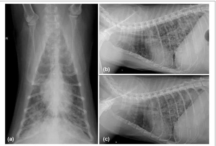
At presentation the cat was bright, alert and respon-sive, with a body condition score of 3/5.1 Bilaterally harsh lung sounds were auscultated. Repeated CBC (Cell-Dyn 3700; Abbott Laboratories) and serum bio-chemistries (Vet Axcel Clinical Chemistry System; Alfa Wassermann Diagnostic Technologies) revealed a mod-erate leukopenia (3.7 K/μl; reference interval [RI] 5.5– 20.0 K/μl) and persistent moderate neutropenia (1702/μl; RI 2500–12,500/μl) with a left shift (bands 222/μl, RI 0–300/μl), mild lymphopenia (1369/μl; RI 1500–7000/μl) and mild hyperglycemia (178 mg/dl; RI 70–160 mg/dl). Repeat, three-view thoracic radio-graphs revealed a moderately progressed severe, diffuse, mixed bronchial-to-structured interstitial (mil-iary-to-nodular) pulmonary pattern in all lung lobes but most severe in the caudodorsal lung fields. Peribronchial cuffing was evident, with multifocal areas of poorly defined alveolar disease (especially in the perihilar and caudal dorsal lung fields) (Figure 1).
Differential diagnoses for a middle-aged-to-elderly cat with dyspnea and the combined radiographic abnor-malities included feline asthma, allergic pneumonitis (secondary to inhaling irritants such as mold, smoke or household chemicals), feline heartworm eosino-philic pneumonitis or other verminous pneumonia (Aelurostrongylus species, Eucoleus species, Filaroides species and Dirofilaria immitis), and neoplasia (such as bronchogenic adenocarcinoma).
Thoracic and abdominal sonography were performed with a BioSoundEsoate MyLab50 ultrasound machine and microconvex ultrasound probe with available fre-quencies from 5 to 8 mHz. Abdominal sonography was normal. Thoracic sonography revealed many B-lines (comet tail artifact, a subtype of reverberation artifact) with intermittent, well-defined, small (2–3 mm), hypo-echoic nodules, intermittent irregular pleural margins and multifocal, intermittent, variously sized (2–4 mm) ‘shred’ signs (Figure 2).2–4 The cat was sedated (dexme-detomidine 4.7 μg/kg IM). Following aseptic prepara-tion of the clipped skin, percutaneous ultrasound-guided
fine-needle aspiration of the sonographically affected lung parenchyma was performed. A 5 ml syringe with 1 ml room air within the syringe chamber was attached to a 22 G × 1.5 inch fine needle (without stylet). Approximately one-half the length of the needle was gently inserted through the skin at the right proximal third of the eighth intercostal space and was directed with ultrasound guidance into the affected pulmonary parenchyma/nodules. Negative pressure (1–4 ml) was applied, while 3–5 to and from movements were per-formed within the lesions. The collected material was sprayed onto glass slides for evaluation by an on-site, board-certified veterinary clinical pathologist. All
4 Journal of Feline Medicine and Surgery Open Reports
and pneumothorax. During the recovery period (approx-imately 30 mins), the cat was monitored closely for clini-cal signs of respiratory distress and pallor.
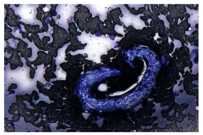
Cytologic evaluation revealed high cellularity aspi-rate samples consisting of innumerable macrophages and non-degenerate neutrophils admixed with fewer eosinophils. Numerous multinucleated giant cells were observed, along with several clusters of respiratory endothelial cells. Widely scattered larvae, measuring
approximately 350 μm, were observed throughout the smears and, occasionally, were seen in concentric coils. Some of these larvae possessed distinctly notched tails (Figure 3). Also present were multiple oval structures measuring approximately 80 μm × 70 μm, with a frothy interior, containing numerous round basophilic structures consistent with blastomeres (Figure 4). Long, beaded, filamentous, extracellular rods were noted on one slide. These findings were consistent with pyogranulomatous-to-eosinophilic inflammation with intralesional A abstrusus larvae and ova, with pos-sible additional concurrent nocardiosis. Additional acid-fast staining was performed to confirm the pres-ence of Nocardia species; in the interim, a Baermann test was performed and confirmed the presence of A abstrusus.
The cat was hospitalized for treatment and observa-tion. Intravenous trimethoprim sulfa for presumed nocardiosis (16.7 mg/kg diluted in 2 ml D5W IV q12h for 7 days) was instituted pending acid-fast staining of cyto-logic samples. In addition, the cat was placed on oral prednisolone to decrease inflammation associated with parasite death (0.5 mg/kg PO q24h for 10 days). Fenbendazole was instituted to eliminate the A abstrusus (50 mg/kg PO q24h for 14 days), in addition to monthly topical selamectin.
A repeat CBC 5 days following initial diagnosis dem-onstrated resolved neutropenia (2829/μl; RI 1500– 7000/μl), improving leukopenia (4.1 K/μl; RI 5.5–20.0 K/μl) and moderate lymphopenia (861/μl; RI 1500– 7000/μl). Acid-fast staining of the filamentous organ-isms identified during cytology was negative for Nocardia species and was obtained on the seventh day of hospi-talization. No episodes of respiratory distress or cough-ing were noted durcough-ing the hospitalization period. The cat’s attitude and appetite improved. A repeated Baermann test 2 weeks following initial hospitalization was negative for A abstrusus larvae. The patient was dis-charged on day 16 of hospitalization, with clinically nor-mal behavior, a nornor-mal resting respiration rate (23–42 breaths per min)7 and normal lung auscultation bilater-ally. Six weeks following initial diagnosis, recheck tho-racic radiographs demonstrated almost complete resolution of the previously described pulmonary pat-tern. A residual, but much improved, mild-to-moderate, mixed bronchial and unstructured interstitial pulmo-nary pattern persisted within the right caudal dorsal and accessory lung lobes (Figure 5). The cat was bright, alert and responsive, with no abnormal lung sounds heard upon auscultation. All coughing and lethargy had resolved.
Discussion
We report herein the first documented case of A abstrusus infection diagnosed by ultrasound-guided lung aspirate Figure 4 Cytologic sample obtained by way of
ultrasound-guided pulmonary fine-needle aspiration showing A abstrusus
ova. At the center of the image is a large, oval egg containing numerous basophilic-staining blastomeres surrounded by abundant non-degenerate neutrophils and alveolar macrophages (characterized by their abundant, vacuolated cytoplasm) on a background of blood and proteinaceous debris. Modified Wright’s stain
Figure 3 Cytologic sample obtained via pulmonary ultrasound-guided fine-needle aspirate containing
in the Americas, with subsequent successful medical management and outcome. Previously, the diagnosis of A abstrusus via fine-needle biopsy is described in two cats.8,9 In people, a broad spectrum of parasitic infections can have thoracic manifestations, but sonography and sonographic diagnosis are seldom used for screening.10 We describe herein the radiographic features, and, for the first time in the veterinary literature, to our knowl-edge, the associated sonographic pulmonary parenchy-mal features of A abstrusus. Image-guided percutaneous fine needle aspiration is less invasive than fine-needle biopsy or excisional biopsy, and may be associated with lower morbidity and mortality, as in the case of image-guided percutaneous biopsy in people and percutane-ous fine-needle aspiration biopsy in dogs and cats.11,12 Indications for image-guided interventions include the establishment of the benign or malignant nature of a lesion, obtaining material for microbiologic analysis in patients with known or suspected infections, staging malignancy in cases of known or suspected malignancy, and establishing the nature and extent of diffuse paren-chymal disease.11
A abstrusus is a sporadically identified metastrongy-loid nematode of cats responsible for cardiorespiratory
disease in Europe and the Americas.13 The parasite is transmitted indirectly in a life cycle that incorporates mollusks and snails as intermediate hosts, and rodents, frogs, lizards, snakes, and birds as paratenic hosts.14,15 Cats are infected when they consume an intermediate or paratenic host containing larvae; these larvae then migrate to the lungs, mature to their adult forms, and produce larvae that migrate to the pharynx and are passed into the feces, thus completing the cycle.13 The hatching of eggs and migration of larvae are responsi-ble for most clinical signs.13 Clinical signs associated with A abstrusus are uncommon but are primarily asso-ciated with parenchymal pulmonary infiltration and the subsequent inflammatory response.13 Respiratory signs can include coughing, sneezing, progressive dyspnea and death.13,15 The Baermann fecal test is con-sidered to be the gold-standard diagnostic technique in living patients to achieve a diagnosis, followed by other copromicroscopic techniques and bronchoalveo-lar lavage cytology.13,16,17 A abstrusus can be easily detected at necropsy via histology of affected lung lobes.18
6 Journal of Feline Medicine and Surgery Open Reports
obtained by lung impression of an affected kitten during necropsy; a female larva was found in the bloodstream of another stray cat during a blood parasite survey.19,20 Radiographic findings in the cat of this report were clas-sic, prompting sonographic pulmonary evaluation and the interventional procedure. At our institution, thoracic sonography combined with fine-needle aspiration is often performed to obtain samples for cytology and cul-ture, especially when radiographs support the presence of severe pulmonary disease such as severe bronchial, interstitial or alveolar opacities. We consider sono-graphic-guided fine-needle pulmonary aspiration to be a viable, quick, relatively safe and easily performed test when severe disease is radiographically present. In addi-tion, it is our experience that mild-to-moderate radio-graphic findings of pulmonary disease can have easily identifiable sonographic lesions that may be amenable to sonographic aspiration, especially when evaluation is performed by trained ultrasound personnel. Pulmonary fine-needle aspiration may be especially useful in unsta-ble patients for which conventional testing may delay diagnosis and treatment. The Baermann technique takes considerable time to complete (at least 6 h) and is unable to diagnose parasitic infections during the prepatent period, while bronchoalveolar lavage requires general anesthesia and has a mortality rate of up to 6% in cats with respiratory compromise.13,21 An additional weak-ness of both transtracheal washes and the Baermann technique is the tendency of A abstrusus to shed variable numbers of ova and larvae during its life cycle, which can result in false negatives when using either or both of these diagnostic tests.13
There is limited literature available regarding the safety of pulmonary aspiration in cats with verminous pneumonia, and a prospective clinical study would be ideal for complete assessment of this procedure’s clinical performance, diagnostic efficacy and safety in patients afflicted with this disease. Multiple studies assessing risk associated with fine-needle aspiration in canine and feline patients with fungal and neoplastic disease revealed minimal risk of clinical signs secondary to aspi-ration and a high diagnostic yield when the aspirate was sonographically guided.12,22 Three veterinary studies of pulmonary aspiration assisted with an imaging modality had only one animal with clinical pneumothorax out of 97 cases (an incidence of 1.03%).12,22,23 Another study evaluating pulmonary aspiration without imaging guid-ance had a higher rate of clinical pneumothorax (6.2%).24 Complications associated with pulmonary fine-needle aspiration biopsy with a 20 G Wescott-type needle include a risk as high as 17% for pneumothorax and a mortality rate of 2.1% secondary to pneumothorax.12 Greater risk was associated with larger needle gauge, and although more tissue may be obtained with a larger needle, blood contamination can hamper the diagnostic quality of the
sample.12 In people there is a 1.6% incidence of dyspnea due to tension pneumothorax secondary to endoscopic sonographically guided fine-needle aspiration of large pulmonary mass lesions.6 Complications were not noted in the cat of this report but can include pneumothorax, pulmonary parenchymal hemorrhage, pulmonary paren-chymal lacerations and inadvertent cardiac puncture. The cat of the current report was sedated, further reduc-ing the risk of associated complications. Motion from breathing, in our opinion, did not hamper the ability to effectively perform the aspirate.
Conclusions
We describe our positive experience with, and the clini-cal utility of, percutaneous ultrasound-guided fine-nee-dle aspiration of the pulmonary parenchyma of a cat with A abstrusus, which resulted in a rapid definitive diagnosis. The safety of pulmonary aspirates in cats with pulmonary parasitism is not well assessed in the veteri-nary literature; owners should be counseled regarding potential complications.
Conlict of interest The authors declared no potential con-flicts of interest with respect to the research, authorship, and/ or publication of this article.
Funding The authors received no financial support for the research, authorship, and/or publication of this article.
References
1 LaFlamme D. Development and validation of a body
con-dition score system for cats: a clinical tool. Feline Pract
1997; 25: 13–17.
2 Lichenstein DA and Meziere GA. Relevance of lung
ultra-sound in the diagnosis of acute respiratory failure. Chest
2008; 134: 117–125.
3 Lisciandro GR, Fosgate GT and Fulton RM. Frequency
and number of ultrasound lung rockets (b-lines) using a regionally based lung ultrasound examination named vet blue (veterinary bedside lung ultrasound exam) in dogs
with radiographically normal lung findings. Vet Radiol
Ultrasound 2014; 55; 315–322.
4 Lisciandro GR, Lagutchik MS, Mann KA, et al. Evaluation
of a thoracic focused assessment with sonography for trauma (TFAST) protocol to detect pneumothorax and
concurrent thoracic injury in 145 traumatized dogs. J Vet
Emerg Crit Care 2008; 18: 258–269.
5 Vasques-Sequeiros E, Levy MJ, Van Domsellar M, et al.
Diagnostic yield and safety of endoscopic ultrasound guided fine needle aspiration of central mediastinal lung
masses. Diagn Ther Endosc 2013; 2013: 150492.
6 Chan SSW. Emergency bedside ultrasound to detect
pneu-mothorax. Acad Emerg Med 2003; 10: 91–94.
7 Jennings DB and Szlyk PC. Ventilation and respiratory
pattern and timing in resting awake cats. Can J Physiol
Pharmacol 1985; 63: 148–154.
8 Ribiero VM, Barcante JMP, Negrao-Correa D, et al.
alteration during Aeulorstrongylus abstrusus infection in cats. Pesq Vet Bras 2014; 34: 990–995.
9 Durr B. Diagnosis of Aelurostrongylus abstrusus infection
by fine needle aspiration of the lungs in two cats.
Kleinti-erpraxis 2009; 54: 88–92.
10 Martinez S, Restrepo S, Carrilo JA, et al. Thoracic
manifes-tations of tropical parasitic infections: a pictorial review.
Radiographics 2005; 25: 135–155.
11 Gupta S, Wallace MJ, Cardella JF, et al. Quality
improve-ment guidelines for percutaneous needle biopsy. J Vasc
Interv Radiol 2010; 21: 969–975.
12 McMillian M. Fluoroscopically guided percutaneous fine
needle aspiration biopsy of thoracic lesions in dogs and cats. Vet Radiol Ultrasound 1988; 29: 194–197.
13 Traversa D and Gugleilmini C. Feline aelurostrongylosis
and canine angiostrongylosis: a challenging diagnosis for
two emerging verminous pneumonia infections. Vet
Para-sitol 2008; 157: 163–174.
14 Giannelli A, Ramos RAN, Annoscia G, et al. Development
of the feline lungworms Aelurostrongylus abstrusus and
Troglostrongylus brevior in Helix aspersa snails.
Parasitol-ogy 2014; 141: 563–569.
15 Dirven M, Szatmari V, van den Ingh T, et al. Reversible
pulmonary hypertension associated with lungworm
infection in a young cat. J Vet Cardiol 2012; 14: 465–474.
16 Gaglio G, Cringoli G, Rinaldi L, et al. Use of the FLOTAC
technique for the diagnosis of Aelurostrongylus abstrusus
in the cat. Parasitol Res 2008; 103: 1055–1057.
17 Lacorcia L, Gasser RB, Anderson GA, et al. Comparison
of bronchoalveolar lavage fluid examination and other diagnostic techniques with the Baermann technique for
detection of naturally occurring Aelurostrongylus
abstru-sus infection in cats. J Am Vet Med Assoc 2009; 235: 43–49.
18 Schnyder M, Di Cesare A, Basso W, et al. Clinical,
labo-ratory and pathological findings in cats experimentally
infected with Aelurostrongylus abstrusus. Parasitol Res
2014; 113: 1425–1433.
19 Kohart NA, Boes KM, Sponenberg DP, et al. What is your
diagnosis? Lung impression smear from a stray kitten. Vet
Clin Path 2014; 43: 113–114.
20 Rassouli M, Ghaderi J, Goudarzi A, et al. Aelurostrongylus
abstrusus in a stray cat’s blood stream. Comp Clin Pathol 2015; 24: 773–775.
21 Johnson LR and Drazenovich TJ. Flexible bronchoscopy
and bronchoalveolar lavage in 68 cats (2001–2006). J Vet
Intern Med 2007; 21: 219–225.
22 Wood EF, O’Brien RT and Young KM. Ultrasound-guided
fine-needle aspiration of focal parenchymal lesions of the
lung in dogs and cats. J Vet Intern Med 1998; 12: 338–342.
23 Zekas LJ, Crawford JT and O’Brien RT. Computed
tomog-raphy-guided fine-needle aspirate and tissue-core biopsy
of intrathoracic lesions in thirty dogs and cats. Vet Radiol
Ultrasound 2005; 46: 200–204.
24 Tseke E, Stokhof AA, van der Ingh TSGA, et al. Thoracic
needle aspiration biopsy of the lung in dogs with