The contribution of fermentative bacteria and methanogenic archaea to
azo dye reduction by a thermophilic anaerobic consortium
Andr´e B. dos Santos
a,b,∗, Marta P. de Madrid
a, Frank A.M. de Bok
d,
Alfons J.M. Stams
d, Jules B. van Lier
a, Francisco J. Cervantes
caSub-department of Environmental Technology, Wageningen University, Bomenweg 2, P.O. Box 8129, 6700EV Wageningen, The Netherlands bDepartamento de Engenharia Hidr´aulica e Ambiental, Universidade Federal do Cear´a, Campus do Pici, Bloco 713, 60455-760 Fortaleza, Cear´a, Brazil
cDepartamento de Ciencias del Agua y del Medio Ambiente, Instituto Tecnol´ogico de Sonora, 5 de Febrero 818 Sur Cd. Obreg´on, Sonora 85000, Mexico dLaboratory of Microbiology, Wageningen University, Hesselink van Suchtelenweg 4, Wageningen 6703 CT, The Netherlands
Received 25 November 2004; received in revised form 1 September 2005; accepted 14 September 2005
Abstract
The contribution of fermentative bacteria and methanogenic archaea to azo dye reduction by a thermophilic anaerobic consortium was stud-ied. Additionally, the effects of different electron-donating substrates and the redox mediator riboflavin on dye reduction were assessed by using either a methanogenic consortium or pure cultures of methanogens. Our results indicate that fermentative bacteria and methanogenic archaea play an important role in this reductive process. The thermophilic methanogensMethanothermobacter thermoautotrophicusHand a
Methanothermobacter-related strain NJ1 were only able to reduce the dye in the presence of riboflavin. This suggests that anaerobic dye reduction is not a universal property among methanogenic archaea and that redox mediators may improve reductive decoulorisations by allowing some microbial groups commonly found in wastewater treatment systems to participate more effectively.
© 2005 Elsevier Inc. All rights reserved.
Keywords: Azo dye; Redox mediators; Methanogens; Granular sludge; Thermophilic
1. Introduction
Although the use of dyes makes the world more beautiful, it represents a serious pollution problem worldwide. The release of coloured compounds into the environment is undesirable not only because of their aesthetic appearance and colour, which may affect photosynthesis of aquatic plants, but also because many dyes from wastewater and their breakdown products may be toxic and/or mutagenic to life[1].
Reductive decolourisation of azo dyes can be readily achieved under anaerobic conditions either chemically or biologically. Chemical decolourisation may involve biogenic reductants, such as sulfide, cysteine, ascorbate or Fe2+[2,3]. In contrast,
micro-bial decolourisation requires an unspecific enzymatic capacity ubiquitously found in a wide diversity of microorganisms[4]. This has been demonstrated with pure cultures of phylogenet-ically distinct microorganisms from the genera Clostridium,
∗Corresponding author. Tel.: +55 85 99916052.
E-mail address:andrebds@deha.ufc.br (A.B. dos Santos).
Salmonella, Bacillus, Sphingomonas,Agrobacterium, Ralsto-nia,Hydrogenophaga, Flexibacter, Rhodococcus, Lactobacil-lus, Eubacterium and Escherichia, which are able to reduce dyes commonly ingested through food, drugs and cosmetics
[5–7]. It is currently accepted that azo dye reduction is due to a co-metabolic reaction, in which the biologically formed reducing equivalents can be chemically transferred to the azo dyes. In anaerobic consortia, reducing equivalents are formed by fermentative bacteria. Methanogens, consume these reduc-ing equivalents to form methane. However, it might well be that some methanogens conduct the reducing equivalents towards dye reduction instead of methanogenesis. Therefore, fermenta-tive bacteria and methanogenic archaea may play an important role in the reduction of azo dyes. However, little is known about these microbiological aspects of anaerobic consortia (e.g. from wastewater treatment plants) on the reductive decolourisation. In other words, the true contribution of the different microbes present in a mixed culture such as granular sludge (e.g. fermenta-tive bacteria and methanogenic archaea) on dye reduction, is still unknown. Even though a large portion of textile wastewaters, mainly from the dyebath and rinsing steps, is discharged at high
temperatures (40–70◦C), azo dye reduction under thermophilic conditions by using either pure cultures or mixed cultures has gained only little attention so far[8].
It was found that redox mediators can increase the shuttling of electrons between the primary electron donor and the azo dye, which is normally the rate-limiting step during anaero-bic azo dye reduction [9]. Flavin-based compounds, such as FAD, FMN and riboflavin, as well as quinone-based compounds such as anthraquinone-2-sulphonate (AQS), anthraquinone-2,6-disulphonate (AQDS) and lawsone are examples of compounds with catalytic properties [10,6,11]. Therefore, the dosage of redox mediators may represent an interesting option to increase the rates of electron transfer and subsequent reductive decolouri-sation in bioreactors. After non-specific enzymes reduce the mediator, the latter can transfer the reducing equivalents chemi-cally to the azo dye, recovering its original form[12]. The reduc-tion of the mediator by the cells, which can occur either intracel-lularly[9]or extracellularly by a membrane-bound enzyme[13], will depend on many factors such as its size, polarity, standard redox potential (E′0) and other physical-chemical characteristics
[10]. In the current investigation the contribution of fermentative bacteria and methanogenic archaea to the reductive decolouri-sation of azo dyes by a thermophilic anaerobic consortium was studied. Additionally, the effect of different electron-donating substrates and the redox mediator riboflavin on dye reduction was assessed with mixed and pure cultures.
2. Materials and methods
2.1. Chemicals
Reactive Red 2 (RR2), Reactive Red 4 (RR4) and Reactive Orange 14 (RO14) were selected as azo dye model compounds for the present study (Fig. 1), because they presented the lowest rates of decolourisation among many azo dyes previously tested[14]. Riboflavin (Vitamin B2) was selected as redox media-tor model compound. Vancomycin and 2-bromoethane sulphonic acid (BES), inhibitors of fermentative bacteria and methanogens, respectively, were used to assess the contribution of the different microbial groups on the reduction of azo dyes[15,16]. Chemicals were purchased from Aldrich (Gillingham, UK), Sigma (Bornem, Belgium) or Acros (Geel, Belgium) and used without additional purification.
2.2. Seed inoculum and basal medium for decolourisation assays
Anaerobic granular sludge was collected from a full-scale mesophilic upflow anaerobic sludge blanket (UASB) reactor treating paper mill wastewater (Eerbeek, The Netherlands). The mesophilic sludge was acclimated at 55◦C
until stable performance was obtained as described elsewhere[17].
2.3. Activity test
In the activity tests 1.3±0.1 g volatile suspended solids (VSS) l−1 of the thermophilic sludge was added to 57 ml serum bottles containing 25 ml basal medium, after which the bottles were sealed with butyl rubber stoppers. The basal medium consisted of (mg l−1): NH
4Cl (280), K2HPO4 (250), MgSO4·7H2O (100) and CaCl2·2H2O (10) and 1 ml l−1of trace elements containing (mg/l): H3BO3(50), FeCl2·4H2O (2000), ZnCl2(50), MnCl2·4H2O (500), CuCl2·2H2O
(38), (NH4)6Mo7O24·4H2O (50), AlCl3·6H2O (90), CoCl2·6H2O (2000), NiCl2·6H2O (92), Na2SeO3·5H2O (162), EDTA (1000) and HCl 36% (1). The medium was buffered 6.2 g l−1sodium bicarbonate, to keep the pH around 7.1. Resazurin was not included in the trace elements solution due to its mediating properties. Anaerobic conditions were established by flushing the headspace with N2/CO2(70%:30%), after which the sludge was pre-incubated for 2–3 days with glucose (0.45 gCOD l−1) as an electron donor. Then, the headspace was exchanged to N2/CO2(70%:30%) again, and the azo dyes (0.3 mM), riboflavin (0.012 mM) and different electron donors (1.5 gCOD l−1) were added. The elec-tron donors were added in excess compared to the amounts required for complete reduction of the dyes. Sterile controls were autoclaved once at 122◦C for 240 min
and again after 5 days incubation period, after which electron donors, mediators and dyes were added from sterile stock solutions. The pH and the amount of VSS were determined at the end of the experiment.
2.3.1. Contribution of fermentative bacteria and methanogenic archaea in a thermophilic consortium to azo dye reduction
To evaluate the contribution of fermentative bacteria and methanogens to azo dye reduction, an experiment in the presence of vancomycin (1.0 g l−1) and BES (10.5 g l−1), was conducted at 55◦C. Three reactive azo dyes RR2, RR4
and RO14 were tested at a concentration of 0.3 mM, either in the presence or in the absence of these specific inhibitors. Glucose (1.5 gCOD l−1) was selected as primary electron donor and the redox mediator, riboflavin (0.012 mM), was added to some of the series to assess its effect on the rates of dye reduction.
To evaluate the effect of substrate concentration on azo dye reduction by mixed cultures at 55◦C, different glucose concentrations were tested
(gCOD l−1): 1.5, 0.75, 0.25, 0.10, 0.05 and 0, either in the presence or in the absence of riboflavin (0.012 mM). The azo dye RO14 (0.3 mM) was selected as a model compound. Additionally, experiments with the inhibitors vancomycin (1.0 g l−1) and BES (10.5 g l−1), were conducted at 55◦C with a low
concentra-tion of glucose (0.05 gCOD l−1).
Abiotic decolourisation was evaluated in sludge-free and autoclaved sludge controls, and endogenous (glucose-free) reduction activity was determined as well.
2.3.2. Effect of different electron-donating substrates on azo dye reduction by a thermophilic consortium
To assess the effect of different electron-donating substrates on azo dye reduction, H2/CO2, acetate, methanol and formate were tested at a concentration of 1.5 gCOD l−1at 55◦C. The electron donors were added in excess compared to
the amount required to completely reduce the dyes. The batches were incubated on a rotary shaker at 50 rpm in the dark. Riboflavin (0.012 mM) was added to some of the bioassays as well. The effect of BES (10.5 g l−1) was also studied, to assess the involvement of methanogens on dye reduction. When H2/CO2 (80%:20%) was tested as the electron donor, a 117 ml bottle with 50 ml basal medium and a headspace of 1.8 bar was used (equivalent to 1.2 gCOD l−1as H
2). In that case, the rotation speed was increased from 50 to 200 rpm to improve the mass transfer of hydrogen to the liquid phase. The depletion of hydrogen and methane formation was followed in time. Abiotic decolourisation was evaluated in sludge-free and autoclaved sludge controls, and endogenous (substrate-free) controls assessed the role of biological activity on dye reduction.
2.3.3. Growth and azo dye reduction by pure cultures of methanogens
Azo dye reduction by pure cultures ofMethanothermobacter thermoau-totrophicus H(DSM 1053, Toptimum 65◦C) and a
Methanothermobacter-relatedstrain NJ1 (Toptimum55◦C) was studied. The ability of the mesophilic pure cultureMethanosarcina Barkeri(DSM 800,Toptimum37◦C) to reduce the dye was also tested. These organisms were selected randomly, but they are commonly found in anaerobic granular sludge[18]. They were inoculated into 117 ml serum bottles with 50 ml of a bicarbonate buffered mineral medium (with N2/CO2in the headspace after flushing) as described elsewhere[19]. The Na2S concentration was decreased from 1.0 to 0.25 mM to minimize the contribution of the chemical decolourisation. Resazurin was not included due to its medi-ating properties. H2/CO2(80%:20%) was applied at 1.8 bar when supplied as electron donor (about 1.2 gCOD l−1as H
2). For decolourisation assays, the azo dye RR2 (0.1 mM) was selected as model compound. Riboflavin (0.012 mM) and BES (5.3 g l−1) were added to certain series from sterile stock solutions.
Sterile controls with autoclaved-cells and series lacking cells were included to evaluate the abiotic decolourisation. Protein content and pH were measured at the end of the experiment.
2.4. Analyses
Azo dye reductionwas determined photometrically (Spectronics 60, Milton-Roy Analytical Products Division, Belgium). Samples were diluted in phosphate buffer (10.86 g/l of NaH2PO4·2H2O and 5.98 g/l of Na2HPO4·2H2O), and then centrifuged for 3 min at 10,000 rpm. The absorbance was read at the wave-lengths corresponding to the maximum absorbance wavelength, i.e. RR2 at 539 nm, RR4 at 521 nm and RO14 at 433 nm. The extinction coefficients used, i.e. a factor to convert absorbance units into concentration, were 33.3, 24.0 and 11.6 AU cm−1mM−1for RR2, RR4 and RO14, respectively.Methane produc-tion was monitored by injecting 100L samples in a Packard-Becker 438/S gas chromatograph (Packard-Becker, Delft, The Netherlands), equipped with a Porapak Q column (2 m×2 mm, 80/100 mesh, Milipore Corp., Bedford, MA). The temperature of column, injection port and flame ionisation detector were 60, 200 and 220◦C, respectively, with nitrogen as carrier gases (20 ml/min). VFA,methanolandethanolwere measured with a Hewlett Packard 5890 gas chromatograph (Palo Alto, USA), equipped with a Supelcoport Fluorad FC 431 columns (2 m×2 mm, 100–120 mesh). Temperatures used for column, injection port and flame ionisation were 130, 200 and 280◦C, respectively. For alcohol
determination the temperature of column was set at 70◦C.Glucose,lactate
andformatewere measured on a High Pressure Liquid Chromatograph (HPLC) equipped with an Ion-300 column and a refractive index detector according to Van Lier[20].VSScontent was analyzed according to APHA standard methods [21].Proteincontent was determined by using the Bradford method[22]with bovine serum albumin as standard. Whole cells were harvested by centrifugation at 15,000 rpm for 10 min, and then protein was extracted by boiling for 15 min in a 1M NaOH solution.
2.5. Calculations
For the calculation of the decolourisation rates, a first-order kinetic with respect to the dye concentration was used, whereas the first-order rate constant “k” was determined using Eq.(1). It is worth to mention that this determination is just an approach to approximate the decolourisation rates.
At=A0e−kt (1)
Atis the absorbance on time “t”,A0is the initial absorbance att= 0, “k” is the first-order rate constant (day−1) and “t” is the time elapsed of the experiment (days). Time is plotted against ln(At/A0) and thek-value is estimated by the slope of a linear regression.
3. Results
3.1. Contribution of fermentative bacteria and
methanogenic archaea in a thermophilic consortium to azo dye reduction
Table 1
Azo dye reduction at 55◦C by anaerobic granular sludge
Azo dye Riboflavin Inhibitor k/VSS (day−1gVSS−1l)
BES Vancomycin
RR2 − − − 0.84 (0.02)
RR2 − + − 1.18 (0.17)
RR2 − − + 0.27 (0.03)
RR2 + − − 1.43 (0.05)
RR2 + + − 1.27 (0.01)
RR2 + + 0.56 (0.03)
RR4 − − − 1.74 (0.19)
RR4 − + − 1.61 (0.18)
RR4 − − + 0.52 (0.02)
RR4 + − − 1.82 (0.03)
RR4 + + − 1.53 (0.01)
RR4 + − + 0.69 (0.03)
RO14 − − − 0.84 (0.01)a/0.16 (0.01)a
RO14 − + − 0.85 (0.01)a/0.23 (0.04)a
RO14 − − + 0.13a,b/a,c
RO14 + − − 2.47 (0.03)a/0.61 (0.04)a
RO14 + + − 2.12 (0.01)a/0.90 (0.07)a
RO14 + − + 0.20a,b/0.11a,b
Vancomycin and BES were used to inhibit fermentative bacteria and methanogens, respectively, to elucidate their contribution in the decolourisa-tion of azo dyes. Glucose (1.5 gCOD l−1) and riboflavin (at 0.012 mM) were added as the electron donor and redox mediator, respectively. The results are means of triplicate incubations and the S.D. is shown in parenthesis.
aValues (k′/k′′) correspondent to glucose concentrations of 1.50 and
0.05 gCOD l−1, respectively.
b Calculated after a lag-phase of about 2 days.
cColour removal almost negligible during the course of the experiment.
and inhibitors (Table 1). In contrast, with RR4, there was no clear effect of the redox mediator on the rates of dye reduction. For batch assays supplemented with riboflavin, the reductive decolourisation of all tested azo dyes was slightly lower in the BES-supplemented bottles (11–16%) than the values found in the absence of inhibitors.
The RO14 reduction rates increased with increasing glu-cose concentrations (Fig. 2). In the absence of riboflavin, the decolourisation rate was up to 4.5-fold higher in the glucose-supplemented bottles than in the controls free of external elec-tron donor. In the presence of riboflavin (0.012 mM), the effect of a glucose concentration gradient on RO14 reduction was more pronounced than in the assays lacking external redox mediator. This effect was most evident at the highest substrate concentra-tions (Fig. 2). The decolourisation rates in the series containing glucose (1.5 gCOD l−1) and supplemented with riboflavin were 7.8-fold higher than the values found in the controls free of glu-cose (endogenous) that also contained riboflavin. For a medium lacking both redox mediator and inhibitors, the decolourisation rate of RO14 achieved at 0.05 gCOD l−1glucose was only
1.1-fold higher than in the glucose-free controls (Table 1). Further-more, at this concentration, the effect of BES on dye reduction was more pronounced. For assays lacking riboflavin, the rate in the BES-supplemented series was 1.4-fold higher than the value found in the controls free of BES (Table 1), which was in con-trast to about equal rates found when glucose was provided at 1.5 gCOD l−1. For the vancomycin-supplemented series,
lack-ing riboflavin, colour removal was almost negligible durlack-ing the
Fig. 2. Azo dye reduction of RO14 at different concentrations of glucose, in both riboflavin-free (䊉) and riboflavin-supplemented () incubations. Sludge-free bottles controlled the stability of the dye at 55◦C. The results are means of
triplicate incubations and the bars indicate the standard deviation.
course of the experiment at 0.05 gCOD l−1 of glucose, but a
complete decolourisation was found in the series containing this mediator (data not shown).
Negligible decolourisation (<5%) was found in the sludge-free and autoclaved sludge controls (data not shown) during the same experimental period. For the endogenous controls (no addition of glucose), also a complete decolourisation was found, but the rates were considerably (3.3–5.8-fold) lower than the values found in the presence of glucose. In the series containing BES, the compounds acetate, propionate, butyrate and ethanol were the main fermentation products, while neg-ligible methane production occurred (data not shown). In the vancomycin-supplemented series, there was a minor oxidation of glucose and formation of fermentation products such as VFA and alcohols, and methane was negligible (data not shown). Only traces of methane were found in the endogenous controls.
3.2. Effect of different electron-donating substrates on azo dye reduction
Azo dye reduction rates at 55◦C varied considerably among the methanogenic substrates acetate, H2/CO2, methanol and
formate (Table 2). Hydrogen and formate had a better electron-donating capacity to reductive decolourisation than acetate and methanol. The redox mediator, riboflavin, accelerated RR2 reduction with all methanogenic substrates tested; the decolouri-sation rates were enhanced up to 7.1-fold compared to the riboflavin-free controls. Interestingly, acetate and methanol became more effective electron donors to sustain dye reduc-tion when riboflavin was added, as indicated by the increase on the first-order rate constant (Table 2).
Table 2
Reduction of different azo dyes at 55◦C by anaerobic granular sludge while testing the methanogenic substrates acetate, H
2/CO2, methanol and formate
Substrate Azo dye Riboflavin Inhibitor BES k/VSS (day−1gVSS−1l)
Acetate RR2 − − 1.08 (0.08)
RR2 − + 1.04 (0.23)
RR2 + − 1.66 (0.09)
RR2 + + 1.68 (0.04)
Acetate RO14 − − 0.17 (0.01)
RO14 − + 0.16 (0.01)
RO14 + − 0.79 (0.04)
RO14 + + 0.56 (0.04)
H2/CO2 RR2 − − 3.99 (0.05)
RR2 − + 5.29 (0.25)
RR2 + − 15.02 (1.08)
RR2 + + 14.74 (0.74)
H2/CO2 RO14 − − 1.00 (0.01)
RO14 − + 1.72 (0.18)
RO14 + − 7.10 (0.39)
RO14 + + 6.47 (0.38)
Formate RR2 − − 1.59 (0.14)
RR2 − + 1.87 (0.09)
RR2 + − 3.11 (0.10)
RR2 + + 3.40 (0.35)
Methanol RR2 − − 0.69 (0.02)
RR2 − −a 0.48 (0.02)
RR2 − +a 0.42 (0.11)
RR2 + − 1.69 (0.01)
RR2 + −a 0.76 (0.03)
RR2 + +a 0.72 (0.07)
Riboflavin (at 0.012 mM) was added as a redox mediator. The results are means of triplicate incubations and the S.D. is shown in parenthesis. aVancomycin was added to assess the contribution of methylotrophic methanogens.
significantly stimulated dye reduction when hydrogen (1.3-fold) and formate (1.2-fold) were used as electron donors (Fig. 4,
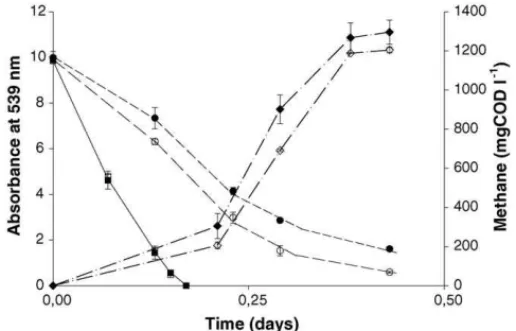
Table 2). A much lower methane production rate was found in the riboflavin-supplemented series with acetate (Fig. 3) and
Fig. 3. Azo dye reduction (left axis) of RR2 when acetate (1.5 gCOD l−1) was the electron donor, with the CH4formation being shown on the right axis. Sym-bols for dye reduction in the series contained: acetate (䊉), acetate + BES (), acetate + riboflavin (), acetate + riboflavin + BES (). Symbols for CH4 for-mation in the series contained: acetate (), acetate + riboflavin (♦). BES and riboflavin were added to some bioassays as methanogens inhibitor and redox mediator, respectively. Sludge-free series controlled the stability of the dye at 55◦C. The results are means of triplicate incubations and the bars indicate the
standard deviation. Only traces of CH4were found in the BES-supplemented bottles and endogenous controls.
methanol (data not shown) in comparison with controls lacking the redox mediator. In contrast, almost no difference in the rates was found when hydrogen (Fig. 4) and formate (data not shown) were the electron donors. For all methanogenic substrates tested,
Fig. 4. Azo dye reduction (left axis) of RR2 when H2/CO2(1.8 bar) was the electron donor, with the CH4formation being shown on the right axis. Symbols for dye reduction in the series contained: H2/CO2 (䊉), H2/CO2+ BES (), H2/CO2+ riboflavin (), H2/CO2+ riboflavin + BES (). Symbols for CH4 for-mation in the series contained: H2/CO2(), H2/CO2+ riboflavin (♦). BES and riboflavin were added to some bioassays as methanogens inhibitor and redox mediator, respectively. Sludge-free series controlled the stability of the dye at 55◦C. The results are means of triplicate incubations and the bars indicate the
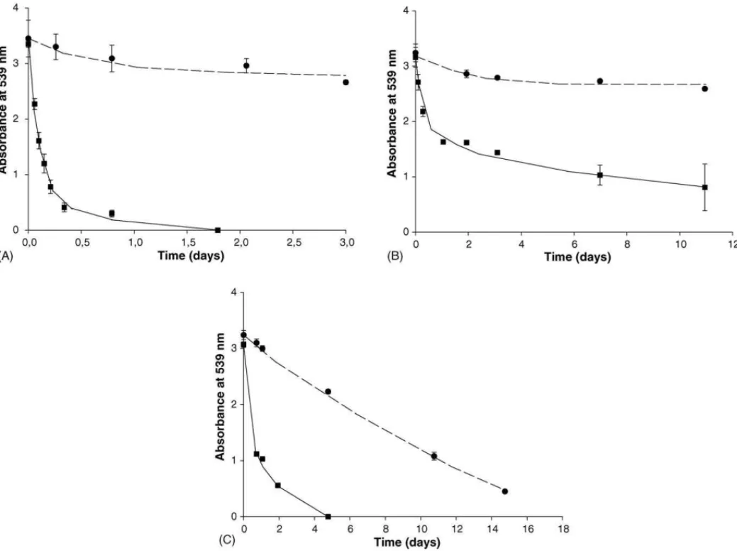
Fig. 5. Azo dye reduction of RR2 by the thermophilic strainsM. thermoautotrophicusH(A) and NJ1 (B), and by the mesophilic strainM. barkeri(C). Symbols for dye reduction in the series contained: strain (䊉), strain + riboflavin (). H2/CO2(1.8 bar) was used as electron donor. Riboflavin was added to some bioassays as a redox mediator. Sludge-free series controlled the stability of the dye. The results are means of triplicate incubations and the bars indicate the standard deviation.
their conversion was not directly coupled to the dye reduc-tion, but to the production of methane (data not shown), there-fore stressing the competition for reducing equivalents between methanogenesis and dye reduction. The concentration of elec-tron donors, in terms of elecelec-tron equivalents, was by far higher than the amount required for reducing the dye RR2.
No methane formation was detected in the headspace in the BES-supplemented series during the course of the experiment. Negligible decolourisation was found in both autoclaved sludge and sludge-free controls (data not shown).
3.3. Azo dye reduction by pure cultures of methanogens
Results of the experiments conducted with the thermophilic methanogens M. thermoautotrophicus H (Fig. 5A) and the strain NJ1 (Fig. 5B) revealed that these strains were unable to reduce the dye RR2 in the series lacking the redox mediator riboflavin. In contrast, the mesophilic methanogenM. barkeri (Fig. 5C) was able to reduce RR2 in both bioassays supple-mented and lacking riboflavin. For M. thermoautotrophicus H, we also tested the effect of re-flushing the headspace with H2/CO2and the addition of riboflavin in batches when no
decolourisation was taking place. We found that once riboflavin was supplemented there was an immediate reduction of the dye (data not shown).
The effect of BES on azo dye reduction varied consider-ably among the strains tested. In the presence of riboflavin, almost no dye reduction was found in the assays containing the strainM. thermoautotrophicusHand BES. In contrast, a complete decolourisation was found with the strain NJ1 and M. barkeri, under equal incubation conditions. Interestingly withM. barkeri, the rates of dye reduction between BES-free and BES-supplemented were comparable, while a consider-able difference was found when riboflavin was not included. No methane formation was detected in the headspace in the BES-supplemented bottles during the course of the experiment. Negligible decolourisation was found in both autoclaved cells and cell-free controls flushed with H2/CO2(data not shown).
4. Discussion
studied. The overall results indicate that although methanogens may contribute, azo dye reduction by the thermophilic anaero-bic consortium is mainly carried out by fermentative bacteria. This conclusive statement is justified by the following specific results: (i) there is a very apparent effect of vancomycin on the rates of decolourisation compared to the effect of BES, using a methanogenic consortium; (ii) reduction of the azo-linkage is not an inherent property of methanogens as some pure cul-tures of methanogens could not directly reduce the dye, and (iii) methanogenic consortium exerts a clear electron-donor depen-dency for dye reduction. The presence of a redox mediator, like riboflavin, not only strongly increased the rates of decolouri-sation by the thermophilic consortium studied, but also allowed pure cultures of methanogens, unable to directly reduce azo dyes, to participate in decolourisation processes.
4.1. The contribution of fermentative bacteria and methanogenic archaea to azo dye reduction
With glucose as electron donor, the rates of dye reduction after inhibition of the methanogenic archaea by BES were com-parable to the rates in incubations without this inhibitor. In some cases, even a higher rate of decolourisation was found when the methanogens were inhibited by BES, stressing that dye reducers and methanogens may compete for the same reducing equiva-lents. In contrast, when the fermentative bacteria were inhib-ited by vancomycin, a clear decrease in decolourisation rates was found, evidencing the important contribution of glucose-fermenting microorganisms on dye reduction.
When riboflavin was supplemented as a redox mediator, the effect of glucose concentration on the reductive decolourisa-tion was more pronounced than in series lacking riboflavin. Previous investigation with acetate, propionate, and butyrate, in the absence of redox mediators, showed that the rates of dye reduction were independent of the electron donors tested[23]. This suggests that the low concentrations of reducing equiv-alents produced during the anaerobic oxidation of propionate and butyrate could not sustain the dye reduction. Apparently in our research, riboflavin was mainly assisting the transfer of reducing equivalents from glucose fermentation to the dye, thus allowing fermentative microorganisms to participate more effectively in the reductive decolourisation. The ability of fer-mentative bacteria to use humic acids as an electron acceptor instead of riboflavin, as well as to reduce other azo dyes, agrees with our results[24,25,17].
4.2. Effect of different electron-donating substrates on azo dye reduction
The addition of electron-donating substrates consider-ably improved the rates of reductive decolourisation in a methanogenic consortium. Hydrogen and formate were the most effective substrates. Riboflavin increased the rates of reduction in all cases. Although acetate is usually a good substrate for methanogenesis, it was a very poor electron donor for reductive decolourisation. With this compound, a remarkable impact of riboflavin was found in the methane production rate (Fig. 3). It
seems that the presence of riboflavin on the acetoclastic incuba-tions helped the dye reducers on the competition for reducing equivalents with methanogens. During a study of different elec-tron donors on carbon tetrachloride reduction, hydrogen was an effective electron donor, whereas, methanol and acetate had a very poor electron donating capacity[26]. In the same study, 20M of the redox mediator, AQDS, considerably increased the reduction rates, which allowed that even poor electron donors became effective for dechlorination. The same distinct catalytic effect of a redox mediator with poor electron donors is in agree-ment with our present findings. A possible explanation for the low decolourisation rates of methanogenic archaea when acetate and methanol are the electron-donating substrates may be related to the enzymes involved in methanogenesis from these com-pounds, which do not utilize NAD+or NADP+as coenzymes. Instead some of the enzymes use a 5-deazaflavin F420as
coen-zyme [27]. It is currently accepted that NADH or NAD(P)H are the primary electron donors for biological azo dye reduction
[2,11,28,29]. However, the real biochemical and/or physiologi-cal reasons to the low electron-donating capacity of acetate and methanol remain unclear.
4.3. Effect of the methanogenic inhibitor BES with mixed and pure cultures
In the absence of riboflavin, the effect of BES on azo dye reduction was negligible with the methanogenic substrates acetate and methanol. In contrast, remarkable results were obtained when hydrogen and formate were included as electron donors; the rates of decolourisation were considerably higher (up to 1.7-fold) in BES-supplemented bottles compared to non-inhibited controls. Apparently, the inclusion of BES stimulated azo dye reduction by chemically out-competing methanogen-esis for the reducing equivalents available in hydrogen- and formate-supplemented cultures. Based on our findings it seems that acetate- and methanol utilizing methanogens do not play a role in the reductive decolourisation. The effect of BES with hydrogen as the electron donor is in agreement with the conclu-sions of Field et al. (2004), who studied the reduction of arsenate by a methanogenic consortium. In this study, methanogens and arsenate reducers were probably competing for the same elec-trons, likely in a similar reduction mechanism as we found for dye reduction [30]. In contrast, no influence of BES on the rate of AQDS reduction, i.e. when the quinone-mediator is the final electron acceptor, was found while testing hydro-gen and acetate as electron donors, which suggested that the methanogens present in the consortia were not involved in the quinone-respiration[31]. However, many methanogenic archaea such asM. barkeri,Methanococcus voltaeiand Methanospiril-lum hungateiJF1 can use AQDS as a terminal electron acceptor
[32,33].
Recently, it was reported that some methanogens preferentially oxidize some electron donors linked to the reduction of Fe(III) instead of the typically producing methane, also indicating the competition for electrons between these physiological processes
[34].
4.4. The catalytic effect of riboflavin on the decolourisation rates of mixed and pure cultures
Experiments with pure cultures of thermophilic methanogens M. thermoautotrophicus H and the strain NJ1 reveal that these strains were unable to reduce the azo dye RR2 in the absence of the redox mediator riboflavin. Stolz (2001) sug-gested that almost all azo dyes tested so far are biologically reduced under anaerobic conditions. For instance, a complete reduction of the sulfonated azo dye Amaranth by␣,and␥ -proteobacteria (Gram negative bacteria), andlowandhigh GC Gram positive bacteria was recently reported[6]. Based on our results with methanogenic archaea, anaerobic azo dye reduc-tion cannot be considered as a universal characteristic, as it has been repetitively reported. Interestingly, only a very small amount of reducing equivalents was required to completely reduce the azo-linkage. Theoretically, four electrons are needed to reduce one molecule of RR2. Thus, to reduce 0.1 mmol of RR2, 0.4 mmol electron equivalents (3.2 mgCOD) are needed, which represents 0.2% of the COD supplemented as electron donor (1200 mgCOD l−1). Therefore, the inability of the
ther-mophilic strains tested to reduce the dye was apparently not due to the lack of reducing equivalents but due to the lack of capability.
In the presence riboflavin, we found that both thermophilic strains were able to reduce RR2. The electrons released from hydrogen oxidation are likely transferred to riboflavin and then are used to reduce the azo dyes extracellularly. Reduc-tive decolourisation of the sulfonated dye model compound RR2 is unlikely to occur intracellularly. In our research, we did not find a relation between reduction rate and molecu-lar weight (Table 1, Fig. 1). Moreover, bacteria and archaea have a very limited membrane permeability, which is affected by the dye polarity[13,35,14]. The redox mediator lawsone, i.e. a quinone-based compound, was reduced intracellularly by Escherichia coli, after which reducing equivalents were trans-ported to the outside of the cell and subsequently reduced the azo dye Amaranth[9]. In contrast, it was shown that the redox mediator anthraquinone-2-sulfonate (AQS) considerably increased the anaerobic reduction rates of azo dyes by Sphin-gomonas xenophaga BN6. For this strain was suggested that a membrane bound NADH: ubiquinone oxidoreductase of the respiratory chain was the responsible for the dye reduction. Therefore, AQS was reduced at the outward-facing part of the cell membrane, and the cell penetration was not needed (13). Because hydrogenases are found in both cytoplasm and periplasm, we still do not know where the reduction of riboflavin was taking place, and therefore enzymatic studies are required. Anyhow, redox mediators are show to be important for allow-ing methanogens to participate more effectively on reductive decolourisation.
Because high rates of decolourisation were found when hydrogen was the electron donor in the bioassays with granular sludge, irrespective of the presence of riboflavin, very likely other hydrogenotrophic methanogens rather than the strains tested could have been directly involved in the azo cleavage. Another possibility for the fast reductive decolourisation of the riboflavin-free series in the methanogenic sludge incubations could be due to the presence of some fermentative bacteria with hydrogenase that coupled the oxidation of hydrogen to the dye reduction, or even due to redox mediators naturally produced in the sludge, which allows methanogens to partici-pate in this reductive process. Quinone-based compounds have not been found in methanogens, but phenazine derivatives have been shown to fulfil a quinone-like role in the electron trans-fer ofMethanosarcina mazei G¨o1[36]. It might well be that riboflavin or related compounds are formed and excreted by anaerobic bacteria in the sludge. Recently, many investigations also demonstrate the ability of either quinones or other redox mediators to transfer reducing power from bacterial cells to var-ious natural or xenobiotic compounds, therefore stimulating the reductive biotransformation of various compounds such as ferric iron, nitroaromatic compounds or polyhalogenated pollutants
[37,38]. Interestingly, an enzyme ofParacoccus denitrificans, called FerA, which catalyzed the reduction of a number of Fe(III) complexes by NADH, required the addition of FMN or riboflavin for activity on Fe(III) substrates[39].
Acknowledgements
The authors would like to thank Bo Jiang and Nico Tan for the assistance with the pure cultures experiment. This work was sup-ported by “Conselho Nacional de Desenvolvimento Cient´ıfico e Technol´ogico – CNPq” (Project no. 200488/01-5), an organiza-tion of the Brazilian Government for the development of Science and Technology.
References
[1] Weisburger JH. Comments on the history and importance of aromatic and heterocyclic amines in public health. Mutat Res 2002;506/507:9–20. [2] Stolz A. Basic and applied aspects in the microbial degradation of azo
dyes. Appl Microbiol Biotechnol 2001;56:69–80.
[3] Yoo ES. Kinetics of chemical decolorisation of the azo dye C.I. Reactive Orange 96 by sulfide. Chemosphere 2002;47:925–31.
[4] Chung KT, Stevens SEJ. Degradation of azo dyes by envi-ronmental microorganisms and helminths. Environ Toxicol Chem 1993;12:2121–32.
[5] Chen H, Wang RF, Cerniglia CE. Molecular cloning, overex-pression, purification, and characterization of an aerobic FMN-dependent azoreductase fromEnterococcus faecalis. Protein Expres Purif 2004;34:302–10.
[6] Rau J, Knackmuss HJ, Stolz A. Effects of different quinoid redox medi-ators on the anaerobic reduction of azo dyes by bacteria. Environ Sci Technol 2002;36:1497–504.
[7] Brown MA, DeVito SC. Predicting azo dye toxicity. Crit Rev Environ Sci Tech 1993;23:249–324.
[9] Rau J, Stolz A. Oxygen-insensitive nitroreductases NfsA and NfsB of Escherichia coli function under anaerobic conditions as lawsone-dependent azo reductases. Appl Environ Microbiol 2003;69:3448–55. [10] Dos Santos AB, Bisschops IAE, Cervantes FJ, Van Lier JB. Effect of
different redox mediators during thermophilic azo dye reduction by anaerobic granular sludge and comparative study between mesophilic (30◦C) and thermophilic (55◦C) treatments for decolourisation of
tex-tile wastewaters. Chemosphere 2004;55:1149–57.
[11] Semd´e R, Pierre D, Geuskens G, Devleeschouwer M, Moes AJ. Study of some important factors involved in azo derivative reduction by Clostrid-ium perfringens. Int J Pharm 1998;161:45–54.
[12] Keck A, Klein J, Kudlich M, Stolz A, Knackmuss HJ, Mattes R. Reduction of azo dyes by redox mediators originating in the naphthale-nesulfonic acid degradation pathway of Sphingomonassp. strain BN6. Appl Environ Microbiol 1997;63:3684–90.
[13] Kudlich M, Keck A, Klein J, Stolz A. Localization of the enzyme system involves in anaerobic reduction of azo dyes bySphingomonassp. strain BN6 and effect of artificial redox mediators on the rate of azo dye reduction. Appl Environ Microbiol 1997;63:3691–4.
[14] Van der Zee FP, Lettinga G, Field JA. Azo dye decolourisation by anaerobic granular sludge. Chemosphere 2001;44:1169–76.
[15] Paulo PL, Stams AJM, Field JA, Dijkema C, van Lier JB, Lettinga G. Pathways of methanol conversion in a thermophilic anaerobic (55◦C)
sludge consortium. Appl Microbiol Biotechnol 2003;63:307–14. [16] Oremland RS, Capone DG. Use of “specific” inhibitors in
biogeochem-istry and microbial ecology. Adv Microb Ecol 1988;10:285–383. [17] Dos Santos AB, Cervantes FJ, Yaya-Beas RE, Van Lier JB. Effect of
redox mediator, AQDS, on the decolourisation of a reactive azo dye containing triazine group in a thermophilic anaerobic EGSB reactor. Enzyme Microbiol Technol 2003;33:942–51.
[18] Stams AJM. Metabolic interactions between anaerobic bacte-ria in methanogenic environments. Antonie van Leeuwenhoek 1994;66:271–94.
[19] De Bok FAM, Roze EHA, Stams AJM. Hydrogenases and formate dehy-drogenases ofSyntrophobacter fumaroxidans. Antonie van Leeuwenhoek 2002;81:283–91.
[20] Van Lier JB, Rebac S, Lens P, Van BF, Oude ES, Stams AJM, et al. Anaerobic treatment of partly acidified wastewater in a two-stage expanded granular sludge bed (EGSB) system at 8◦C. Water Sci Technol
1997;36:317–24.
[21] APHA, Standard methods for the examination of water and wastewater, 20th ed. Washington, DC: American Public Health Association; 1998. [22] Bradford MM. A rapid and sensitive method for the quantification of
microgram quantities of protein utilizing the principle of protein-dye binding. Anal Biochem 1976;72:248–54.
[23] Van der Zee FP, Bouwman RHM, Strik DPBTB, Lettinga G, Field JA. Application of redox mediators to accelerate the transformation of reactive azo dyes in anaerobic bioreactors. Biotechnol Bioeng 2001;75:691–701.
[24] Benz M, Schink BAB. Humic acid reduction by Propionibacterium freudenreichiiand other fermenting bacteria. Appl Environ Microbiol 1998;64:4507–12.
[25] Yoo ES, Libra J, Wiesmann U. Reduction of azo dyes byDesulfovibrio desulfuricans. Water Sci Technol 2000;41:15–22.
[26] Cervantes FJ, Vu-Thi-Thu L, Lettinga G, Field JA. Quinone-respiration improves dechlorination of carbon tetrachloride by anaerobic sludge. Appl Microbiol Biotechnol 2004;64:702–11.
[27] Berk H, Thauer RK. Function of coenzyme F420-dependent NADP reductase in methanogenic archaea containing and NADP-dependent alcohol dehydrogenase. Arch Microbiol 1997;168:396–402.
[28] Zimmermann T, Kulla H, Leisinger T. Properties of purified Orange II azoreductase, the enzyme initiating azo dye degradation by Pseu-domonasKF46. Eur J Biochem 1982;129:197–203.
[29] Blumel S, Stolz A. Cloning and characterization of the gene coding for the aerobic azoreductase fromPigmentiphaga kullaek24. Appl Micro-biol Biotechnol 2003;62:186–90.
[30] Field JA, Sierra-Alvarez R, Cortinas IGF, Moreira MT, Kopplin M, Gan-dolfi AJ. Facile reduction of arsenate in methanogenic sludge. Biodegra-dation 2004;15:185–96.
[31] Cervantes FJ, van der Velde S, Lettinga G, Field JA. Competi-tion between methanogenesis and quinone respiraCompeti-tion for ecologically important substrates in anaerobic consortia. FEMS Microbiol Ecol 2000;34:161–71.
[32] Cervantes FJ, De Bok FAM, Duong-Dac T, Stams AJM, Let-tinga G, Field JA. Reduction of humic substances by halorespiring, sulfate-reducing and methanogenic microorganisms. Environ Microbiol 2002;4:51–7.
[33] Bond DR, Lovley DR. Reduction of Fe(III) oxide by methanogens in the presence and absence of extracellular quinones. Environ Microbiol 2002;4:115–24.
[34] Van Bodegom PM, Scholten JCM, Stams AJM. Direct inhibition of methanogenis by ferric iron. FEMS Microbiol Ecol 2004;49:261–8. [35] Russ R, Rau J, Stolz A. The function of cytoplasmic flavin
reduc-tases in the reduction of azo dyes by bacteria. Appl Environ Microbiol 2000;66:1429–34.
[36] Abken HJ, Tietze M, Brodersen J, Bauemer S, Beifuss U, Deppenmeier U. Isolation and characterization of methanophenazine and function of phenazines in membrane-bound electron transport of Methanosarcina mazeiGo1. J Bacteriol 1998;180:2027–32.
[37] Lovley DR, Fraga JL, BluntHarris EL, Hayes LA, Phillips EJP, Coates JD. Humic substances as a mediator for microbially catalyzed metal reduction. Acta Hydroch Hydrob 1998;26:152–7.
[38] Dunnivant FM, Schwarzenbach RP, Macalady DL. Reduction of sub-stituted nitrobenzenes in aqueous solutions containing natural organic matter. Environ Sci Technol 1992;26:2133–41.
[39] Mazoch J, Tesarik R, Sedlacek V, Kucera I, Turanel J. Isolation and biochemical characterization of two soluble iron(III) reductases from