C
a s eR
e p o Rt1 9 9 Arq Bras Oftalmol. 2017;80(3):199-201 http://dx.doi.org/10.5935/0004-2749.20170049
ABSTRACT
We describe a unique complication during primary posterior continuous cur-vilinear capsulorhexis (PCCC) in a patient with megalocornea scheduled for phacoemulsification with toric multifocal intraocular lens (IOL) implantation. After nucleus emulsification and cortex removal, the capsular bag was filled with cohesive viscoelastic in preparation for PCCC to achieve reverse optic capture of the IOL, thus ensuring stability. However, as soon as the initial puncture was made using a 27-gauge needle to start the capsulotomy, the posterior capsule opening extended peripherally from 0º-180º. This capsule extension was similar to the Argentinean-flag sign in hypermature cataracts, and both are caused by excessive intracapsular pressure. Careful bimanual manipulation was performed to implant the IOL on the desired axis, which occurred uneventfully. At a postoperative visit, the patient exhibited excellent uncorrected visual acuity with a well-aligned IOL.
Keywords: Cornea/abnormalities; Cataract; Phacoemulsification; Capsulorhexis; Humans; Case reports
RESUMO
Os autores demonstram uma complicação durante a realização de uma capsulo-tomia circular contínua posterior (CCCP) em um paciente com megalocórnea programado facoemulsificação com implante de lente intraocular (LIO) tórica multifocal. Após a remoção do núcleo e córtex, o saco capsular foi preenchido por viscoelástico coesivo com finalidade de prepará-lo para realização da CCCP e com isso assegurar o correto alinhamento do implante no eixo desejado. Entretanto, assim que a agulha de 27-gauge foi utilizada para confecção puntura inicial da capsulotomia, imediatamente a cápsula posterior se abriu até periferia de 0-180 graus, similar à lesão capsular vista no sinal da Bandeira Argentina em cataratas hipermaduras, ambos causados por pressão excessiva intracapsular. Manipulação cuidadosa foi realizada para implantação da lente no eixo correto, a qual aconte-ceu sem intercorrências. No pós-operatório, a paciente apresentou uma excelente acuidade visual sem correção com LIO corretamente alinhada no eixo desejado.
Descritores: Córnea/anormalidades; Catarata; Facoemulsificação; Capsulorrexe; Hu -manos; Relatos de casos
Posterior argentinean flag-like sign during primary posterior continuous
curvilinear capsulorhexis
Sinal da bandeira argentina-
like
durante capsulorrexis posterior primária
Frederico França Marques1, daniela Mieira Villano Marques1, Fabio Kenji MatsuMoto1
Submitted for publication: November 21, 2016 Accepted for publication: February 22, 2017
1 Department of Ophthalmology and Visual Sciences, Universidade Federal de São Paulo, São Paulo,
SP, Brazil.
Funding: No specific financial support was available for this study.
Disclosure of potential conflicts of interest: None of the authors have any potential conflicts of interest to disclose.
Corresponding author: Frederico França Marques. Rua Indiana, 527/81 - São Paulo, SP - 04562-000 Brazil - E-mail: [email protected]
INTRODUCTION
The Argentinean-flag sign is a known complication during curvilinear continuous capsulorhexis (CCC) in hypermature or in-tumescent cataracts characterized by a spontaneous tear in the anterior capsule extending into the periphery as a result of high in tracapsular pressure. This complication can compromise the final visual result by causing lens decentration, tilting, or even aphakia because of the absence of capsule support.
Many techniques have been proposed to prevent the develop-ment of this complication, such as use of a small CCC, cortex aspi-ration using a 27-gauge cannula for the initial puncture, or use of a femtosecond laser to perform CCC. However, none of these methods is capable of preventing this complication during primary posterior curvilinear continuous capsulorhexis (PCCC).
In this paper, we describe a spontaneous posterior capsule tear after the initial puncture during PCCC in a patient with megalocor-nea scheduled for toric multifocal intraocular lens (IOL) implanta-tion with reverse optic capture to prevent intracapsular IOL rotaimplanta-tion.
CASE REPORT
A 53-year-old man presented to our clinic with blurred vision in both eyes. His best corrected visual acuity was 20/25 and 20/30
with -3.50 sphere (sph) -3.50 cylinder (cyl) @ 170o in the oculus dextrus
(OD) and -6.00 sph -0.75 cyl @ 160o in the oculus sinister (OS).
Slit-lamp examination revealed a large corneal diameter, Descemet’s membrane with no sign of tears, a deep anterior chamber depth (ACD), and a 1+ nuclear cataract in the OD and 2+ nuclear cataract in the OS with no signs of phacodonesis or iridodonesis. Gonios-copy revealed an open angle with no abnormalities. Intraocular pressure was 13 mmHg in both eyes. The indings of fundoscopy were unremarkable.
The simulated keratometry readings were 41.02 × 43.56 @ 93o
in the OD and 41.61 × 42.5 @ 104o in the OS (iTrace; Tracey
Po s t e r i o r a r g e n t i n e a n f l a g-l i k e s i g n d u r i n g P r i m a ry P o s t e r i o r c o n t i n u o u s c u rv i l i n e a r c a P s u l o r h e x i s
2 0 0 Arq Bras Oftalmol. 2017;80(3):199-201
of optical coherence tomography of the maculae were normal in both eyes.
After thorough discussion of the oversized capsular bag and possible IOL decentration with the patient, we decided to implant bilateral multifocal IOLs. For the first eye (OS), after performing an uneventful phacoemulsiication, we implanted a non-toric +12.0 Tecnis ZMA00 (Abbott Medical Optics Inc., Santa Ana, CA, USA) into the capsular bag with no complications regarding IOL centration. The patient achieved excellent postoperative visual acuity on Day 1 with an uncorrected visual acuity (UCVA) of 20/20 and J1. Regarding the other eye, because of its corneal astigmatism, a Tecnis ZMT300 (Abbott Medical Optics Inc.) toric 13.0 D multifocal IOL was placed at 87º.
Based on the high possibility of lens rotation because of the large capsular bag and single-piece design of the IOL, we planned to perform phacoemulsiication with primary PCCC to achieve reverse capture of the IOL optic zone, thus ensuring IOL stability.
After confirming the 14-mm W-W diameter with calipers, pha-coemulsification and cortical aspiration were performed without complications. To perform PCCC, the capsular bag was filled with
a cohesive viscoelastic device (ProVisc®; Alcon Laboratories, Inc.,
Fort Worth, TX, USA). A 27-gauge needle mounted in a dispersive
ophthalmic viscosurgical device (OVD; Viscoat®, Alcon
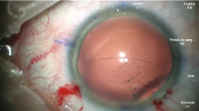
Laborato-ries, Inc.) was used to perform the initial puncture of the central posterior capsule and inject dispersive viscoelastic into Berger’s space to avoid rupture of the anterior hyaloid membrane (Figure 1). However, as soon as the posterior capsule was punctured, a tear extended spontaneously to the periphery in a similar fashion to an Argentinean-flag sign in the anterior capsule (Figure 2). Because the eye was well pressurized with a stable anterior chamber, the anterior hyaloid membrane remained intact and no vitreous humor
prolapsed. IOL insertion into the capsular bag was performed using a bimanual technique with extreme caution to avoid extending the tear, vitreous humor prolapse, and dropping the IOL into the vitreous cavity.
After gentle sequential maneuvers, the IOL was centered and aligned on the desired axis with satisfactory stability. The OVD in the anterior segment was carefully washed out with light depression of the main incision during irrigation, taking care to preserve anterior chamber stability. A miotic agent was used to bring the pupil down, and a spatula through the side port was used to conirm the absence of vitreous. The incisions were hydrated, and topical antibiotic was instilled.
On Day 1, his UCVA was 20/30 and J2 p, and the IOL was on the correct axis (Figure 3). One week later, the patient was very pleased
with his UCVA of 20/25 J2, with a manifest refraction of +0.25 -0.50 @ 10o
achieving the same visual acuity because of some degree of am-blyopia probably originating from the higher corneal astigmatism in this eye.
DISCUSSION
Capsule extension during capsulotomy can occur at any time during capsulorhexis maneuvers. Usually, it occurs because increa-sed intracapsular pressure directs the vector forces of the capsule tear toward the zonules. This situation can be abrupt, and it is com-monly found in hypermature cataracts, leading to the well-known Argentinean-lag sign.
Our patient had megalocornea, which is a developmental ano-maly of unknown etiology characterized by enlargement of the cornea to 13 mm or greater. The condition can be associated with anterior megalophthalmos and/or dysgenesis of the iris, lens, and
ciliary body, and exhibits X-linked recessive inheritance(1). Fortunately,
the zonules were intact and no capsular bag abnormalities, which can make toric alignment a real challenge, were found besides the large corneal size.
The main factors leading to postoperative IOL rotation away from the intended vertical meridian are eyes with an axial length greater
than 25 mm and with-the-rule astigmatism(2). Many techniques have
been described to avoid this rotation or realign the IOL, such as in-sertion of a regular capsular tension ring (CTR) and/or Henderson’s CTR to increase the area of contact within the IOL haptic and optic zone by stretching the posterior capsule or even suturing the IOL to a CTR, or PCCC to achieve reverse optic capture performed manually
or using a femtosecond laser(3-6). In our case, another option was
to insert the IOL inside the capsular bag and capture just the optic anteriorly, leaving the haptics inside the bag; however, because of the increased chance of pigment-dispersion glaucoma secondary to chaing between the posterior surface of the iris and the square edge
of the IOL optic, we decided against this procedure(7).
Because of the large W-W diameter of 14 mm, the IOL diameter of 13 mm, and the intended axis of 87º, we decided to perform PCCC
with reverse optic capture as described by Gimbel(4) for correct IOL
alignment and centration instead of using CTRs because, in most patients with megalocornea, the CTR alone is insufficient. To per-form PCCC, the anterior chamber must be stable and the posterior capsule concave after the initial puncture to create a small tear; dis-persive viscoelastic must be injected into Berger’s space to protect the anterior hyaloid membrane; and once the tear is created, Utrata forceps must be used to create a circular opening.
In our opinion, the cause of posterior capsule extension during the primary PCCC, although inverse, was the same the that en-countered in eyes with the Argentinean-lag sign: the capsular bag was overilled within cohesive viscoelastic, leading to an extremely concave posterior capsule and extending the intentional tear imme-diately after the initial puncture toward the periphery, similarly to an Argentinean-lag sign. To our knowledge, this inding has never been demonstrated before.
Figure 1. The 27-gauge needle puncturing the central posterior capsule.
Ma r q u e s FF, e t a l.
2 0 1 Arq Bras Oftalmol. 2017;80(3):199-201
In summary, when planning a PCCC, the surgeon must consider the possibility of this complication, which can be avoided by using a small amount of cohesive viscoelastic to induce minimal distention of the posterior capsule. Avoiding overilling the capsular bag may prevent peripheral extension of the tear.
REFERENCES
1. Saatci AO, Soylev M, Kavukcu S, Durak I, Saatci I, Memisoglu B. Bilateral megalocornea with unilateral lens subluxation. Ophthalmic Genet. 1997;18(1):35-8.
2. Miyake T, Kamiya K, Amano R, Iida Y, Tsunehiro S, Shimizu K. Long-term clinical outcomes of toric intraocular lens implantation in cataract cases with preexisting astigmatism. J Cataract Refract Surg. 2014;40(10):1654-60.
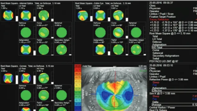
Figure 3. iTrace (Tracey Technologies LLC, Houston, TX, USA) aberrometry of the internal optics, revealing an astigmatism of 177º corresponding to an intraocular lens axis of 87º as planned.
3. Rękas M, Pawlik R, Klus A, Rozycki R, Szalik JP, Oldak M. Phacoemulsiication with corneal astigmatism correction with the use of a toric intraocular lens in a case of me galocornea. J Cataract Refract Surg. 2011;37(8):1546-50.
4. Gimbel HV, Amritanand A. Reverse optic capture to stabilize a topic intraocular lens. Case Rep Ophthalmol. 2013;4(3):138-43.
5. Scott WJ, Owsiak RR. Femtosecond laser-assisted primary posterior capsulotomy for toric intraocular lens ixation and stabilization. J Cataract Refract Surg. 2015;41(8): 1767-71.
6. Sagiv O, Sachs D. Rotation stability of a toric intraocular lens with a second capsular tension ring. J Cataract Refract Surg. 2015;41(5):1098-9.
7. Hong Y, Sun YX, Qi H, Zhou JC, Hao YS. Pigment dispersion glaucoma induced by the chaing efect of intraocular lens haptics in Asian eyes. Curr Eye Res. 2013;38(3): 358-62.