175
Cruz JMN et al. Pelvic lipomatosis: a case report and literature review
Radiol Bras. 2012 Mai/Jun;45(3):175–177
Pelvic lipomatosis: a case report and literature review
*
Lipomatose pélvica: relato de caso e revisão da literatura
José Marcílio Nicodemos da Cruz1, Márcio Antonio Araruna Vieira Filho2, Larisse Vieira Mendes2,
Assuero Florentino Bezerra Junior2, Breno Levi de Oliveira Aragão2, José Marcílio Nicodemos
da Cruz Filho3
Pelvic lipomatosis is a rare disorder where fat tissue deposition is observed in spaces of the pelvic area, causing extrinsic compression of the bladder, rectum and blood vessels. In the present report, the authors describe the case of a 48-year-old male patient presenting low back pain, dysuria and pollakiuria. Findings at excretory urography, computed tomography and magnetic resonance imaging are described.
Keywords: Pelvic lipomatosis; Urography; Computed tomography; Magnetic resonance imaging.
A lipomatose pélvica é uma doença rara, na qual se observa acúmulo de tecido adiposo na pelve causando compres-são extrínseca da bexiga, reto e vasos sanguíneos. Neste estudo relatamos o caso de um paciente do sexo masculino, 48 anos de idade, que apresentou dores lombares e síndrome disúria-polaciúria. São descritos os achados na urogra-fia excretora, tomograurogra-fia computadorizada e ressonância magnética.
Unitermos: Lipomatose pélvica; Urografia; Tomografia computadorizada; Ressonância magnética.
Abstract
Resumo
* Study developed at Universidade Federal do Ceará (UFC) – Campus Cariri, Barbalha, CE, Brazil.
1. Post-graduation in Radiology, Professor of Human Anato-my and Radiology, Universidade Federal do Ceará (UFC) – Cam-pus Cariri, Barbalha, CE, Brazil.
2. Graduate Students of Medicine, Universidade Federal do Ceará (UFC) – Campus Cariri, Barbalha, CE, Brazil.
3. Graduate Student of Medicine, Faculdade de Medicina Estácio de Juazeiro do Norte, Juazeiro do Norte, CE, Brazil
Mailing Address: Dr. José Marcílio Nicodemos da Cruz. Avenida Padre Cícero, 2085, Salesiano. Juazeiro do Norte, CE, Brazil, 63010-000. E-mail: [email protected]
Received September 28, 2011. Accepted after revision March 1st, 2012.
Cruz JMN, Vieira Filho MAA, Mendes LV, Bezerra Jr AF, Aragão BLO, Cruz Filho JMN. Pelvic lipomatosis: a case report and literature review. Radiol Bras. 2012 Mai/Jun;45(3):175–177.
0100-3984 © Colégio Brasileiro de Radiologia e Diagnóstico por Imagem
CASE REPORT
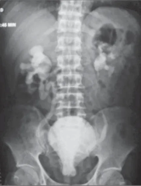
tion of “tear- or pear-shaped bladder” (Fig-ure 1). Post-micturition image did not dem-onstrate the presence of any urinary residue (Figure 2). Computed tomography demon-strated large amounts of fatty tissue occu-pying the pelvis from the bottom of the pelvic cavity, causing extrinsic compres-sion and elevation of the bladder with anterocranial deformity, besides occupa-tion of the prostatic compartment. Addi-The present report describes a case of
pelvic lipomatosis, considering the scarce presence of articles approaching this dis-ease in the scientific literature, and high-lighting the imaging findings at excretory urography, computed tomography and magnetic resonance imaging.
CASE REPORT
A 48-year-old male, white patient born in Barro, CE, Brazil, reported low back pain for three years, with progressive wors-ening in the last two months, particularly on his left side, without irradiation, of com-pressive nature and in stitches, with mild to moderate intensity and post-micturition relief. Besides the pain, the patient pre-sented dysuria and pollakiuria. No relevant past medical history was reported. At physi-cal examination, atrophic testicle was ob-served and the prostate was nonpalpable at digital rectal examination. The patient was 1.65 m in height and had 60 kg of weight. Ultrasonography demonstrated bilateral hydronephrosis, with no other findings. Excretory urography confirmed the pres-ence of hydronephrosis and demonstrated extrinsic compression of the bladder caus-ing deformity and elevation of the bladder floor compatible with the typical
presenta-INTRODUCTION
Pelvic lipomatosis is a rare disease that was first described in late 1950’s by Engles(1). The condition is characterized by deposition of mature fat tissue in the pel-vic cavity and absence of delimitation by a capsule. Intermingled inflammatory reac-tions may be observed, causing compres-sion of the bladder, rectum and blood ves-sels. Clinical manifestations include signs and symptoms of extrinsic obstruction of such structures. The exact incidence of this disease is still remains undetermined, but it is known that it is most prevalent in men and individuals with dark-skinned pheno-type, most frequently at the third or fourth decade of life, despite the existence of re-ports describing the disease in the elderly and in children(2,3).
176
Cruz JMN et al. Pelvic lipomatosis: a case report and literature review
Radiol Bras. 2012 Mai/Jun;45(3):175–177 Figure 3. Computed tomography. Axial (A) and sagittal (B) images showing the development of hypodense fat in the pelvis, causing extrinsic compression of the bladder and of the rectum.
tionally, signs of extrinsic rectosigmoid compression with widening of the recto-sacral space (Figure 3). Magnetic reso-nance imaging was also performed, dem-onstrating deformation of the bladder and rectum caused by fatty tissue both in the sagittal and coronal planes (Figure 4).
DISCUSSION
Taking the unknown etiology of pelvic lipomatosis into consideration, some
au-thors have proposed some theories in an at-tempt to explain the development of this disease whose genesis would be associated with a localized manifestation of general-ized obesity(4); other authors have raised the hypothesis of a response to repeated uro-logical infections, attributing the origin of the fatty tissue deposition to the inflamma-tory process. Additionally, other authors suggest that pelvic lipomatosis might rep-resent a variant of Dercum’s disease, or other who raise the hypothesis of a
local-ized response to hormonal and metabolic phenomena, as well as those who based on the observation of the highest incidence in black men, suggest the possibility of a ge-netic basis for the disease(5).
The clinical manifestations result from the extrinsic compression of the structures comprising the urinary system, the lower intestinal tract and the vascular system. Thus, the occurrence of dysuria, pollakiuria, nocturia, hematuria (less frequently), ur-gency, urinary incontinence and retention, besides repeated urological infections may be observed. Equally, constipation, tenes-mus, diarrhea, lower limbs edema and thrombophlebitis, low back pain, suprapu-bic and perineal pain, painful ejaculation, epidydimitis and orchitis may be observed. Additionally, there is the possibility of re-nal failure, also with development of arte-rial hypertension(6).
At physical examination, one may ob-serve pain at abdominal palpation, pres-ence of a palpable mass in the hypogastric region, urinary retention, elevation of the prostate at digital rectal examination, lower limbs edema and arterial hypertension.
The diagnosis is done by means of im-aging methods which demonstrate deposi-tion of fatty tissues causing compression of adjacent structures, possibly resulting in relevant morphological deformities. It is important to highlight the relevant role of Figure 2. Excretory urography demonstrating a “tear- or pear-shaped bladder” with elevation of its floor,
177
Cruz JMN et al. Pelvic lipomatosis: a case report and literature review
Radiol Bras. 2012 Mai/Jun;45(3):175–177 excretory urography in the demonstration of the typical presentation of tear- or pear-shaped bladder, besides uretero-hydro-nephrosis. Computed tomography defines the cause of the extrinsic compression of the bladder, demonstrating the perivesical and perirectal involvement by fat, thus be-ing sufficient for establishbe-ing the diagno-sis of pelvic lipomatodiagno-sis. Magnetic reso-nance imaging presents the advantage of not utilizing ionizing radiation, constitut-ing a highly accurate method for studyconstitut-ing affected anatomic structures, contributing with accurate high-definition images.
Treatment options are limited. Conser-vative approaches utilizing dietary manage-ment, use of antimicrobial agents and cor-ticoids and even attempts with radiotherapy have not demonstrated to be eminently ef-ficacious. Surgery faces difficulties be-cause of the absence of clearly defined dis-section planes to allow the release of
pel-vic organs. Some authors have reported achievement of therapeutic success with lipoaspiration techniques(7). Management of symptoms can be achieved with urinary derivation, but the potential risk for malignization must be taken into consider-ation e further treatment modalities must be studied.
Additionally to the consequences of the obstructive phenomena, the chronic vesi-cal wall irritation or even the compression of lymph chains caused by the deposition of fat tissue may explain the frequent find-ing of proliferative processes involvfind-ing the bladder mucosa in patients affected by this disease, such as cystic and glandular cysti-tis. In spite of being considered benign le-sions, the possibility of development of vesical adenocarcinoma should be consid-ered, justifying the periodic follow-up with cystoscopy and biopsy of suspicious le-sions, besides urinary cytology(8).
REFERENCES
1. Engels EP. Sigmoid colon and urinary bladder in high fixation: roentgen changes simulating pelvic tumor. Radiology. 1959;72:419–22.
2. Zaman W, Singh V, Kumar B, et al. Pelvic lipoma-tosis in a child. Urol Int. 2002;69:238–40. 3. Buitrago Sivianes S, Tallada Buñuel M, Vicente
Prados FJ, et al. Pelvic lipomatosis. Diagnostic and therapeutic considerations apropos of 3 cases. Arch Esp Urol. 2002;55:900–6.
4. Sacks SA, Drenick EJ. Pelvic lipomatosis: effect of diet. Urology. 1975;6:609–15.
5. Tong RS, Larner T, Finlay M, et al. Pelvic lipoma-tosis associated with proliferative cystitis occurring in two brothers. Urology. 2002;59:602.
6. Hudolin T, Kastelan Z, Goluza E, et al. Pelvic and retroperitoneal lipomatosis: case report. Acta Clin Croat. 2010;49:465–8.
7. Halachmi S, Moskovitz B, Calderon N, et al. The use of an ultrasonic assisted lipectomy device for the treatment of obstructive pelvic lipomatosis. Urology. 1996;48:128–30.
8. Heyns CF, De Kock ML, Kirsten PH, et al. Pelvic lipomatosis associated with cystitis glandularis and adenocarcinoma of the bladder. J Urol. 1991;145: 364–6.