ABSTRACT
Gastro-intestinal carcinoids are slow growing tumors arising from ente-rochromaffin or Kulchitsky cells. Their clinical presentation depends on what combination of bioactive substances is secreted. Midgut carcinoid can present with the carcinoid syndrome in the presence of liver metas-tases. Its most typical clinical manifestations include cutaneous flushing and diarrhea. A nonspecific biochemical tumor marker for carcinoid tumors is serum chromogranin A and a specific marker for the carcinoid syndrome is the increased urinary excretion of 5-hydroxy indole acetic acid (5-HIAA). Localizing studies in carcinoid tumors/syndrome are: transabdominal ultrasonography (US), endoscopy, endoscopic US, videocapsule endoscopy, computerized tomography, magnetic reso-nance imaging, selective abdominal angiography,111In-pentetreotide scintigraphy (and intraoperative radionuclide probe), 1 2 3I (1 3 1I ) -metaiodobenzylguanidine (MIBG) scintigraphy, bone scintigraphy and 11C-5-HT positron emission tomography (PET). Therapies for carcinoid tumors/syndrome are: surgery, somatostatin analogs, interferon-alpha, radiotherapy, liver dearterialization, liver (chemo, or radio)-emboliza-tion, alcohol sclerotherapy of liver metastases, radiofrequency ablation of liver metastases, cryosurgery of liver metastases, occasionally liver transplantation, radiotherapy-coupled somatostatin analogs,131I-MIBG and occasionally chemotherapy. (Arq Bras Endocrinol Metab 2005;49/5:850-860)
Keywords:Carcinoid; Neuroendocrine; Tumor; Imaging; 5-HIAA; Chro-mogranin A
RESUMO
Síndrome Carcinóide: Diagnóstico e Manejo Clínico.
Carcinóides gastro-intestinais são tumores de crescimento lento ori-ginários das células enterocromafínicas ou de Kulchitsky. Sua apresen-tação clínica depende das combinações de substâncias bioativas que são secretadas. Carcinóides de intestino delgado podem se apresen-tar com síndrome carcinóide na presença de metástases hepáticas. A manifestação clínica típica incluiflushingcutâneo e diarréia. A cromo-granina-A é um marcador bioquímico tumoral inespecífico de tumores carcinóides e o aumento da excreção urinária de ácido 5-hidroxiin-dolacético (5-HIAA), um marcador específico para a síndrome carcinóide. Estudos de localização nos tumores/síndrome carcinóide são: ultrassonografia (US) abdominal, endoscopia, US endoscópica, endoscopia com vídeo-cápsula, tomografia computadorizada, ressonância magnética, angiografia abdominal seletiva, cintilografia com 111In-pentetreotide (e sonda radioisotópica intraoperatória), cin-tilografia com123I (131I) -metaiodobenzilguanidina (MIBG), cintilografia óssea e tomografia por emissão de pósitron (11C-5-HT). Tratamento para tumores/síndrome carcinóide são: cirurgia, análogos de somatostati-na, interferon-alfa, radioterapia, embolização arterial hepática, quimioembolização hepática, escleroterapia alcoólica de metástases hepáticas (MH), ablação por radiofreqüência de MH, criocirurgia de
Aart J. van der Lely
Wouter W. de Herder
Department of Internal Medicine, Section of Endocrinology, Erasmus MC, Rotterdam, the Netherlands.
MH, ocasionalmente transplante hepático, radioter-apia associada a análogos de somatostatina,131 I-MIBG e ocasionalmente quimioterapia.(Arq Bras Endocrinol Metab 2005;49/5:850-860)
D e s c r i t o r e s : Carcinóide; Neuroendócrino; Tumor; Imagens; 5-HIAA; Cromogranina A
HISTORICAL OVERVIEW
G
ASTRO-INTESTINAL CARCINOIDSare slow growing neoplasms as compared with adenocarcinomas, but they can also behave aggressively. They are derived from neoplastic proliferation of e n t e r o c h r o m a f f i n (ECL) or Kulchitsky cells (1).In 1888, Lubarsch first described a patient with multiple carcinoids of the ileum but regarded them as carcinomas (2). Two years later, Ransom first described the classical symptomatology of the carcinoid syn-drome in a patient with an ileal carcinoid tumor and hepatic metastasis (3). However, it was Oberndorfer in 1907, who coined the term “karzinoide” to describe these tumors, which he believed to behave in a more benign fashion than adenocarcinomas (4). In 1963, Williams and Sandler classified carcinoids according to their embryologic site of origin as foregut carcinoids (respiratory tract, stomach, duodenum, biliary system, and pancreas), midgut carcinoids (small intestine, appendix, cecum, and proximal colon), and hindgut carcinoids (distal colon and rectum) (5). However, these lesions exhibit a high degree of morphologic and biologic heterogeneity and a more generic term, neu-roendocrine tumor (NET) has been introduced to replace the term carcinoid. Such lesions are currently referred to as gastroenteropancreatic (GEP) NETs (GEP-NETs). According to the WHO classification, distinction was made between well-differentiated NETs (benign behavior or uncertain malignant potential), well-differentiated neuroendocrine carcinomas (low-grade malignancy), and poorly differentiated (usually small cell) neuroendocrine carcinomas of high-grade malignancy. Nevertheless, the term carcinoid was not abandoned and for GEP-NETs, it is used synonymous-ly with the term “well-differentiated NET”. The term “malignant carcinoid” is used synonymously with the term well-differentiated neuroendocrine carcinoma (6). The differentiation is based on tumor morphology, tumor size (in general larger tumors are more aggres-sive), and the presence or absence of local invasion and/or metastasis, thus reflecting biological behavior. Most NETs are well-differentiated tumors that are characterized by a solid trabecular or glandular s t r u
c-ture, tumor cell monomorphism with absent or low c y t o l o g i c a l atypia, and a low mitotic (< 2 mitoses/ m m2) and proliferative status (< 2% Ki-67 positive
cells). Only in the presence of metastasis and/or inva-s i v e n e inva-s inva-s iinva-s the tumor defined ainva-s a well-differentiated neuroendocrine carcinoma. Poorly differentiated NETs are invariably malignant, are defined as poorly differen-tiated neuroendocrine carcinomas, and are character-i z e d by a predomcharacter-inantly solcharacter-id structure wcharacter-ith abundant necrosis, cellular atypia with a high mitotic index (≥ 10 m i t o s e s / m m2) and proliferative status (> 15% Ki-67
positive cells), diffuse reactivity for cytosolic markers, and scant or weak reactivity for granular markers or neurosecretory products (1).
Carcinoid lesions are the most common NETs and compose approximately 50% of all NETs of the gastrointestinal tract. In most instances, they are dis-covered incidentally at the time of surgery for other abdominal disorders. Their presence may be unde-tectable for years without obvious signs or symptoms. Evidence for this observation is supported by their rel-atively high incidence in large autopsy series (7). When symptoms do occur, they are due either to local tumor mass effects, the effects of tumor-engendered fibrosis, or to the secreted bioactive products from the neo-plasm. Symptoms caused by local tumor effects include vague abdominal pain (invasion, intussuscep-tion, fibrous adhesions, hypermotility), which is often undiagnosed or leads to erroneous diagnoses like irri-table bowel syndrome (8,9).
and do not usually cause any systemic signs or symp-toms. However, when liver metastases are present or when the primary lesions are found in the bronchus and/or ovaries, the systemic features of the carcinoid syndrome become more evident. This classical syn-drome occurs in fewer than 10% of patients, and its most typical clinical manifestations include cutaneous flushing most commonly of the face, neck, and upper chest and diarrhea, occurring in up to 75%. Less fre-quent manifestations include cardiac valvular abnor-malities (plaque-like, fibrous endocardial thickening that principally involves the right side of the heart (causing tricuspid regurgitation tricuspid stenosis, pul-monary regurgitation, and pulpul-monary stenosis), bron-choconstriction and (as already mentioned) pellagra. Foregut carcinoids can secrete 5-HTP, histamine and polypeptide hormones like ACTH. The can produce a characteristic clinical syndrome known as “atypical” carcinoid syndrome. Midgut carcinoids release 5-HT and other vasoactive compounds such as kinins, prostaglandins, and substance P and they are more likely to cause the classic carcinoid syndrome with the development of hepatic metastases. Hindgut carcinoid tumors rarely contain 5-HT and usually do not present with the carcinoid syndrome; however. The symptoms of the carcinoid syndrome can be both of variable intensity as well as paroxysmal, responding intermit-tently to a particular “trigger” agent, such as alcohol, cheese, coffee (these are serotonin-rich foods), or exercise (8,10,11).
Many carcinoid tumors exhibit a significant association with other non-carcinoid tumors of various histological types. A relatively large percentage of car-cinoids are multicentric.
Because carcinoid tumors frequently present with obscure clinical manifestations, numerous investi-gatory procedures are often undertaken prior to estab-lishing the correct diagnosis. Although clinical diagno-sis is based on symptoms, biochemical confirmation is necessary. The diagnostic strategies employed usually depend on the individual clinical presentation (8,10,11).
Biochemical markers
24-hour urinary excretion of 5-hydroxy indole acetic acid (5-HIAA)
The measurement of 24-hour urinary excretion of 5-HIAA is useful because it provides a summation of tumor secretory activity that may occasionally be missed by random plasma peptide sampling if secretion is paroxysmal. The test specificity is approximately 88%.
Certain serotonin-rich foods (bananas, avocados, plums, eggplant, tomatoes, plantain, pineapples, kiwis and wal-nuts) can increase urinary 5-HIAA levels and should be avoided during specimen collection (12-14).
Chromogranin A
Chromogranin A (CgA) is a member of the chromo-granin family, which is stored in the secretory granules of neuroendocrine cells. Because CgA is a constitutive secretory product of most NETs, its detection in plas-ma can be utilized as a general tumor plas-marker for carci-noids and even for “non-functioning” tumors. In car-cinoid tumors, the highest concentrations of CgA were noted in metastatic midgut lesions with CgA elevation in 87% of lesions, whereas 5-HIAA increases was noted in 76%. CgA concentration correlated with tumor bur-den. Plasma CgA levels are sensitive but nonspecific markers of carcinoid tumors because they are also ele-vated in pancreatic NETs, as well as in other types of NETs. False-positive increased CgA concentrations can be seen in renal impairment, liver failure, atrophic gas-tritis, and inflammatory bowel disease (15).
If biochemical results are equivocal, these tests should be repeated and plasma CgA measured because it is the most sensitive and reliable screening test.
Localization studies
If one of the peptides/amines or its breakdown pro-ducts are initially elevated, the precise localization of the primary lesion and its metastases should be under-taken, starting with 1 1 1In-labelled pentetreotide
scintigraphy (16).
Table 1.Localizing studies in carcinoid tumors/the carci-noid syndrome.
Morphological
Transabdominal ultrasonography Endoscopy
Endoscopic ultrasonography Videocapsule endoscopy Computerized Tomography (CT) Magnetic Resonance Imaging (MRI) Selective abdominal angiography
Functional
111In-pentetreotide scintigraphy
123I (131I) - metaiodobenzylguanidine (MIBG) scintigraphy
Bone scintigraphy
Positron emission tomography (PET) Intraoperative radionuclide probe
Miscellaneous
Nuclear medicine
111In-labelled pentetreotide scintigraphy
111In-labelled pentetreotide shares the
receptor-bind-ing profile of Octreotide and Lanreotide (see later), rendering it an ideal radiopharmaceutical for imaging of somatostatin receptor subtype (sst) 2- (and 5) (sst2
and sst5)-positive (see later) tumors (17). The
sensitiv-ity of the study can be enhanced by the simultaneous use of single positron emission computed tomography (SPECT) imaging (17). The overall sensitivity of 111
In-labelled pentetreotide scintigraphy is approximately 80% to 90%, and it is effective in detecting primary and metastatic lesions not apparent by conventional radio-logic-imaging techniques) (18,19).111In-labelled
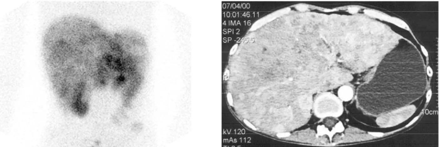
pen-tetreotide scintigraphy should be used as the initial imaging method in patients with carcinoid tumors (figure 1). Of particular advantage is the fact that one scan images the entire body; thus covert metastases may be identified. Intraoperative g detection has been considered as theoretically additional to external 111
In-labelled pentetreotide scintigraphy in the detection of small endocrine lesions, but high-background uptake (kidneys, liver, and spleen) and inadequate collimators have considerably limited its general utility (20).
Bone scintigraphy
Bone scintigraphy with99mTcMDP is the mainstay for
identifying bone metastases associated with NETs, with reported detection rates above 90%. Two studies that utilized111In-labeled pentetreotide demonstrated
similar diagnostic rates, ranging between 60% and 100% (21,22).
Radiolabelled metaiodobenzylguanidine ( M I B G )
Metaiodobenzylguanidine (MIBG) is a guanidine derivative that exploits the specific type 1 amine uptake mechanism at the cell membrane and storage within the intracellular storage vesicles. Several NETs including carcinoids exhibit this specific uptake mech-anism and can thus accumulate MIBG. Scanning with radiolabelled metaiodobenzylguanidine (123I-, or131
I-MIBG) has an overall sensitivity ranging from 55% to 70%, with a specificity of 95%. 111In-pentetreotide
scintigraphy is generally more sensitive than123I-, or 131I-MIBG scintigraphy (21,23).
Positron emission tomography (PET)
Positron emission tomography (PET) is a relatively novel, noninvasive technique that facilitates biochemical and metabolic studies of human tumors. Because neo-plastic cells are characterized by a higher glycolytic rate than normal cells, the use of [1 8F]
fluoro-2-deoxy-glu-cose (FDG) was initially used in biochemical imaging for the diagnosis and staging of cancer. However, as already stated, most NETs (and carcinoids) are well differentiat-ed and slow growing, they have a low metabolic rate and cannot be visualized efficiently with this tracer, as evi-denced by detection rates ranging between 25% and 73%. Because carcinoid tumors characteristically synthe-size serotonin, the administration of radioactive sero-tonin precursor1 1C-5-HT has been shown to provide
excellent tumor visualization, with high detection rates (24,25). More recently,6 8Ga coupled to Octreotide has
been used as tracers for PET imaging, also achieving high detection rate (26). PET should, therefore, be con-sidered still as an investigational yet very promising
Figure 1.Planar111In-pentetreotide scintigraphy (left) and transverse abdominal CT (right) images showing extensive
method for carcinoid imaging.
Endoscopy, endoscopic ultrasound and videocapsule endoscopy
Upper and lower gastrointestinal endoscopy Upper gastrointestinal endoscopy can identify lesions as far as the ligament of Treitz and lower and lower gastrointestinal endoscopy can detect some terminal ileal tumors as well as colon and rectal carcinoids.
Endoscopic ultrasound
Endoscopic ultrasound is a highly sensitive method for detecting carcinoid tumors of the stomach and duode-num and is superior to conventional ultrasound, par-ticularly in the detection of small lesions localized to the bowel wall because it can detect luminal lesions as small as 2 to 3mm in size (27).
Videocapsule endoscopy
(Video)capsule endoscopy has obvious potential for surveillance of the small intestine for carcinoid tumors (28,29).
Radiology
Ultrasonography, computerized tomography (CT), magnetic resonance imaging (MRI) and angiography
Additional studies such as transabdominal ultrasonog-raphy, triple-phase helical computerized tomography (CT), magnetic resonance imaging (MRI), and selec-tive mesenteric angiography may identify an additional 10% to 15% of primaries but are probably only justified if surgery is contemplated and more precise topograph-ic delineation considered necessary to define resection. Transabdominal ultrasound identifies approxi-mately one third of small bowel carcinoids and two thirds of liver metastases, and may also be used to guide percutaneous biopsies of liver tumors (30,31).
Mass lesions and evidence of calcification and fibrosis define CT scan and MRI findings associated with carcinoid tumors. Radiating strands of fibrosis and spiculation are characteristic hallmarks, especially in conjunction with a mass lesion. The degree of radi-ating strands detected by CT tends to increase with the degree of fibrosis seen histopathologically, and mesenteric fibrosis may lead to traction or fixation of the bowel (32). Mesenteric lymph node metastases are evident on CT scans in 91% (33). MRI and CT pro-vide important means of initial localization of carci-noid tumors or their metastases; however, their detec-tion rates and sensitivities are lower than imaging with
1 1 1In-pentetreotide scintigraphy. Median detection
rate and sensitivity of CT and/or MRI are about 80%, in contrast to 89% detection rate and 84% sensitivity with111In-pentetreotide scintigraphy. The diagnostic
efficacy of either CT or MR does not differ much. The reported detection rates of CT alone range between 76% and 100%, whereas MRI alone reported rates are between 67% and 81%.
Angiographic changes are distinctive, with nar-rowing or occlusion of the distal ileal arcade and stenosis of the intramesenteric arteries being a charac-teristic finding.
Importantly, patients with equivocal biochem-istry, negative nonspecific markers, and negative 1 1 1I n
-pentetreotide scintigraphy should probably not be fur-ther investigated but instead followed up annually.
Therapy
Surgery
Surgery is generally regarded as the most effective treatment for both local tumor effects (obstruction, bleeding, and perforation) and symptoms caused by the secretory agents because it removes the primary lesion and decreases levels of bioactive agents (16,34-37). In essence, surgery may be categorized as:
Adequate resection with curative or palliative intent for primary and regional lesions;
Surgical resection of regional or distant metastatic disease with cytoreductive intent; and
Resection of disease for symptom palliation without cytoreductive intent (35-37).
If residual tumor is present after surgery (liver, lymph nodes, peritoneal), long-acting somatostatin analogs (see later) have proven efficacious in the man-agement of carcinoid syndrome symptomatology (16,36-38).
Hepatic metastases can be resected because debulking (cytoreductive surgery) may reduce the symptoms, facilitate pharmacologic management, and improve survival (39). Liver transplantation is occa-sionally successful, but should only be performed when the presence of extrahepatic tumors has been ruled out (16,34,36).
(Chemo- and radio-)embolization
lesions. Hepatic artery embolization combined with sequential chemotherapy has been more encouraging, resulting in a reduction of tumor size in 78% of patients (40). Similarly, embolization with Yttrium-labeled microspheres has been useful in some circum-stances (41).
Cryosurgical debulking or radiofrequency ablation
Cryosurgical debulking or radiofrequency ablation (RFA) of hepatic carcinoid metastases have been described as of some benefit for palliation of carcinoid syndrome, but their efficacy remains to be rigorously evaluated (35,42,43).
Chemotherapy
Conventional chemotherapeutic agents such as strep-tozotocin, 5-FU, doxorubicin, and cyclophosphamide alone have used in the pasted and have yielded disap-pointing results, with an overall 20%–40% response rate. Etoposide may be marginally more effective either alone or in combination with cisplatin. Current-ly, the indications for chemotherapy need to be very carefully reconsidered, because biotherapy with somatostatin analogs and/or interferon alpha can con-trol the symptoms of humoral syndromes and may also affect tumor growth. However, chemotherapy may be beneficial for selected cases of advanced tumors that do not respond to other forms of therapy, and for
poorly differentiated NETs (16,44,45).
Medical therapy
Somatostatin analogs
Somatostatin is a small cyclic peptide. It circulates in the blood in two biologically active forms: somato-statin-14 and somatostatin-28. Somatostatin inhibits a variety of physiological functions in the gastrointesti-nal tract, like gastrointestigastrointesti-nal motility, gastric acid pro-duction, pancreatic enzyme secretion, and bile and colonic fluid secretion. It inhibits the secretion of pan-creatic and intestinal hormones like insulin, glucagon, secretin, and vasoactive intestinal polypeptide (46). However, the multiple simultaneous effects of phar-macological concentrations of somatostatin in differ-ent organs, the need for intravenous administration, the short duration of action and the post-infusion rebound hypersecretion of hormones considerably hampered its clinical use (47). Somatostatin acts through high-affinity G protein-coupled membrane receptors.
Five somatostatin receptor (sst) subtype genes have been cloned and characterized. They were code-named sst1, sst2, sst3, sst4, and sst5. Tumors arising
from somatostatin-target tissues, frequently express a high density of ssts. The five sst subtypes all bind somatostatin with high affinity. The sst1 and sst4
receptors do not bind the currently available octapep-tide somatostatin-analogs Octreooctapep-tide and Lanreooctapep-tide (see later), whereas sst2A, sst3, and sst5 receptors
dis-play a high, low, and moderate affinity, respectively, toward these octapeptide somatostatin-analogs. The predominant expression of sst2receptors on carcinoid
tumors forms the basis for the successful clinical appli-cation of these octapeptide somatostatin-analogs in controlling symptoms related to hormonal hypersecre-tion (47-49). The high density of ssts on these tumors further allows the use of radiolabelled somatostatin-analogs like111In-pentetreotide to visualize these
sst-positive tumorsin vivo(see earlier). Octreotide (San-dostatin) was the first octapeptide somatostatin analog that was synthesized. Its elimination half-life after sub-cutaneous administration is two hours, and rebound
hypersecretion of hormones does not occur.
Octreotide binds only with a high affinity to sst2and
sst5. Other cyclic analogs with almost similar affinity
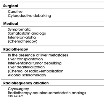
and activity profiles, like Lanreotide (Somatuline) have been developed subsequently (47). Octreotide and Lanreotide have been registered in most countries for the control of hormonal symptoms in patients with carcinoids. Lanreotide does not differ significantly from Octreotide in treatment of carcinoid symptoms. Table 2.Therapy of carcinoid tumors/the carcinoid
syn-drome.
Surgical
Curative
Cytoreductive debulking
Medical
Symptomatic Somatostatin analogs Interferon-alpha (Chemotherapy)
Radiotherapy
In the presence of liver metastases Liver transplantation
Interventional tumor debulking Liver dearterialization
(Chemo, or radio)-embolization Alcohol sclerotherapy
Radiofrequency ablation
Cryosurgery
Radiotherapy-coupled somatostatin analogs
Octreotide can be administered by multiple subcuta-neous injections or by continuous subcutasubcuta-neous infu-sion as well as by the intravenous route, either as a sin-gle injection or as a continuous infusion over many hours or days. The slow-release depot intramuscular formulation of Octreotide (Sandostatin LAR) has to be administered once every 4 weeks and that of Lan-reotide (LanLan-reotide-PR) has to be administered once every 2 weeks. A new slow-release depot preparation of Lanreotide, Somatuline Autogel, has been intro-duced in several European countries. This drug has to be administered deep subcutaneously once every 4 weeks. The introduction of these long-acting somato-statin analogs and depot administration has facilitated the control of most carcinoid syndrome symptoms and greatly improved quality of life. Dosages of Octreotide and Lanreotide can be adjusted in accordance with clinical successful control (8,16,38,50).
Somatostatin analogs decrease the release of bioactive secreted products with effective resolution of flushing and diarrhea in between 70% and 80% of patients. Although biochemical response rates ranged from 0% to 77%, tumor response rates were very low (0%–9%) (8,16,37,50-54).
Only modest adverse effects have been reported like: nausea, cramps, loose stools, mild steatorrhea, bil-iary sludge, or cholelithiasis (in up to 50% of patients but only 1% with acute symptoms warranting chole-cystectomy), impaired glucose tolerance, local pain and erythema at injection site and very rarely gastric atony (37,50-54). Intravenous Octreotide is particu-larly effective in the management of a “carcinoid cri-sis,” which is usually engendered by anesthesia, surgi-cal, or radiologic intervention. This life-threatening clinical condition is characterized by profound hypotension and tachycardia often associated with mortality without rapid preemptive pharmacologic intervention (55).
Interferon alpha
Recombinant leukocyte interferon-a may be of some use in the treatment of disseminated carcinoid tumors and carcinoid syndrome. The precise mechanism of action is not well understood but may include direct inhibition of cell proliferation, immune cell-mediated cytotoxicity, inhibition of angiogenesis, and induction of differentiation via cell cycle block (56). Although these agents are more toxic than somatostatin analogs, they may exhibit greater antitumor activity, but sub-stantial adverse effects include fever, fatigue, anorexia, and weight loss as well as alopecia, autoimmune dis-eases and myelosuppression. In patients with the
carci-noid syndrome, biochemical response rates ranged from 7% to 53%, and objective tumor response rates ranged from 7% to 20% (57,58).
There was little advantage in the use of a com-bination of Octreotide and interferon-a in patients in whom Octreotide alone or interferon-a produced no benefit. Although biochemical responses were report-ed in 72–77%, no objective tumor regression was observed. It is debatable whether somatostatin analogs and interferon-a exhibit a synergistic effect in carcinoid syndrome symptom management (56,58).
Supportive care
Supportive care of carcinoid tumors or carcinoid syn-drome includes avoiding stress and conditions or sub-stances that precipitate symptoms; dietary supplemen-tation with nicotinamide is also recommended. Mild diarrhea responds to antidiarrheal agents, such as lop-eramide, or opiates and bronchoconstriction to bron-chodilators that interact with-adrenergic receptors. Cyproheptadine decreases diarrhea in 50%, but adverse effects (20%) can be prohibitive. Cardiac failure may require diuretics and even valve replacement. Some brief relief with prednisone has been reported. Overall somatostatin analog therapy has supplanted most other medication (8,16,37,38,59).
Peptide receptor radionuclide therapy (prrt)
In general, carcinoids are resistant to radiotherapy, although external beam therapy has been used for palli-ation of bone metastases and the management of spinal cord compression and brain metastases. More recently, systemic peptide receptor radionuclide therapy (prrt) has been introduced for inoperable or metastasized GEP NETs (60). In general, sst-agonist complexes follow the mechanism and route of internalization as described for many other G protein-coupled receptor complexes. The predominant expression of sst2receptors in most
sst-pos-itive endocrine tumors and the efficiency of sst2r e c e p t o r s
to undergo agonist-induced internalization are very important for the radiotherapeutic application of radio-labelled octapeptide somatostatin-analogs. However, [1 1 1I n - D T P A0] Octreotide may not be the most suitable
compound to carry out radiotherapy because the Auger electrons emitter1 1 1In has a low tissue penetration. In
addition, a stable coupling of- or-emitting isotopes to [DTPA0]Octreotide could not be achieved, which
ini-tiated the development of a novel compound, like [ D O T A0, T y r3]Octreotide, allowing a stable binding
with the -emitter yttrium-90 (9 0Y) [9 0Y - D O T A0,
T y r3]Octreotide (9 0Y-DOTATOC; OctreoTher®), and
Fur-thermore, [1 1 1I n - D O T A0]Lanreotide and [9 0Y
-D O T A0]Lanreotide can also be used for radiotherapy of
s s t2- and sst5-positive advanced, or metastatic endocrine
tumors (61-64).
Initial studies with high dosages of 111
In-pente-treotide in patients with metastasized NETs were encouraging but partial remissions exceptional. On average, response rates were between 13% and 20% The subsequent use of [90Y-DOTA0,Tyr3]Octreotide
have suggested increased efficacy with some partial remissions (10% and 30%). The effects of radionuclide therapy are better at maintaining the status quo, with 53% and 79% of patients achieving biochemical or tumor size stability, respectively. The newest radiola-belled somatostatin analog [1 7 7L u - D O T A0, T y r3]
octreotate, which has a higher affinity for sst2 has
resulted in complete or partial responses in 28% of patients and tumor responses in 38% of patients, respectively. In these studies, tumor regression was positively correlated with a high uptake on the111
In-pentetreotide scintigraphy, limited hepatic tumor mass, and high Karnofsky performance score. Overall symptomatic improvement as well as improvement in quality of life (65) may occur with either 111In,90Y, or
177Lu-labeled somatostatin analogs that have been
used for prrt, but the results obtained with [90
Y-D O T A0, T y r3]Octreotide and [1 7 7L u - D O T A0, T y r3]
octreotate are more encouraging in terms of tumor regression. Issues of concern are renal damage (but this can be decreased by a pre-therapy amino acid infu-sion, which produces an added degree of kidney pro-tection) and induction of myeloproliferative disorders (which is more prevalent in patients pre-treated with chemotherapy) (66). Because the acute adverse effects of this type of therapy are few and mild and the dura-tion of the therapy response for radiopharmaceuticals more than 2 years, this therapeutical modality is increasingly accepted as standard therapy.
131I-MIBG therapy
As already mentioned, more than 70% of carcinoids concentrate MIBG. The use of 131I-MIBG therapy can
be considered early in an adjuvant setting, after surgery to eradicate occult disease, or later for treat-ment of disseminated disease. Using this radiopharma-ceutical an objective tumor response was recorded in 15%, with a symptomatic response in 65% of the patients (67).
New therapeutical developments
In recent years, many new sst selective analogs have been synthesized. A new so-called “universal”
somato-statin analog, named SOM230, with high affinity for sst1, sst2, sst3, and sst5receptors has been is currently
under evaluation in phase II-III trials (68,69). New fundamental insights in receptor physiology also opened the concept of multi-receptor family cross talk, like between somatostatin and dopamine receptors and focus has also been addressed to the development of new drugs interacting with these phenomenons (70). In the near future it will become clear whether new bispecific or more universal somatostatin-analogs are indeed effective in tumors resistant to the current clinically available octapeptide analogs Octreotide and Lanreotide and can prevent endocrine tumors from tachyphylaxis to treatment (71). Like peptide receptor radionuclide therapy, peptide receptor-targeted chemotherapy to deliver the chemotherapeutic com-pounds selectively to tumor cells might be a promising approach as well (72,73). Newer radiopharmaceuticals as well as combinations will also be tested for peptide receptor radionuclide therapy. Radiolabelled agonists and antagonists of peptide receptors other than the somatostatin receptors [like: vasoactive intestinal polypeptide (VIP) receptor subtype VPAC1, cholecys-tokinin (CCK) and gastrin receptor subtypes CCK2 (CCK-B) and CCK1 (CCK-A), bombesin and gastrin-releasing peptide (GRP) receptor subtypes (BB1, BB2, BB3 and BB4), neuromedin B receptors, neurotensin receptors (like the receptor subtype NRT1), substance P (like the receptor subtype NK1) and neuropeptide Y receptors] are currently being investigated and might become available as well as therapies for carcinoid tumors and the carcinoid syndrome (74).
REFERENCES
1. Rindi G, Bordi C. Highlights of the biology of endocrine tumours of the gut and pancreas. Endocr Relat Cancer 2003;10(4):427-36.
2. Lubarsch O. Über den primären Krebs des Ileum nebst Bemerkungen über das gleichzeitige Vorkommen von Krebs und Tuberkulose.Virchows Arch 1888;3:280-317.
3. Ransom WB. A case of primary carcinoma of the ileum.
Lancet 1890;2:1020-3.
4. Oberndorfer S. Karzinoïde Tumoren des Dunndarms.
Frankfurter Zeitschrift fur Pathologie 1907;1:426-9.
5. Williams ED, Sandler M. The classification of carcinoid tumours.Lancet 1963;1:238-9:238-9.
6. Solcia E, Kloppel G, Sobin LH.Histological typing of endocrine tumours. Berlin/Heidelberg/New York: Springer Verlag,2000.
Pathol Microbiol Scand [A] 1976;84(4):322-30.
8. Modlin IM, Kidd M, Latich I, Zikusoka MN, Shapiro MD. Current status of gastrointestinal carcinoids. Gastroen-terology 2005;128(6):1717-51.
9. van der Horst-Schrivers AN, Wymenga AN, Links TP, Willemse PH, Kema IP, de Vries EG. Complications of midgut carcinoid tumors and carcinoid syndrome.
Neuroendocrinology 2004;80(suppl. 1):28-32.
10. Caplin ME, Buscombe JR, Hilson AJ, Jones AL, Watkin-son AF, Burroughs AK. Carcinoid tumour. L a n c e t 1998;352(9130):799-805.
11. Kulke MH, Mayer RJ. Carcinoid tumors.N Engl J Med 1999;340(11):858-68.
12. Zuetenhorst JM, Korse CM, Bonfrer JM, Peter E, Lamers CB, Taal BG. Daily cyclic changes in the urinary excre-tion of 5-hydroxyindoleacetic acid in patients with car-cinoid tumors.Clin Chem 2004;50(9):1634-9.
13. Tormey WP, FitzGerald RJ. The clinical and laboratory correlates of an increased urinary 5-hydroxyin-doleacetic acid.Postgrad Med J 1995;71(839):542-5.
14. Feldman JM, Lee EM, Castleberry CA. Catecholamine and serotonin content of foods: effect on urinary excre-tion of homovanillic and 5-hydroxyindoleacetic acid.J Am Diet Assoc 1987;87(8):1031-5.
15. Nobels FR, Kwekkeboom DJ, Coopmans W, Schoen-makers CH, Lindemans J, de Herder WW, et al. Chro-mogranin A as serum marker for neuroendocrine neo-plasia: comparison with neuron-specific enolase and the alpha-subunit of glycoprotein hormones. J Clin Endocrinol Metab 1997;82(8):2622-8.
16. Plockinger U, Rindi G, Arnold R, Eriksson B, Krenning EP, de Herder WW, et al. Guidelines for the diagnosis and treatment of neuroendocrine gastrointestinal tumours.
Neuroendocrinology 2005;80(6):394-424.
17. Balon HR, Goldsmith SJ, Siegel BA, Silberstein EB, Kren-ning EP, Lang O, et al. Procedure guideline for somato-statin receptor scintigraphy with (111)In-pentetreotide. J Nucl Med 2001;42(7):1134-8.
18. Krenning EP, Kwekkeboom DJ, Oei HY, de Jong RJ, Dop FJ, Reubi JC, et al. Somatostatin receptor scintigraphy in carcinoids, gastrinomas and Cushing’s syndrome.
Digestion 1994;55(suppl. 3):54-9.
19. Kwekkeboom DJ, Krenning EP. Somatostatin receptor scintigraphy in patients with carcinoid tumors.World J Surg 1996;20(2):157-61.
2 0 . Benjegard SA, Forssell-Aronsson E, Wangberg B, Skan-berg J, Nilsson O, Ahlman H. Intraoperative tumour detection using1 1 1I n - D T P A - D - P h e1-octreotide and a
scin-tillation detector.Eur J Nucl Med 2001; 2 8 ( 1 0 ) : 1 4 5 6 - 6 2 .
21. Zuetenhorst JM, Hoefnagel CA, Boot H, Valdes Olmos RA, Taal BG. Evaluation of111In-pentetreotide,131I-MIBG
and bone scintigraphy in the detection and clinical management of bone metastases in carcinoid disease.
Nucl Med Commun 2002;23(8):735-41.
2 2 . Meijer WG, van der Veer E, Jager PL, van der Jagt EJ, Pires BA, Kema IP, et al. Bone metastases in carcinoid tumors: clinical features, imaging characteristics, and markers of bone metabolism. J Nucl Med 2 0 0 3; 4 4 ( 2 ) : 1 8 4 - 9 1 .
23. Hanson MW, Feldman JM, Blinder RA, Moore JO, Cole-man RE. Carcinoid tumors: iodine-131 MIBG scintigra-phy.Radiology 1989;172(3):699-703.
24. Eriksson B, Orlefors H, Oberg K, Sundin A, Bergstrom M, Langstrom B. Developments in PET for the detection of endocrine tumours. Best Pract Res Clin Endocrinol Metab 2005;19(2):311-24.
25. Orlefors H, Sundin A, Garske U, Juhlin C, Oberg K, Skog-seid B, et al. Whole-body 1 1C - 5 - h y d r o x y t r y p t o p h a n
positron emission tomography as a universal imaging technique for neuroendocrine tumors — comparison with somatostatin receptor scintigraphy and computed tomography. J Clin Endocrinol Metab 2005;90(6):3392-400.
26. Maecke HR, Hofmann M, Haberkorn U. (68)Ga-labeled peptides in tumor imaging.J Nucl Med 2005;46(suppl. 1):172S-8S.
27. Gibril F, Jensen RT. Comparative analysis of diagnostic techniques for localization of gastrointestinal neuroen-docrine tumors.Yale J Biol Med 1997;70(5-6):509-22.
28. Forner A, Mata A, Puig M, Varela M, Rodriguez F, Llach J, et al. Ileal carcinoid tumor as a cause of massive lower-GI bleeding: the role of capsule endoscopy. Gas-trointest Endosc 2004;60(3):483-5.
29. Coates SW Jr, DeMarco DC. Metastatic carcinoid tumor discovered by capsule endoscopy and not detected by esophagogastroduodenoscopy. Dig Dis Sci 2004;49(4):639-41.
30. Rioux M, Langis P, Naud F. Sonographic appearance of primary small bowel carcinoid tumor. Abdom Imaging 1995;20(1):37-43.
31. Andersson T, Eriksson B, Lindgren PG, Wilander E, Oberg K. Percutaneous ultrasonography-guided cutting biop-sy from liver metastases of endocrine gastrointestinal tumors.Ann Surg 1987;206(6):728-32.
32. Pantongrag-Brown L, Buetow PC, Carr NJ, Lichtenstein JE, Buck JL. Calcification and fibrosis in mesenteric car-cinoid tumor: CT findings and pathologic correlation.
AJR Am J Roentgenol 1995;164(2):387-91.
33. Hellman P, Lundstrom T, Ohrvall U, Eriksson B, Skogseid B, Oberg K, et al. Effect of surgery on the outcome of midgut carcinoid disease with lymph node and liver metastases.World J Surg 2002;26(8):991-7.
34. Ahlman H, Nilsson O, Olausson M. Interventional treat-ment of the carcinoid syndrome.Neuroendocrinology 2004;80(suppl. 1):67-73.
35. de Vries H, Verschueren RC, Willemse PH, Kema IP, de Vries EG. Diagnostic, surgical and medical aspect of the midgut carcinoids. Cancer Treat Rev 2002;28(1):11-25.
36. Akerstrom G, Hellman P, Hessman O, Osmak L. Man-agement of midgut carcinoids. J Surg Oncol 2005;89(3):161-9.
38. Oberg K, Kvols L, Caplin M, Delle Fave G, de Herder WW, Rindi G, et al. Consensus report on the use of somato-statin analogs for the management of neuroendocrine tumors of the gastroenteropancreatic system. A n n Oncol 2004;15(6):966-73.
39. Sarmiento JM, Heywood G, Rubin J, Ilstrup DM, Nagor-ney DM, Que FG. Surgical treatment of neuroendocrine metastases to the liver: a plea for resection to increase survival.J Am Coll Surg 2003;197(1):29-37.
40. Ruszniewski P, O’Toole D. Ablative therapies for liver metastases of gastroenteropancreatic endocrine tumors.Neuroendocrinology 2004;80(suppl. 1):74-8. 41. Herba MJ, Thirlwell MP. Radioembolization for hepatic
metastases.Semin Oncol 2002;29(2):152-9.
42. Gulec SA, Mountcastle TS, Frey D, Cundiff JD, Mathews E, Anthony L, et al. Cytoreductive surgery in patients with advanced-stage carcinoid tumors. Am Surg 2002;68(8):667-71.
43. Berber E, Flesher N, Siperstein AE. Laparoscopic radiofre-quency ablation of neuroendocrine liver metastases.
World J Surg 2002;26(8):985-90.
44. Moertel CG. Karnofsky memorial lecture. An odyssey in the land of small tumors.J Clin Oncol 1987 ;5(10):1502-22.
45. Rougier P, Mitry E. Chemotherapy in the treatment of neuroendocrine malignant tumors. D i g e s t i o n 2000;62(suppl. 1):73-8.
46. Lamberts SW, Krenning EP, Reubi JC. The role of somato-statin and its analogs in the diagnosis and treatment of tumors.Endocr Rev 1991;12(4):450-82.
47. Lamberts SW, van der Lely AJ, de Herder WW, Hofland LJ. Octreotide.N Engl J Med 1996;334(4):246-54.
48. de Herder WW, van der Lely AJ, Lamberts SW. Somato-statin analogue treatment of neuroendocrine tumours.
Postgrad Med J 1996;72(849):403-8.
49. de Herder WW, Lamberts SW. Somatostatin and somato-statin analogues: diagnostic and therapeutic uses. Curr Opin Oncol 2002;14(1):53-7.
50. de Herder W, Lamberts S. Somatostatin analog therapy in treatment of gastrointestinal disorders and tumors.
Endocrine 2003;20(3):285-90.
51. Ruszniewski P, Ish-Shalom S, Wymenga M, O’Toole D, Arnold R, Tomassetti P, et al. Rapid and sustained relief from the symptoms of carcinoid syndrome: Results from an open 6-month study of the 28-day prolonged-release formulation of Lanreotide. Neuroendocrinolo-gy 2004;80(4):244-51.
52. Arnold R. Medical treatment of metastasizing carcinoid tumors.World J Surg 1996;20(2):203-7.
53. Rubin J, Ajani J, Schirmer W, Venook AP, Bukowski R, Pommier R, et al. Octreotide acetate long-acting for-mulation versus open-label subcutaneous octreotide acetate in malignant carcinoid syndrome. J Clin Oncol 1999;17(2):600-6.
54. Wymenga AN, Eriksson B, Salmela PI, Jacobsen MB, van Cutsem EJ, Fiasse RH, et al. Efficacy and safety of pro-longed-release lanreotide in patients with gastrointesti-nal neuroendocrine tumors and hormone-related symptoms.J Clin Oncol 1999;17(4):1111-7.
55. Kvols LK, Martin JK, Marsh HM, Moertel CG. Rapid rever-sal of carcinoid crisis with a somatostatin analogue.N
Engl J Med 1985;313(19):1229-30.
56. Pape UF, Wiedenmann B. Adding interferon-alpha to octreotide slows tumour progression compared with octreotide alone but evidence is lacking for improved survival in people with disseminated midgut carcinoid tumours.Cancer Treat Rev 2003;29(6):565-9.
57. Oberg K. Interferon in the management of neuroen-docrine GEP-tumors: a review. Digestion 2000;62(suppl. 1):92-7.
58. Faiss S, Pape UF, Bohmig M, Dorffel Y, Mansmann U, Golder W, et al. Prospective, randomized, multicenter trial on the antiproliferative effect of lanreotide, interfer-on alfa, and their combinatiinterfer-on for therapy of metastat-ic neuroendocrine gastroenteropancreatmetastat-ic tumors — the International Lanreotide and Interferon Alfa Study Group.J Clin Oncol 2003;21(14):2689-96.
59. Kaltsas GA, Besser GM, Grossman AB. The diagnosis and medical management of advanced neuroendocrine tumors.Endocr Rev 2004;25(3):458-511.
60. Krenning EP, Valkema R, Kwekkeboom DJ, de Herder WW, van Eijck CH, de Jong M, et al. Molecular imaging as in vivo molecular pathology for gastroenteropan-creatic neuroendocrine tumors: implications for follow-up after therapy.J Nucl Med 2005;46(suppl. 1):76S-82S.
61. Anthony LB, Woltering EA, Espenan GD, Cronin MD, Mal-oney TJ, McCarthy KE. Indium-111-pentetreotide pro-longs survival in gastroenteropancreatic malignancies.
Semin Nucl Med 2002;32(2):123-32.
62. Virgolini I, Britton K, Buscombe J, Moncayo R, Paganelli G, Riva P. In- and Y-DOTA-lanreotide: results and impli-cations of the MAURITIUS trial. Semin Nucl Med 2002;32(2):148-55.
63. Kwekkeboom DJ, Teunissen JJ, Bakker WH, Kooij PP, de Herder WW, Feelders RA, et al. Radiolabeled somato-statin analog [177Lu-DOTA0,Tyr3]octreotate in patients
with endocrine gastroenteropancreatic tumors.J Clin Oncol 2005;23(12):2754-62.
64. Kwekkeboom DJ, Mueller-Brand J, Paganelli G, Antho-ny LB, Pauwels S, Kvols LK, et al. Overview of results of peptide receptor radionuclide therapy with 3 radiola-beled somatostatin analogs. J Nucl Med 2005;46(suppl. 1):62S-6.
65. Teunissen JJ, Kwekkeboom DJ, Krenning EP. Quality of life in patients with gastroenteropancreatic tumors treat-ed with [1 7 7L u - D O T A0, T y r3]octreotate. J Clin Oncol
2004;22(13):2724-9.
66. Valkema R, Pauwels SA, Kvols LK Kwekkeboom DJ, Jamar F, de Jong M, et al. Long-term follow-up of renal function after peptide receptor radiation therapy with (90)Y-DOTA(0),Tyr(3)-octreotide and (177)Lu-DOTA(0), Tyr(3)-octreotate.J Nucl Med 2005;46(suppl. 1):83S-91.
67. Taal BG, Zuetenhorst H, Valdes Olmos RA, Hoefnagel CA. [131I]MIBG radionuclide therapy in carcinoid
syn-drome.Eur J Surg Oncol 2002;28(3):243.
68. Lamberts SW, van der Lely AJ, Hofland LJ. New somato-statin analogs: will they fulfill old promises? Eur J Endocrinol 2002;146(5):701-5.
somato-statin analog, SOM230, in patients with metastatic car-cinoid tumors refractory or resistant to octreotide LAR.
ASCO 2005, abstract number 8024.
70. Rocheville M, Lange DC, Kumar U, Patel SC, Patel RC, Patel YC. Receptors for dopamine and somatostatin: formation of hetero-oligomers with enhanced function-al activity.Science 2000;288(5463):154-7.
71. Hofland LJ, Lamberts SW. The pathophysiological con-sequences of somatostatin receptor internalization and resistance.Endocr Rev 2003;24(1):28-47.
72. Kiaris H, Schally AV, Nagy A, Szepeshazi K, Hebert F,
Hal-mos G. A targeted cytotoxic somatostatin (SST) ana-logue, AN-238, inhibits the growth of H-69 small-cell lung carcinoma (SCLC) and H-157 non-SCLC in nude mice.
Eur J Cancer 2001;37(5):620-8.
73. Reubi JC, Macke HR, Krenning EP. Candidates for pep-tide receptor radiotherapy today and in the future.J Nucl Med 2005;46(suppl. 1):67S-75S.
74. Reubi JC. Peptide receptors as molecular targets for cancer diagnosis and therapy.Endocr Rev 2003;24(4): 389-427.
Endereço para correspondência:
A.J. van der Lely
Department of Internal Medicine, Section of Endocrinology Erasmus MC Dr Molewaterplein 40
3015 GD Rotterdam, Netherlands Fax: (31) (10) 463-3268