GABA and glutamate transporters: new events and function
in the vertebrate retina
José Luiz Martins do Nascimento, Luis Armando Sawada, Karen Renata Matos Oliveira, Maria
Elena Crespo-López, Anderson Manoel Herculano Oliveira da Silva, Moisés Hamoy, Consuelo
Yumiko Yoshioka e Silva, Gilmara Nazareth Tavares Bastos, and Wendell Mauro Soeiro-Pantoja
Universidade Federal do Pará, Belém, PA, Brazil
Abstract
The neural retina is a highly complex tissue composed of excitatory and inhibitory neurons and glial cells. Glutamate, the main excitatory neurotransmitter, mediates information transfer from photoreceptors, bipolar cells, and ganglion cells, whereas interneurons, mainly amacrine and horizontal cells, use γ-aminobutyric acid (GABA), the main inhibitory neurotransmitter. In this review we place an emphasis on glutamate and GABA transporters as highly regulated molecules that play fundamental roles in neurotransmitter clearance, neurotransmitter release, and oxidative stress. We pharmacologically characterized glutamate transporters in chicken retina cells and identified two glutamate transporters: one Na+-dependent transporter and one Na+ -independent transporter. The Na+-dependent uptake system presented characteristics related to the high-affinity x
AG
- system (EAAT1), and the Na+-independent uptake system presented characteristics related to the x
CG
- system, which highly contributes to glutamate transport in the retina. Glutamate shares the xCG- system with another amino acid, L-cysteine, suggesting the possible involvement of glutathione. Both transporter proteins are present mainly in Müller glial cells. GABA transporters (GATs) mediate high-affinity GABA uptake from the extracellular space and terminate the synaptic action of GABA in the central nervous system. GABA transporters can be modulated by molecules that act on specific sites to promote transporter phosphorylation and dephosphorylation. In addition to a role in the clearance of GABA, GATs may also release GABA through a reverse transport mechanism. In the chicken retina, a GAT-1 blocker, but not GAT2/3 blocker, was shown to inhibit GABA uptake, suggesting that GABA release from retina cells is mainly mediated by a GAT-1-like transporter. Keywords: neuroretina cells, culture, glutamate transporter, GABA transporter.
Received 26 July 2012; received in revised form 08 January 2013; accepted 09 January 2013. Available online 18 November 2013.
José Luiz Martins do Nascimento, Luis Armando Sawada, Karen Renata Matos Oliveira, Maria Elena Crespo-López, Anderson Manoel Herculano Oliveira da Silva, Moisés Hamoy, Consuelo Yumiko Yoshioka e Silva, Gilmara Nazareth Tavares Bastos, and Wendell Mauro Soeiro-Pantoja, Instituto de Ciências Biológicas, Universidade Federal do Pará, Belém, Brazil. Correspondence regarding this article should be directed to: Do Nacimento, JLM. Laboratório de Neuroquímica Molecular e Celular, Instituto de Ciências Biológicas, Universidade Federal do Pará, Belém, Pará, 66075-99, Brazil. Fax: 55 91 3201601. E-mail: [email protected]
Introduction
The neural retina is a highly complex tissue composed of excitatory and inhibitory neurons and glial cells. Knowledge of retinal neurochemistry has helped to understand the organization of the receptive
ield and a highly integrated network that is critical for
spatial, temporal, and chromatic information processing in the visual system. Glutamate and γ-aminobutyric acid (GABA) are generally regarded as excitatory and inhibitory neurotransmitters, respectively. However, the outer retina is one place where glutamate also
plays an inhibitory role in a subpopulation of bipolar
cells, speciically ON bipolar cells. Glutamate is the
main excitatory neurotransmitter in the outer and inner plexiform layers, mediating direct information transfer from photoreceptors, bipolar cells, and ganglion cells, whereas interneurons, mainly amacrine and horizontal cells, use GABA. In this review we emphasize studies of glutamate and GABA transporters as highly regulated molecules that play fundamental roles as sensors of both GABA and glutamate uptake and important roles in neurotransmitter clearance, neurotransmitter release, and oxidative stress.
Glutamate transporter
Once released at glutamatergic synapses, glutamate is cleared from the extracellular space by several
high-afinity transport mechanisms. Glutamate uptake by astroglia also regulates the eficiency of glutamatergic
synapses by modulating the amount of glutamate within the synaptic cleft and its diffusion from the synapse (Danbolt, 2001; Robinson, 2006). Glutamate
transport deiciencies often appear to be associated
with neuropathologies that depend on different centers in the central nervous system (CNS) (Doble, 1999; Obrenovitch, Urenjak, Zilkha, & Jay, 2000). Thus, treatment of these diseases could be pharmacologically based on the development of compounds that modulate the activity of these transporter proteins.
In the CNS, excitatory amino acid transporters (EAATs) are the main transporters responsible for removal of the glutamate neurotransmitter, maintaining its extracellular concentration below excitotoxic levels (Pines et al., 1992; Danbolt, 2001; Balcar, 2002; Kanai & Hediger, 2004). Glutamate transport by the EAAT is electrogenic, in which it is thermodynamically coupled to the cotransport of at least two sodium ions (Na+) and
one proton (H+) and the countertransport of a potassium
ion (Tanaka et al., 1997; Amara & Fontana, 2002; Bridges & Esslinger, 2005). These transporters are known as Na+-dependent high-afinity glutamate transporters
(Shigeri, Seal, & Shimamoto, 2004; Bridges & Esslinger, 2005). These transporters are also coupled to a Cl- channel, but its function has not yet been described.
Furthermore, numerous membrane transporters have
been characterized that regulate the lux of glutamate
in distinct areas of the CNS including the retina. These transporters are commonly differentiated based on their ionic dependence and include:
• Sodium-dependent xAG- system. This system is
represented by EAATs (Palacin, Estevez, Bertran, &
Zorzano, 1998; McBean, 2002). In vertebrates, ive
different isoforms of glutamate transporters have
been identiied: EAAT1, EAAT2, EAAT3, EAAT4,
and EAAT5. These transporters exhibit distinct regional and cellular localization and different pharmacological and molecular characteristics (Danbolt et al., 1998; Seal & Amara, 1999). The availability of inhibitors that show a high degree of selectivity for only one of the EAATs is still limited, thus hindering the characterization of these transporters isolated in different areas of the CNS. However, the appearance of new compounds with greater selectivity is intensifying with the advancement of new techniques in molecular modeling (Bridges & Esslinger, 2005).
• Sodium-independent xCG- system. This system is mainly represented by a cystine-glutamate exchanger, a member of the glycoprotein-associated amino acid transporter family, and has been described in hepatocytes, alveolar type II cells, human endothelial cells, and macrophages (Ishii, Sato, Miura, Sagara, & Bannai, 1992). In the
CNS, this system was identiied in primary cultures
of neurons (Sagara, Miura, & Bannai, 1993) and astrocytes (Allen, Shanker, & Aschner, 2001; Gochernauer & Robinson, 2001), C6 glioma cells (Cho & Bannai, 1990), microglia (Piani & Fontana, 1994), and human glioma cells (Ye, Rothstein, & Sontheimer, 1999). Under physiological conditions, the xCG- system transports cystine into cells coupled to the eflux of intracellular glutamate through a
Na+-independent mechanism (Bender, Reichelt, &
Norenberg, 2000). Once taken up, cystine is rapidly and spontaneously reduced to cysteine, which is required for the synthesis of glutathione (GSH), an endogenous antioxidant essential for cellular defense (McBean, 2002; Tomi et al., 2003).
In the retina, glutamate is stored and released by photoreceptors, bipolar cells, and ganglion cells. Glutamate is the major excitatory neurotransmitter responsible for phototransduction (Kalloniatis & Napper, 1996). In the outer layer of the retina, photoreceptors continuously release glutamate. This release is modulated by a light membrane of bipolar and horizontal cells. In the inner plexiform layer, two types of bipolar cells release glutamate – ON bipolar cells and OFF bipolar cells – that release the neurotransmitter in the presence and absence of light, respectively. Amacrine and ganglion cells are targets of glutamate release from the inner plexiform layer (Copenhagen & Jahr, 1989; Rauen, Rothstein, & Wassle, 1996).
High-afinity glutamate transporters (EAATs) have been
well characterized in retinal tissue. Immunocytochemical studies demonstrated the presence of these transporters in different retina cells (Eliasof, Arriza, Leighton, Kavanaugh, & Amara, 1998; Pow & Barnett, 2000). However, little is known about the activity of different EAAT subtypes in chicken retinal tissue and the contribution of other transport systems for glutamate uptake.
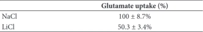
We characterized the involvement of different transport systems for glutamate in chicken retinal cells. The results showed that removal of the Na+ ion changed
the value of the uptake of this neurotransmitter, thus characterizing the presence of two glutamate mechanisms in the retina: Na+-dependent and Na+-independent. These
components accounted for 52% and 48% of the total transport of glutamate, respectively (Table 1).
Table 1. Glutamate transporters in the central nervous system: Na+-dependent and -independent transport
Glutamate uptake (%)
NaCl 100 ± 8.7%
LiCl 50.3 ± 3.4%
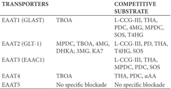
The activity and participation of each of the different transporter proteins (EAAT1, EAAT2, EAAT3, EAAT4, and EAAT5) in Na+-dependent glutamate
uptake was characterized by observing the effects of
various pharmacological antagonists that are speciic
functionality of these transporters is directly related to the presence of Na+ ions. The pharmacological action of speciic EAAT inhibitors is summarized in Table 2.
Table 2. Pharmacology of glutamate transporters in the central nervous system: Na+-dependent
TRANSPORTERS COMPETITIVE
SUBSTRATE
EAAT1 (GLAST) TBOA L-CCG-III, THA, PDC, 4MG, MPDC, SOS, T4HG EAAT2 (GLT-1) MPDC, TBOA, 4MG,
DHKA; 3MG, KA7
L-CCG-III, PD, THA, T4HG, SOS
EAAT3 (EAAC1) L-CCG-III, THA,
MPDC, PDC, SOS
EAAT4 TBOA THA, PDC, αAA
EAAT5 No speciic blockade No speciic blockade
DHKA (a speciic antagonist of EAAT2), SOS (a speciic antagonist of EAAT3), and αAA (a speciic
inhibitor of EAAT4) showed no inhibitory effect on
total glutamate uptake. However, 4MG, a speciic
antagonist of EAAT1, inhibited the uptake of glutamate in chicken retina cells. EAAT5, a carrier expressed on photoreceptors and bipolar cell terminals, has glutamate-gated Cl- conductance but transports substrates poorly,
limiting its potential contribution to glutamate clearance in the retina. Although some overlap was found with the compounds tested, the main Na+-dependent glutamate
transporter in chicken retina cells appears to be mediated mainly by EAAT1.
Strong evidence suggests the existence of a sodium-independent xCG- system,represented mainly by a
cystine-glutamate exchanger in chicken retina cells. Under physiological conditions, the xCG-system transports cystine into cells coupled to the eflux of intracellular
glutamate through a Na+-independent mechanism
(Bender et al., 2000). Once taken up by cells, cystine is rapidly and spontaneously reduced to cysteine, which is required for the synthesis of glutathione (GSH), an endogenous antioxidant essential for cellular defense (McBean, 2002; Tomi et al., 2003). The tripeptide GSH is produced from the amino acids glutamate, cysteine, and glycine by the consecutive actions of two enzymatic reactions (Yoneyama et al., 2008), and its role as a free radical scavenger is particularly important in the retina because this tissue is extremely vulnerable to oxidation because of its high oxygen consumption, high unsaturated fatty acid content, and exposure to light (Handelman & Dratz, 1986; Ahuja, Caffé, Ahuja, Ekstrom, & Van Veen, 2005). Furthermore, the xCG-system can also mediate the inlux of glutamate when its concentration outside the
cell is higher (Bringmann et al., 2009). Thus, two main glutamate transporter proteins in the chicken retina and the majority of glutamate uptake occur mainly in Müller glial cells.
GABA transporter in the retina
GABA is the main inhibitory neurotransmitter in the CNS, including the retina. In this tissue, the activation
of most interneurons promotes GABA release, which binds to its postsynaptic receptors. The two main receptor types are GABAA and GABAC. These receptors are ionotropic ligand-activated chloride channels that promote Cl- inlux. GABA
B receptors are metabotropic
ligand-activated K+ channels that cause K+ eflux. The
activation of GABA receptors promotes inhibitory postsynaptic potentials (IPSPs), which can lead to three forms of inhibition in the cell: (1) generation of hyperpolarization, which lowers the excitation limit in the inhibited neuron and lowers excitatory postsynaptic potentials (EPSPs); (2) takes the cell at rest and leads to a membrane potential close to the potential of K+,
preventing it from reaching the threshold, and (3) an increase in membrane conductance, which reduces the EPSP amplitude (Kandel, Schwartz, & Jessel, 1991).
GABA transport occurs through cellular transmembrane transporter proteins. These proteins have the ability to remove the neurotransmitter from
the synaptic cleft, thereby inluencing neuronal
transmission. Transporter activity can be modulated by hormones (Cushman & Wardzala, 1980), ion channels (Cammack & Schwartz, 1993), and molecules that
promote phosphorylation/dephosphorylation at speciic
sites (Corey, Davidson, Lester, Brecha, & Quick, 1994). Guastella et al. (1990) used molecular biology
techniques to generate the irst GABA transporter (GAT)
clone from rat brains. Pharmacological and kinetic studies suggested the presence of various GAT subtypes including GAT-1, GAT-2, GAT-3, and GAT-4. GAT-1
has the highest afinity for GABA in rodents and humans
(Brecha & Weigmann, 1994). It is a transmembrane protein with approximately 599 amino acids that form 12 membrane domains (Guastella et al., 1990).
GABA uptake occurs via the GAT through Na+ and Cl- ion co-transport driven by the [Na+]
electrochemical gradient (Cammack & Schwartz, 1993; Santos, Gonçalves, & Carvalho, 1990). Over the years, several synaptic physiologists considered that GATs constantly operate in the uptake mode at their maximum rate and are able to eliminate nearly all extracellular GABA. However, this premise is not compatible with transporter thermodynamics. More current conceptualizations indicate that these transporters have the ability to reverse uptake (Attwell, Barbour, & Szatkowski, 1993; Cammack, Rakhilin, & Schwartz, 1994; Levi & Raiteri, 1993; Lu & Hilgemann, 1999; O’Malley, Sandell, & Masland, 1992; Pin & Bockaert, 1989; Schwartz, 1987). Indirect evidence indicates that GATs are near equilibrium under resting conditions and are thus relatively inactive (Richerson & Wu, 2003). A theoretical limit may also exist with regard to how much the GAT can reduce ambient GABA (Attwell et al., 1993; Cavelier, Hamann, Rossi, Mobbs, & Attwell, 2005; Richerson & Wu, 2003).
GABA can be stored and released by a classic Ca2+
relecting membrane transporter reversal (Wu, Wang, &
Richerson 2001, 2003). Transporter-mediated GABA release can be blocked by the presence of intracellular Ca2+, indicating that Ca2+ can modulate transport activity
(Gonçalves & Carvalho, 1994; Gonçalves, Carvalho, & Vale, 1997). Inhibitory postsynaptic neuron activation induced by the binding of glutamate to non-N -methyl-D-aspartate receptors promotes Na+ and Ca2+ inlux and
subsequent depolarization. The activation of
voltage-dependent channels also promotes the inlux of cations
and activation of the GAT (do Nascimento, Ventura, & Paes De Carvalho, 1998; Santos et al., 1990).
Changes in the Na+ gradient can inluence GABA
transport, independent of changes in membrane potential (Do Nascimento & de Mello, 1985; Do Nascimento et al., 1998). Each GABA molecule is co-transported with two Na+ ions in which the driving force for GAT-1 is
much more strongly dependent on the Na+ gradient
than on GABA or Cl- gradients or membrane potential.
Therefore, even modest changes in neuronal activity would be expected to alter the driving force for
GAT-1 and may favor GAT-GAT-1 reversal. The inluence of Na+
levels on tonic GABA inhibition may be even greater under pathological conditions.
Other forms of GAT-1 control can also be attributed to the transporter phosphorylation/dephosphorylation conformation. Protein phosphorylation is an
important posttranslational modiication that regulates
several biological functions including the transport of neurotransmitters like GABA. Transporter phosphorylation is directly related to normal inhibitory neurotransmission, prevention of psychiatric disorders,
and modulation of synaptic eficiency (Santos et al.,
1990).
Tian, Knaus, & Shipston (1998) suggested that uptake regulation by the GAT occurs because of phosphorylation, which is not observed for most transporters (e.g., the dopamine transporter), demonstrating differential posttranslational regulation by the GAT.
Previous studies have provided evidence of the
inluence of phosphorylation and dephosphorylation in
GAT activity through the use of phosphatase inhibitors and stimulators. Gonçalves, Meireles, & Vale (1999) found that okadaic acid and Caliculin A, both inhibitors of PP1 and PP2A, inhibited the uptake of GABA in synaptosomes. Cyclosporine A, a PP2B inhibitor, had a stimulatory effect on uptake. These results differ from those found in chicken retina cells in which the PP1 and PP2A inhibitors okadaic acid and Caliculin A
did not inluence GABA uptake. Only PP2B inhibition inluenced both uptake and glutamate-induced release
(Soeiro Pantoja, 2002). The stimulatory effect was much greater when the drugs were administrated in the same medium, indicating that GAT activity may be related to the activity of the enzyme calcineurin (Perrino, Ng, & Soderling, 1995).
Soeiro Pantoja (2002) demonstrated possible second messengers that regulate GAT phosphorylation
and dephosphorylation. The results indicated that endogenous Ca2+ variations appear to partially interfere
with GAT-1 activity and the participation of the Ca2+/
calmodulin complex, which is capable of modulating other signaling pathways such as protein kinase A (PKA) and protein kinase C (PKC). The use of PKA inhibitors and activation agents supported the hypothesis of PKA-dependent adenylate cyclase regulation of GABA uptake (Soeiro Pantoja, 2002). However, staurosporine and phorbol ester, a PKC inhibitor and activator, respectively, did not affect GAT-1 in chicken retina cells. These data are summarized in Table 3.
Table 3. Effects of PKA, PKC, and Ca2+ pathways on [3H] GABA uptake from cultured chick retina cells
Drugs Mechanism of
action
Uptake (%)
PKA
Control 100 ± 2
BrAMPc AMPc analog
100 µM 98.2 ± 6
200 µM 111.4 ± 5
Forskolin Adenylyl cyclase activator
20 µM 120.8 ± 6
PKI Protein kinase
inhibitor
1 µM 96.3 ± 5
10 µM 60.1 ± 4
50 µM 12.4 ± 2
H89 PKA inhibitor
1 µM 95.2 ± 7
10 µM 81.6 ± 3
50 µM 58.4 ± 2
100 µM 23.3 ± 3
PKC and Ca2+
Ca2+ free medium 50.2 ± 5.6
EGTA (2 µM) Ca2+ chelator 20.4 ± 3.1
W7 (10 µM) Calmodulin inhibitor
60 ± 4.2
BAPTA AM (10 µM) Ca2+ chelator 20 ± 5.2
Phorbol ester (10 nM-10 µM)
PKC activator 98.2 ± 3.5
Stauroporine (10 nM-10 µM)
PKC inhibitor 97.1 ± 5.3
Ca2+ ions and GAT phosphorylation greatly inluence GABA uptake. The presence of Ca2+ and
absence of protein phosphatase inhibitors favor the effect of calcineurin, corresponding to uptake inhibition when dephosphorylation sites are activated. The presence of Ca2+ ions and PP2B inhibitors corresponds to an
increase in GABA uptake via GAT-1. Phosphorylation in the absence of Ca2+ decreases GABA uptake. These
in both cellular phosphorylation and dephosphorylation processes (Soeiro Pantoja, 2002).
The regulation of the GAT appears to be determined by internally localized Ca2+/calmodulin-dependent
phosphatase activity (calcineurin). Other phosphorylation sites that are sensitive to PP1 and PP2A inhibitors either positively or negatively potentiate the effects when the GAT is in phosphorylated and dephosphorylated states, respectively (Perrino et al., 1995).
Speciic sites in the GAT-1 protein allow the
transporter to present two main conformations in terms of transport, substrate, and operability, thus seemingly related to its state of phosphorylation or dephosphorylation (Gonçalves et al., 1999; Corey et al., 1994; Gomeza, Casado, Giménez, & Aragón, 1991; Sitges, Dunkeley, & Chiu, 1995). When the GAT is phosphorylated by the biochemical cascade that is activated by Ca2+/calmodulin or even by PKA
activation accompanied by calcineurin inhibition, its
conformation favors GABA inlux. When the GAT is
dephosphorylated by calcineurin, the conformation
favors GABA eflux, suggesting that phosphorylation
sites sensitive to calcineurin are required for GAT-1 activity (Soeiro Pantoja, 2002).
In summary, cumulative evidence suggests two main GABA transport regulation processes (i.e., Na+ gradient
and GAT-1 phosphorylation/dephosphorylation), although other forms of modulation exist that control
GAT distribution, traficking, and substrates. Ongoing
studies are elucidating the multiple mechanisms that
inluence GAT-1 transporter function and expression in
the chicken retina.
Acknowledgements
Research was inancially supported by FINEP “Rede
Instituto Brasileiro de Neurociência” (IBN Net), CNPQ, and FAPESPA. JLMN is a CNPq research fellow.
References
Ahuja, P., Caffé, A. R., Ahuja, S., Ekstrom, P., & Van Veen, T. (2005). Decreased glutathione transferase levels in rd1/rd1 mouse retina: Replenishment protects photoreceptors in retinal explants.
Neuroscience, 131, 935-943.
Allen, J. W., Shanker, G., & Aschner, M. (2001). Methylmercury inhibits the in vitro uptake of the glutathione precursor, cystine, in astrocytes, but not in neurons. Brain Research, 894, 131-140. Amara, S. G., & Fontana, A. C. (2002). Excitatory amino acid
transporters: Keeping up with glutamate. Neurochemistry International, 41, 313-318.
Attwell, D., Barbour, B., & Szatkowski, M. (1993). Nonvesicular release of neurotransmitter. Neuron, 11(3), 401-407.
Balcar, V. J. (2002). Molecular pharmacology of the Na+-dependent
transport of acidic amino acids in the mammalian central nervous system. Biological and Pharmaceutical Bulletin, 25, 291-301. Bender, A. S., Reichelt, W., & Norenberg, M. D. (2000).
Characterization of cystine uptake in cultured astrocytes.
Neurochemistry International, 37, 269-276.
Brecha, N. C., & Weigmann, C. (1994). Expression of GAT-1, a high afinity gamma- aminobutyric acid plasma membrane transporter in the rat retina. Journal of Comparative Neurology, 345(4), 602-611. Bridges, R. J., & Esslinger, C. S. (2005). The excitatory amino acid
transporters: Pharmacological insights on substrate and inhibitor speciicity of the EAAT subtypes. Pharmacology and Therapeutics,
107(3), 271-285.
Bringmann, A., Pannicke, T., Biedermann, B., Francke, M., Landiev, I., Grosche, J., Wiedemann, P., Albrecht, J., & Reichenbach, A. (2009). Role of retinal glial cells in neurotransmitter uptake and metabolism. Neurochemistry International, 54, 143-160.
Cammack, J. N., & Schwartz, E. A. (1993). Ions required for the electrogenic transport of GABA by horizontal cells of the catish retina. Journal of Physiology, 472, 81-102.
Cammack, J. N., Rakhilin, S. V., & Schwartz, E. A. (1994). A GABA transporter operates asymmetrically and with variable stoichiometry. Neuron, 13(4), 949-960.
Cavelier, P., Hamann, M., Rossi, D., Mobbs, P., & Attwell, D. (2005). Tonic excitation and inhibition of neurons: Ambient transmitter sources and computational consequences. Progress in Biophysics and Molecular Biology, 87(1), 3-16.
Cho, Y., & Bannai, S. (1990). Uptake of glutamate and cysteine in C-6 glioma cells and in cultured astrocytes. Journal of Neurochemistry,
55, 2091-2097.
Copenhagen, D. R., & Jahr, C. E. (1989). Release of endogenous excitatory amino acids from turtle photoreceptors. Nature, 341, 536-539.
Corey, J. L., Davidson, N., Lester, H. A., Brecha, N., & Quick, M. W. (1994). Protein kinase C modulates the activity a cloned γ-aminobutyric acid transporter expressed in Xenopus oocytes via regulated subcellular redistribution of the transporter. Journal of Biological Chemistry, 269(20), 14759-14767.
Cushman, S. W., & Wardzala, L. J. (1980). Potential mechanism of insulin action on glucose transport in the isolated rat adipose cell: Apparent translocation of intracellular transport systems to the plasma membrane. Journal of Biological Chemistry, 255(10), 4758-4762.
Danbolt, N. C. (2001). Glutamate uptake. Progress in Neurobiology,
65(1), 1-105.
Danbolt, N. C., Chaudhry, F. A., Dehnes, Y., Lehre, K. P., Levy, L. M., Ullensvang, K., & Storm-Mathisen, J. (1998). Properties and localization of glutamate transporters. Progress in Brain Research,
116, 23-43.
Do Nascimento, J. L. M., & De Mello, F. G. (1985). Induced release of
γ-aminobutyric acid by a carrier-mediated, high-afinity uptake of
L-glutamate in culture chick retina cells. Journal of Neurochemistry,
45(6), 1820-1827.
Do Nascimento, J. L. M., Ventura, A. L., & Paes De Carvalho, R. (1998). Veratridine and glutamate-induced release of [3H]-GABA
from cultured chick retina cells: Possible involvement of a GAT-1-like subtype of GABA transporter. Brain Research, 798(1-2), 217-222.
Doble, A. (1999). The role of excitotoxicity in neurodegenerative diseases: Implications for therapy. Pharmacology and Therapeutics,
81(3), 163-221.
Eliasof, S., Arriza, J. L., Leighton, B. H., Kavanaugh, M. P., & Amara, S. G. (1998). Excitatory amino acid transporters of the salamander
retina: Identiication, localization, and function. Journal of
Neuroscience, 18(2), 698-712.
Gochenauer, G. E., & Robinson, M. B. (2001). Dibutyryl-cAMP (dbcAMP) up-regulates astrocytic chloride-dependent L-[3H]
glutamate transport and expression of both system Xc
- subunits.
Journal of Neurochemistry, 78, 276-286.
Gomeza, J., Casado, M., Giménez, C., & Aragón, C. (1991). Inhibition of high afinity γ-aminobutyric acid uptake in primary astrocyte cultures by phorbol esters and phospholipase C. Biochemical Journal, 275, 435-439.
Gonçalves, P. P., & Carvalho, A. P. (1994). Effects of anions on the uptake and release of γ-aminobutyric acid by isolated synaptic plasma membranes. Neurochemistry, 25(25), 483-492.
Gonçalves, P. P., Carvalho, A. P., & Vale, M. G. P. (1997). Regulation of [γ-3H]aminobutyric acid transport by Ca2+ in isolated synaptic
plasma membrane vesicles. Molecular Brain Research, 51(1-2), 106-114.
Gonçalves, P. P., Meireles, S. M., & Vale, M. G. P. (1999). Regulation of the γ-aminobutyric acid transporter activity by protein phosphatases in synaptic plasma membranes. Neuroscience Research, 33, 41-47.
Handelman, G., & Dratz, E. (1986). The role of antioxidants in the retina and retinal pigment epithelium and the nature of prooxidant-induced damage. Free Radical Biology and Medicine, 2, 1-89. Ishii, T., Sato, H, Miura, K., Sagara, J., & Bannai, S. (1992). Induction
of cystine transport activity by stress. Annals of the New York Academy of Sciences, 663, 497-498.
Kalloniatis, M., & Napper, G. A. (1996). Glutamate metabolic pathways in displaced ganglion cells of the chicken retina. Journal of Comparative Neurology, 367(4), 518-536.
Kanai, Y., & Hediger, M. A. (2004). The glutamate/neutral amino acid transporter family SLC1: Molecular, physiological and pharmacological aspects. Pflugers Archives, 447(5), 469-479. Kandel, E. R., Schwartz, J. H., & Jessel, T. M. (1991). Principles of
neural science, 3rd edition. Norwalk, CT: Appleton & Lange. Levi, G., & Raiteri, M. (1993). Carrier-mediated release of
neurotransmitters. Trends in Neurosciences, 16(10), 415-419. Lu, C. C., & Hilgemann, D. W. (1999). GAT1 (GABA:Na+:Cl-)
cotransport function: Kinetic studies in giant Xenopus oocyte membrane patches. Journal of General Physiology, 114(3), 445-457.
McBean, G. J. (2002). Cerebral cystine uptake: A tale of two transporters. Trends in Pharmacological Sciences, 23, 299-302. O’Malley, D. M., Sandell, J. H., & Masland, R. H. (1992). Co-release
of acetylcholine and GABA by the starburst amacrine cells. Journal of Neuroscience, 12(4), 1394-1408.
O’Shea, R. D. (2002). Roles and regulation of glutamate transporters in the central nervous system. Clinical and Experimental
Pharmacology and Physiology, 29(11), 1018-1023.
Obrenovitch, T. P., Urenjak, J., Zilkha, E., & Jay, T. M. (2000). Excitotoxicity in neurological disorders: The glutamate paradox.
International Journal of Developmental Neuroscience, 18(2-3), 281-287.
Palacin, M., Estevez, R., Bertran, J., & Zorzano, A. (1998). Molecular biology of mammalian plasma membrane amino acid transporters.
Physiological Reviews, 78, 969-1054.
Perrino, B. A., Ng, L. Y., & Soderling, T. R. (1995). Calcium regulation of calcineurin phosphatase activity by its B subunit and calmodulin: Role of the autoinhibitory domain. Journal of Biological Chemistry, 270(1), 340-346 (erratum: 270, 7012). Piani, D., & Fontana, A. (1994). Involvement of the cystine transport
system xc- in the macrophage-induced glutamate-dependent
cytotoxicity to neurons. Journal of Immunology, 152, 3578-3585. Pin, J. P., & Bockaert, J. (1989). Two distinct mechanisms, differentially
affected by excitatory amino acids, trigger GABA release from fetal mouse striatal neurons in primary culture. Journal of Neuroscience,
9(2), 648-656.
Pines, G., Danbolt, N. C., Bjoras, M., Zhang, Y., Bendahan, A., Eide, L., … Kanner, B. I. (1992). Cloning and expression of a rat brain L-glutamate transporter. Nature, 360(6403), 464-467.
Pow, D. V., & Barnett, N. L. (2000). Development expression of excitatory amino acid transporter 5: A photoreceptor and bipolar cell glutamate transporter in rat retina. Neuroscience Letters,
280(1), 21-24.
Rauen, T., Rothstein, J. D., & Wassle, H. (1996). Differential expression of three glutamate transporter subtypes in the rat retina.
Cell and Tissue Research, 286, 325-336.
Richerson, G. B., & Wu, Y. (2003). The dynamic equilibrium of neurotransmitter transporters: Not just for reuptake anymore.
Journal of Neurophysiology, 90(3), 1363-1374.
Robinson, M. B. (2006). Acute regulation of sodium-dependent glutamate transporters: A focus on constitutive and regulated
traficking. Handbook of Experimental Pharmacology, 175, 251-275.
Sagara, J. I., Miura, K., & Bannai, S. (1993). Maintenance of neuronal glutathione by glial cells. Journal of Neurochemistry, 61, 1672-1676. Santos, M. S., Goncalves, P. P., & Carvalho, A. P. (1990). Effect of
ouabain on the γ-[3H]aminobutyric acid uptake and release in the
absence of Ca++ and K+-depolarization. Journal of Pharmacology
and Experimental Therapeutics, 253(2), 620-627.
Schwartz, E. A. (1987). Depolarization without calcium can release
γ-aminobutyric acid from a retinal neuron. Science, 238, 350-355. Seal, R. P., & Amara, S. G. (1999). Excitatory amino acid transporters:
A family in flux. Annual Review of Pharmacology and Toxicology,
39, 431-456.
Shigeri, Y., Seal, R. P., & Shimamoto, K. (2004). Molecular pharmacology of glutamate transporters, EAATs and VGLUTs.
Brain Research Reviews, 45, 250-265.
Sitges, M., Dunkley, P. R., & Chiu, L. M. (1995). A role for calcium/ calmodulin kinase(s) in the regulation of GABA exocytosis.
Neurochemistry Research, 20(3), 245-252.
Soeiro Pantoja, W. M. (2002). Regulação da captação e Liberação de [3H]-GABA por atividade do transportador em células de retina
de embrião de pinto: Influência da fosforilação/defosforilação. Unpublished Masters Thesis. Belém: Universidade Federal do Pará. Tanaka, K., Watase, K., Manabe, T., Yamada, K., Watanabe,
M., Takahashi, K., ... Wada, K. (1997). Epilepsy and exacerbation of brain injury in mice lacking the glutamate transporter GLT-1.
Science, 276(5319), 1699-1702.
Tian, L., Knaus, H. G., & Shipston, M. J. (1998). Glucocorticoid regulation of calcium-activated potassium channels mediated by serine/threonine protein phosphatase. Journal of Biological Chemistry, 273, 13531-13536.
Tomi, M., Funaki, T., Abukawa, H., Katayama, K., Kondo, T., Ohtsuki, S., … Hosoya, K. (2003). Expression and regulation of L-cystine transporter, system xc-, in the newly developed rat retinal
Muller cell line (TR-MUL). Glia, 43, 208-217.
Wu, Y., Wang, W., & Richerson, G. B. (2001). GABA transaminase inhibition induces spontaneous and enhances
depolarization-evoked GABA eflux via reversal of the GABA transporter. Journal of Neuroscience, 21(8), 2630-2639.
Wu, Y., Wang, W., & Richerson, G. B. (2003). Vigabatrin induces tonic inhibition via GABA transporter reversal without increasing vesicular GABA release. Journal of Neurophysiology, 89, 2021-2034. Ye, Z. C., Rothstein, J. D., & Sontheimer, H. (1999). Compromised
glutamate transport in human glioma cells: Reduction-mislocalization of sodium-dependent glutamate transporters and enhanced activity of cystine-glutamate exchange. Journal of Neuroscience, 19, 10767-10777.
Yoneyama, M., Nishiyama, N., Shuto, M., Sugiyama, C., Kawada, K., Seko, K., … Ogita, K. (2008). In vivo depletion of endogenous glutathione facilitates trimethyltin-induced neuronal damage in the dentate gyrus of mice by enhancing oxidative stress.
![Table 3. Effects of PKA, PKC, and Ca 2+ pathways on [ 3 H]](https://thumb-eu.123doks.com/thumbv2/123dok_br/19011469.467607/4.892.460.789.373.976/table-effects-pka-pkc-ca-pathways-h.webp)
