A
RTIGOC
IENTÍFICO Revista Brasileira de FisioterapiaClinical analysis of the effect of laser
photobiomodulation (GaAs – 904 nm)
on temporomandibular joint dysfunction
Análise clínica do efeito da fotobiomodulação laser (GaAs – 904 nm) sobre a
disfunção temporomandibular
Frare JC1, Nicolau RA2
Abstract
Introduction: Over the last few years, there has been great interest in studying new methods for treating temporomandibular joint dysfunction (TMD). Pain, described as facial pain, headache or earache, usually exacerbated by jaw use, is generally the patients’ main complaint. Laser photobiomodulation has been used for treating pain in cases of TMD. Objective: The aim of this study was to investigate the pain levels in TMD patients treated with laser photobiomodulation. Material and methods: Eighteen female patients of mean age was 27 years (± 7), with a diagnosis of TMD, were studied. They were randomly divided into two groups: Placebo (Control) and Experimental Groups. The Experimental Group (n= 10) received treatment twice a week, for four consecutive weeks (totaling eight applications). The GaAs laser (904 nm) was used, with 6 J/cm2, 0.38 mW/cm2, beam area of 0.039cm2 and continuous emission mode. The laser was applied at four pre-auricular points and one in the external auditory meatus. The Placebo Group (n= 8) was manipulated in the same way as the treated group, but with the laser switched off. To analyze the patients’ pain levels, a visual analog scale (VAS) was used, before and after the therapy. To analyze the data, Student’s t test was used, with a significance level of 5% (p< 0.05). Results:
A significant reduction (p< 0.05) in the pain level was observed in the Treated Group. Conclusions: The tested laser photobiomodulation (GaAs, 904 nm) demonstrated positive results regarding the relief of painful symptoms in patients with TMD.
Key words: temporomandibular joint; laser photobiomodulation; pain.
Resumo
Introdução: Nos últimos anos, tem sido notado grande interesse no estudo de novas modalidades para o tratamento das disfunções temporomandibulares (DTM). A dor descrita como dor facial, cefaléia ou dor de ouvido, comumente exacerbada pela função da mandíbula, é, em geral, a principal queixa do paciente. A fotobiomodulação laser vem sendo utilizada no tratamento da dor em DTM.
Objetivo: O objetivo deste estudo foi verificar o nível de dor de pacientes com DTM tratados com fotobiomodulação laser. Materiais e métodos: Foram estudados 18 pacientes do sexo feminino, com idade média de 27 anos (± 7), com diagnóstico de DTM, os quais foram separados aleatoriamente em dois grupos: Placebo (Controle) e Tratado. O Grupo Tratado (n= 10) recebeu atendimento duas vezes por semana, por quatro semanas consecutivas (totalizando oito aplicações). Utilizou-se o laser de GaAs (904 nm), 6 J/cm2, 0,38 mW/cm2, área do feixe de 0,039 cm2, com modo de emissão contínua. Realizou-se a aplicação do laser em quatro pontos pré-auriculares e um em meato acústico externo. O Grupo Placebo (n= 8) foi manipulado como o tratado, porém com o laser desligado. Para análise do nível de dor dos pacientes, empregou-se a Escala Visual Analógica (EVA) de dor, antes e após a terapia. Para a análise dos dados, empregou-se o teste t de Student, com nível de significância de 5% (p< 0,05). Resultados: Observou-se redução significativa (p< 0,05) do nível de dor do Grupo Tratado. Conclusões: A fotobiomodulação laser (GaAs, 904 nm) testada demonstrou ser positiva para o alívio da sintomatologia dolorosa em pacientes com DTM.
Palavras-chave: articulação temporomandibular; fotobiomodulação laser; dor.
Recebido: 5/02/2007 – Revisado: 29/06/2007 – Aceito: 8/11/2007
1 Physical Therapy Course, Universidade Estadual do Oeste do Paraná (Unioste) – Cascavel (PR), Brazil
2 Dentistry Program, School of Health Sciences, Universidade do Vale do Paraíba (Univap), São José dos Campos (SP), Brasil
Correspondence to: Juliana Cristina Frare, Departamento de Fisioterapia, Rua Universitária, 1.619, Jardim Universitário, CEP 85819-110, Cascavel (PR), Brazil, e-mail: [email protected]
38
Introduction
According to the American Academy of Orofacial Pain, tem-poromandibular dysfunction (TMD) is deined as a collective term that covers a large number of clinical problems that afect the masticatory muscles, the temporomandibular joint (TMJ) and associated structures, or both1. TMD is considered to be
a subclassiication of musculoskeletal disorders, and typically demonstrates a recurring or chronic course, with substantial luctuations in its signs and symptoms over time. Common TMD signals and symptoms include noises in the TMJ, limited capacity to open the joint, deviations in the mandible and mas-ticatory muscles movement patterns and/or orofacial pain2.
Dysfunctions in the masticatory muscles are the main originating cause of non-dental pain in the orofacial region. The pain described as facial pain, headache or earache is commonly exacerbated by mandible function, and it is ge-nerally the patients’ main complaint3-5 and the most
com-mon reason for seeking treatment. TMDs are frequently accompanied by recurring headaches and pain in the neck area, showing a high incidence and large quantities of asso-ciated signs and symptoms, such as muscle spasms, reflex pain, difficulty in joint movement, crepitation, headache and hearing disorders6. The physical therapy approach and
appropriate treatment plans for TMD must necessarily be based on the diagnosis7.
Laser photobiomodulation is a low-cost noninvasive type of treatment that has been widely used for controlling a diver-sity of conditions, among them muscle-joint conditions. It is frequently used in clinical physical therapy practice for pain relief and tissue regeneration, and has been certiied as be-neicial in treating temporomandibular dysfunctions. Among the therapeutic efects are anti-inlammatory, analgesic and cell activity modulating actions, which have been proven in various studies5,8-11.
Laser photobiomodulation activates the components of the mitochondrial respiratory chain, resulting in the start of a cascade of cellular events12. Once absorbed by the tissues, the
laser radiation causes the release of substances like histamine, serotonin, bradykinin and prostaglandins that are related to pain. It is also capable of modifying cell and enzymatic activi-ties, to inhibit or stimulate them13.
he main bioelectric efect of the laser photobiomodula-tion is to maintain the cell membrane potential, which stops painful stimulation from reaching the nervous centers. his is due to the eiciency of the sodium-potassium pump, which is due to the higher availability of ATP resulting from the bio-chemical efects14.
The analgesic effects of this therapeutic method are due to its action at different levels. Locally there is a
reduction of inflammation through reabsorption of exu-dates and the elimination of pain-generating substances. There is also interference in the electrical message while transmitting the stimulus, thereby maintaining the ion gradient on both sides of the cell membrane and avoiding or reducing its depolarization. Furthermore, the laser acts on the thick nervous fibers and this stimulation causes blockages of the thin fibers15. Considering the importance
of and the need for controlled clinical studies on the effects of laser therapy on TMD, this study had the aim of evalu-ating the states of pain in patients with TMD after laser photobiomodulation.
Materials and methods
This study was conducted in the Physical Therapy Clinic of the Universidade Estadual do Oeste do Paraná (Unioeste), Cascavel Campus. It was approved by the Research Ethics Committee of Unioeste, procedure no. 036/2006 – CEP. A double-blind, randomized and controlled study was carried out, in which the applicators used material previously pre-pared by the investigator.
For the study, 20 female patients aged 18 to 45 years were selected, who had been referred by dentists at the Dentistry Clinic of Unioeste, Occlusion Sector. hese patients had been diagnosed with TMD, through a speciic physical examination and a screening questionnaire for orofacial pain and TMD re-commended by the American Academy of Orofacial Pain1. he
patients were randomly divided in two equal groups according to the order of attendance at the service: control group (pla-cebo, n= 10) and treated group (n= 10). However, two patients from the control group abandoned the study, and their data were disregarded for this study.
During the selection procedure, the patients underwent clinical assessment using a dental and physical therapy eva-luation form developed for the study and follow-up, based onOkeson1. his form included identiication of the patient,
anamnesis, history of the disease, inspection, palpation of the musculature involved and functional examination of the TMJ.
To evaluate the pain, a visual analog scale (VAS) for pain was used, which was firstly explained to the patients and then applied in the initial evaluation and immediately after the laser application in all sessions. For the treatment, low-power gallium arsenide (GaAs) laser equipment was used. It had previously been calibrated, with a wavelength of 904 nm (Laserplus), energy density of 6 J/cm2, mean power density
of 0.38 mW/cm2, beam area of 0.039 cm2 , mean power of
39
For the control group the same protocol mentioned above was followed, except that the laser equipment remained swi-tched of during the applications. At the end of the study, all the patients in this group received the proposed assistance.
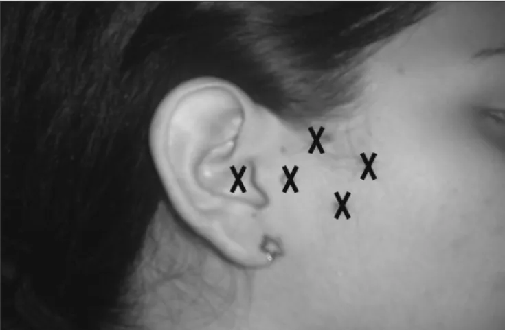
The laser application was performed bilaterally point-by-point, in contact with the surface, perpendicular to the skin16,17,(Figure 1): with four points in the shape of a cross
in the pre-auricular region; and one point in the external auditory meatus.
he medical care was provided twice a week, for four weeks, totaling eight sessions for each patient. No adverse efects were observed or reported by the patients partici-pating in the study. he patients were extensively informed about the nature of the study and they signed an informed consent statement in order to participate in the study. For the statistical analysis of the pain levels, the Student t test and the Kruskal-Wallis test were used, with a signiicance level set at 5% (p< 0.05).
Results
It was observed that the patients’ main complaint was pain, which could be a single complaint (61%) or was associa-ted with other complaints (39%) such as joint cracking (16%), muscular tension (11%), tooth wear/fracturing (6%) and joint rigidity (6%). he location of the patients’ pain varied and, in most cases (88%), it was in more than one location at the same time. he places that were most mentioned by the patients studied were the auricular (83%), temporal (72%), masseter muscle (66%) and neck regions (50%).
Correlations between the symptom duration in months and the factors related to worsening of the symptomatology
showed that emotional tension was mentioned by all the patients as a modifying factor, regardless the duration of the symptomatology, and was accompanied by the func-tional activity performed (especially mastication of hard foods and physical work activities) or by temperature mo-difications, in which cold appeared to be the main modi-fying factor.
Regarding the frequency of painful symptomatology, a large proportion of the population studied reported complaints of pain on a daily (56%) or weekly (39%) basis. he time until the onset of symptoms ranged from seven up to more than 60 months in the selected sample.
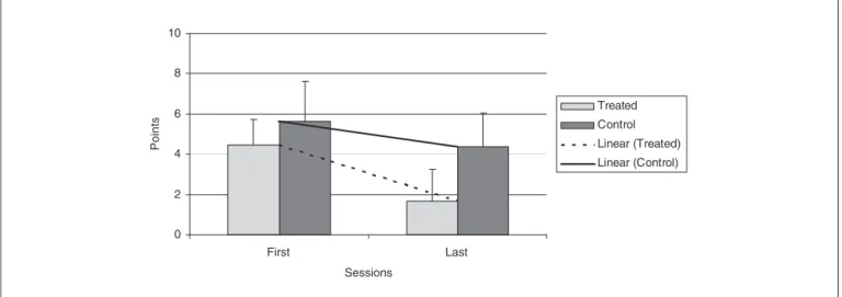
In Figure 2, it can be seen that the painful symptoma-tology of the treated group, initially with a mean of 4.6 points, which ranged from light and occasional to strong and constant pain. A significant statistical reduction was found (p< 0.05) from the fifth session onwards, and this was maintained until the end of the treatment, when the mean
Figure 1. Laser therapy application points in pre-auricular region and
external auditory meatus (X).
0 1 2 3 4 5 6 7 8 9
1 2 3 4 5 6 7 8
Sessions
Points
Treated Control Linear (Treated)
Linear (Control)
40
score from the VAS was 2, and the painful symptomatology ranged from strong and occasional to absence of painful symptomatology.
The control group initially had a mean of 5.6 points, with painful symptomatology varying from unbearable and occasional to light and occasional. They demonstrated a slight reduction of the painful conditions in the first sessions, without statistical significance (p> 0.05). Oscillations in the pain levels were recorded throughout the treatment, reaching the last session with a mean VAS score of 4.4, with painful symptomatology ranging from strong and constant to very light and constant.
Figure 3 shows the comparisons between the control group and the treatment group, for the mean VAS scores that were obtained, between the irst and last sessions. It can be seen that by the end of the treatment, both groups reported reductions in the painful symptomatology, but only the experimental group presented statistically signiicant results (p< 0.05).
Discussion
Low-power laser radiation is a widely used research source in many fields of health studies, since the effects of this therapy are dose dependent18, yet the action of this
ra-diation on different biological tissues and in different patho-logical conditions is still unclear. Through this study, it was sought to clarify the usage of laser photobiomodulation for treating pain in patients with TMD, which is an extremely common pathological condition seen in physical therapy consultation offices.
Studies have shown that pain shows an accentuated in-cidence in TMD, in addition to a large quantity of associated signs and symptoms, such as headaches and neck pain3-5. In
the present study, it was observed that the main complaint of the patients under investigation was pain, which was ma-nifested alone or in associated with other complaints such as joint cracking, muscle tension, tooth wear/fracturing and joint rigidity. Its location in the patients varied and it was generally found in more than one location at the same time. he most frequent locations in this study were the auricular, temporal, masseter muscle and cervical regions.
hrough the VAS, the evolution of the painful symptoma-tology could be observed in both groups. It was seen that in the treated group, there was a statistically signiicant reduc-tion (p< 0.05) from the ifth session onwards, which was then maintained until the end of the treatment. In the control group there was also an improvement of the pain level, although wi-thout statistical signiicance (p> 0.05), demonstrating oscilla-tions in pain levels over the course of the treatment.
The radiation was emitted in the near infrared region (904 nm), indicating a low absorption coefficient and, con-sequently, maximum tissue penetration, which promoted its interaction with molecular and cell structures8,12. Once
absorbed by the tissues, laser radiation causes release of substances like histamine, serotonin, bradykinin and pros-taglandins that are related to pain. It is also able to modify cell and enzymatic activities, through inhibiting or stimula-ting them13. For this to occur, the energy density is the most
important parameter in the technique. If the dose applied is too low, the expected result is not obtained, and if it is too high, an inhibitory effect might be obtained, instead of a stimulative effect18.
Since TMD is a chronic disease, 6 J/cm2 were used. Laser
irradiation exerts a stimulus on the cell mitochondria, cau-sing increased ATP production inside the cells and conse-quent acceleration of mitosis. his leads to increased oxygen consumption and cell respiration activation, thus eliminating
Figure 3. Comparison of the mean VAS scores between the first and last sessions.
0 2 4 6 8 10
Sessions
P
o
in
ts
Treated
Control
Linear (Treated)
Linear (Control)
41
the anaerobic activities that occur in an inlammatory process12,19,20. Other mechanisms that have been reported
for the beneicial efects induced by laser radiation include modulation of prostaglandin levels, increased ibroblastic activity, alterations to the somatosensory evoked potential and velocity of nervous conduction, as well as, improvements in the local circulation of the treated tissues, thereby causing pain relief and tissue repair9,21.
In the present study, 18 female patients of mean age of 27 ± 7 years with a diagnosis of TMD were analyzed. Studies have shown the predominance of dysfunction in this group of patients, i.e., women of the reproductive age22-24. There is
generalized flaccidity in female tissues that may be due to increased estrogen levels1. Women’s joints are usually more
flexible and looser than men’s. TMDs present their highest prevalence among women of the reproductive age. The pat-tern of onset of the disease following puberty and reduction of prevalence rates in the postmenopausal period suggests that reproductive hormones may have an important role in its etiology25.
A study carried out through a literature review on ar-ticles published between 1975 and 2002 states that the prevalence of TMD is from two to five times greater among women than among men in the study samples, because of female reproductive hormones23. Another important reason
for the predominance of TMDs among women is that wo-men show higher stress levels than do wo-men, thus reporting a higher incidence of diseases with psychosomatic invol-vement26. High stress levels not only may increase muscle
tonicity in the head and neck, but also may raise the levels of parafunctional muscle activity1.
he results obtained from the present study showed the complexity of TMD, both in relation to the patients’ symptoma-tology (since it is subject to the inluences of diferent factors, among which are emotional factors and activities preformed) and in relation to its treatment. Individuals’ muscle activity behavior is diicult to standardize, thus making it diicult to develop protocols for eicient treatments.
he clinical evaluation of pain was carried out through a double-blind study, because of the investigator’s possible inluence on the opinions of the patients undergoing the treat-ment. Although the present study was not carried out with a long-term follow-up, the pain reduction in the treatment group, which started after the third session and became accentuated after the fifth session, suggested a gradual improvement in symptomatology. he diferences found in the VAS relected an immediate response to the laser applica-tions, independent of the kind of treatment (real or placebo). In a placebo-controlled clinical trial on the effectiveness of laser photobiomodulation, Kulekcioglu et al.8 observed
a significant pain reduction both in the treated and in the placebo group. The placebo effect was discussed in a study by Gam, Thorsen and Lonnberg, which through meta-analysis observed the effects of laser photobiomodulation on musculoskeletal pain, finding minimal differences be-tween the treated and the placebo groups27. According to
these authors, a good relationship between the professional and the patient, associated with the “high-tech” appearance of the laser, may explain the improvements in the patients’ symptomatology. Moreover, it can be suggested that the limiting and chronic aspects of TMD symptoms, with pe-riods of symptom reduction, might partly explain the pain reduction in the placebo group28.
A systematic review by McNeely et al.3, published in
2006, examined the efficiency of different physical therapy interventions including laser photobiomodulation, for treating TMD. Based on their findings, no differences between the studied groups were observed regarding pain relief. No evidence was found regarding the effects of electrotherapy, including the laser, for pain reduction. However, there were significant improvements in the active and passive mouth opening and in the amplitude of lateral deviations following the laser treatment. Based on preliminary findings, these authors concluded that, although the technique may be potentially effective for TMD, more rigorously controlled clinical trials are needed in order to confirm this conclusion.
Kato et al.5 carried out a comparative study between
transcutaneous electrical nerve stimulation (TENS) and laser photobiomodulation in patients with TMD and observed, through VAS, that there was a reduction in the pain levels and improvements in the maximum mouth opening in both treated groups. Muscle palpation showed signiicant diferences for the laser group.
Gur et al.9 carried out a prospective, double-blind,
rando-mized and controlled study among patients with myofascial pain syndrome in the neck, with the use of GaAs (904 nm). They observed a reduction in pain levels and improvements in functional capacity and quality of life among their pa-tients. Considering the differences that were found in the VAS in the present study between the treatment and control groups, it can be suggested that the cumulative effects of the laser were responsible for pain reduction5,9,10,27,29,30.
42
1. Okeson JP. Fundamentos de oclusão e desordens temporomandibular. 4ª ed. São Paulo: Artes Médicas; 2000.
2. Dworkin SF, LeResche L. Research diagnostic criteria for temporomandibular disorders: review, criteria, examinations and specifications, critique. J Craniomandib Disord. 1992;6:301-55.
3. McNeely ML, Olivo SA, Magee DJ. A systematic review of the effectiveness of physical therapy interventions for temporomandibular disorders. Phys Ther. 2006;86(5):710-25.
4. Milan A, Fava ELF, Lino HE, Milam JRT, Lino Jr HL. Levantamento de incidência de DTMS e análise da efetividade da placa de mordida plana como terapia. Semina. 2004;25:23-38.
5. Kato MT, Kogawa EM, Santos CN, Conti PCR. TENS and low-level laser therapy in the management of temporomandibular disorders. Journal of Applied Oral Science: Revista FOB. 2006;14(2):130-5.
6. Detamore MS, Athanasiou KA. Structure and function of the temporomandibular joint disc: implications for tissue engineering. J Oral Maxillofac Surg. 2003;61(4):494-506.
7. Grossi DB, Chaves TC. Physiotherapeutic treatment for temporomandibular disorders (TMD). Brazilian Journal of Oral Science. 2004;3(10):492-7.
8. Kulekcioglu S, Sivrioglu K, Ozcan O, Parlak M. Effectiveness of low-level laser therapy in temporomandibular disorder. Scand J Rheumatol. 2003;32:114-8.
9. Gur A, Sarac AJ, Cevik R, Altindag O, Sarac S. Efficacy of 904 nm GaAs low level laser therapy in the management of chronic myofascial pain in the neck: a double-blind and randomize-controlled trial . J Clin Laser Med Surg. 2004;35(3):229-35.
10. Bjordal JM. Photoradiation in acute pain: a systematic review of possible mechanisms of action and clinical effects in randomized placebo-controlled trials. Photomed Laser Surg. 2006;24(2):158-68.
11. Fikackova H, Dostalova L, Vosicka R, Peterova V, Navratil L, Lesak J. Arthralgia of the temporomandibular joint and low-level laser therapy. Photomed Laser Surg. 2006;24(4):522-7.
12. Karu T. Low-power laser therapy. In: Vo Dinh, T. Biomedical photonics handbook. North Carolina: Taylor and Francis Group; 2003.
13. Campana VR, Moya M. The relative effects He-Ne laser and meloxican on experimentally induced inflammation. Laser Therapy. 1999;11(2):36-48.
14. Ricci R. Estudo in vitro da bioestimalação de células endoteliais em resposta a diferentes dosimetrias do laser de semicondutor fosfeto de índio-gálio-alumínio. [dissertação]. São José dos Campos: Universidade do Vale do Paraíba. Instituto de Pesquisa e Desenvolvimento; 2003.
15. Chavantes MC, Janete AD. Aplicação de laser na área cardiovascular. Arq Bras Cardiol. 1990;54(1):63-8.
16. Bradley S, Ghabban SN, Songra A. The maxilofacial region: recent research and clinical practice in low intensity laser therapy. In: Simunovic Z. Lasers
References
in medicine and dentistry: basic science and up-to-date clinical application of low level laser therapy – LLTT. Rijieka: Vitagraf; 2000.
17. Brugnera Jr A. Atlas de laserterapia aplicada à clínica odontológica. São Paulo: Santos; 2003.
18. Rigau J. Bioenergia e propriedades ópticas dos tecidos. In: Brugnera Jr A, Pinheiro ALB. Laseres na odontologia moderna. São Paulo: Pancast; 1998. p. 63-78.
19. Wilden L, Karthein R. Import of radiation phenomena of electrons and therapeutic low-level-laser in regard to the mitochondrial energy transfer. J Clin Laser Med Surg. 1998;16(3):159-65.
20. Karu T. Primary and secondary mechanisms of action of visible-to-near IR radiation on cells. J Photochem Photobiol. 1999;49(1):1-17.
21. Enwemeka CS. Therapeutic light. Rehab management. [Acesso em: 2004 Jan/Fev]. Disponível em: http://www.rehabpub.com/features/1022004/2.asp
22. Nekora-Azak A. Temporomandibular disorders in relation to female reproductive hormones: a literature review. J Prosthet Dent. 2004;91(5):492-3.
23. Cauás M, Alves IF, Tenório K, HC Filho JB, Guerra CMF. Incidências de hábitos parafuncionais e posturais em pacientes portadores de disfunção da articulação craniomandibular. Rev Cir e Traumatol Buco-maxilo-facial. 2004;4(2):121-9.
24. Pereira KNF, Andrade LLS, Costa MLG, Portal TF. Sinais e sintomas de pacientes com disfunção temporomandibular. Rev CEFAC: Actualização Científica em Fonoaudiologia. 2005;7(2):221-8.
25. LeResche L, Saunders K, von Korff MR, Barlow W, Dworkin SF. Use of exogenous hormones and risk of temporomandibular disorder pain. Pain. 1997;69:153-60.
26. Penna PP, Gil C. Estudo de um dos aspectos psicossomáticos relacionados com as desordens craniomandibulares. Revista da Pós-Graduação: RPG. 2006;13(2):181-5.
27. Gam AN, Thorsen H, Lonnberg F. The effect of low-level laser therapy on musculoskeletal pain: a meta-analysis. Pain. 1993;52(1):63-6.
28. Venancio AR, Camparis CM, Zanirato FLR. Low intensity laser therapy in the treatment of temporomandibular disorders: a double-blind study. J Oral Rehabil. 2005;32(11):800-7.
29. Sanseverino NTM, Sanseverino CAM, Ribeiro MS. Clinical evaluation of the low intensity laser antialgic action of GaAlAs (785 nm) in the treatment of the temporomandibular disorders. J Clin Laser Med Surg. 2002;31 Suppl. 14:18.