A
RTIGOC
IENTÍFICO Revista Brasileira de FisioterapiaInfluence of complex descongestive
physical therapy associated with intake
of medium-chain triglycerides for treating
upper-limb lymphedema
Influência da fisioterapia complexa descongestiva associada à ingestão de
triglicerídeos de cadeia média no tratamento do linfedema de membro superior
Oliveira J1, César TB2
Abstract
Objective: To investigate the influence of complex decongestive physical therapy (CDP) in association with diet therapy using medium-chain triglycerides (MCT), as an intervention in cases of upper-limb lymphedema. Methods: The lymphedema was evaluated by measuring circumferences, volumes, skin folds and whole-body water content. Feelings of discomfort, pain and heaviness in the arms were evaluated using a visual analog scale. Ten women who had undergone mastectomy and presented upper-limb lymphedema homolateral to the surgery participated in this study. Their mean age was 65.9 ± 10.4 years and their mean body mass index (BMI) was 26.8 ± 3.0kg/m². After nutritional evaluation, they were randomly divided into two groups: the Control Group (n= 5), which underwent physical therapy treatment consisting of CDP (classical massage, manual lymphatic drainage, compression taping and skincare) three times a week, for four weeks; and the MCT Group (n= 5), which underwent the same physical therapy protocol with the addition of daily diet therapy consisting of intake of MCT, for four weeks. Results: At the end of the intervention, analysis of the circumference and volume measurements showed significant differences between the groups (p≤ 0.05), with a greater reduction in lymphedema in the MCT Group. There were no significant differences in the skin fold measurements or whole-body water content. The feeling of heaviness in the arms after the intervention was significantly less in the MCT Group (p≤ 0.05), compared with before the intervention. Conclusion: Physical therapy treatment together with diet therapy with intake of MCT in women with upper-limb lymphedema following surgery and breast cancer treatment was effective in reverting this condition.
Key words: lymphedema; complex decongestive physical therapy; diet therapy; medium-chain triglycerides.
Resumo
Objetivo: Verificar a influência da utilização da fisioterapia complexa descongestiva associada à dietoterapia com triglicerídeos de cadeia média (TCM) como forma de intervenção no linfedema de membro superior (MS). Métodos: Para a avaliação do linfedema, foram utilizadas cirtometria, volumetria, pregas cutâneas e quantidade de água corporal total. A Escala Visual Análoga (EVA) foi utilizada para avaliar as sensações de desconforto, peso e dor no MS. Participaram deste estudo dez mulheres mastectomizadas com linfedema de MS homolateral à cirurgia, com idade média de 65,9 ± 10,4 anos e índice de massa corpórea (IMC) de 26,8 ± 3,0kg/m² que, após avaliação nutricional, foram divididas aleatoriamente em dois grupos: Grupo Controle (n= 5), submetido ao tratamento fisioterapêutico constando da terapia complexa descongestiva (massagem clássica, drenagem linfática manual, bandagem compressiva e cuidados com a pele) três vezes na semana, durante quatro semanas; Grupo TCM (n= 5), submetido ao mesmo protocolo fisioterapêutico somado ao tratamento dietético diário com ingestão de TCM, por quatro semanas. Resultados: Ao final da intervenção, a análise da cirtometria e da volumetria mostraram diferenças significativas entre os grupos (p≤ 0,05), com maior redução do linfedema no Grupo TCM. Não houve diferença significativa nos valores das pregas cutâneas e da quantidade de água corporal total. A sensação de peso no membro superior, antes e após a intervenção, foi significativamente menor (p≤ 0,05) no Grupo TCM. Conclusões: O tratamento fisioterapêutico somado à dietoterapia com ingestão de TCM em mulheres portadoras de linfedema de MS pós-cirurgia e tratamento de câncer de mama foi efetivo na involução desta condição.
Palavras-chave: linfedema; fisioterapia complexa descongestiva; dietoterapia; triglicerídeos de cadeia média.
Recebido: 30/01/2007– Revisado: 30/07/2007 – Aceito: 20/11/2007
This study was presented in the form of a poster at the First Physical Therapy Meeting: New Trends in Physical Therapy, promoted by the Physical Therapy course of the School of Philosophy and Sciences, State University of São Paulo (UNESP), Marília Campus, held on March 29 and 30, 2006.
1 Physical Therapy course, Centro Universitário de Araraquara, Araraquara (SP), Brazil
2 Department of Food and Nutrition, School of Pharmaceutical Sciences, Universidade Estadual Paulista (UNESP), Araraquara (SP), Brazil
Correspondence to: Jussara de Oliveira, Avenida Cônego Jerônimo César, 1.190, Carmo, CEP 14800-470, Araraquara (SP), e-mail: [email protected]
Introduction
Follow-up for breast cancer patients after adjuvant treat-ment is important because complications may occur, such as chest wall adherences, restrictions on shoulder movements, pain, hypoesthesia and, particularly, upper-limb lymphedema1.
When the lymphatic vessels are removed or damaged, the lymphatic transportation is damaged and the lymphatic luid accumulates in the interstitial spaces of the tissue around the afected site. his extends to the upper limbs on the afected side and characterizes lymphedema2.
To evaluate lymphedema, a variety of measurements are used, such as circumference measurements3, volumetry4 and
multiple frequency electrical bioimpedance5. hese enable
pre-dictions regarding the degree of lymphedema in the afected limb6 and make it possible to choose the most appropriate
intervention7. Electrical bioimpedance is used to estimate
the quantities of liquid in body compartments and has been applied in investigating luid volumes in limbs presenting lymphedema8,9. he triceps and biceps skinfolds are
nutritio-nal indicators that can be used to investigate the malleability of the skin and the consistency of lymphedema10.
Among the interventions for treating lymphedema, com-plex decongestive physical therapy4 stands out. his includes
procedures such as manual lymphatic drainage (MLD)11,
compressive bandaging (CB)12 or elastic restraint, pelvic loor
exercise programs and skin care2.
Biochemical analyses of human lymphedema show the pre-sence of high proportions of long-chain triglycerides (LCTs) with a high content of kilomicrons13. Changes in the quantities of LCTs in
the lymph luid may alter the composition of the luid, thus leading to decreased low and pressure in the lymphatic system, thereby diminishing its overload13,14. Medium-chain triglycerides (MCTs)
with six to 12 carbons are absorbed directly into the bloodstream because, diferently from long-chain fatty acids, they are not incor-porated into kilomicrons15. After passing through the enterocytes,
they reach the portal circulation and are transported to the liver by albumin, without going through the lymphatic system16.
Soria et al.13 described the use of MCTs, corresponding
to 58% of the total fat consumed, as a substitute for LCTs for patients with idiopathic lymphedema. hey showed that there was a signiicant reduction in the circumference measurement of the afected limb after four months of an LCT-free diet in association with physical therapy treatment.
Because lymphedema is a frequent complication following breast cancer and results in the loss of functional abilities and esthetic deformities17, new forms of interventions in
associa-tion with the convenassocia-tional techniques are needed in order to reduce the presence of lymphedema. herefore, the present study had the objective of investigating the inluence of using
classical massage, manual lymphatic drainage and compres-sive bandaging in association with consumption of MCTs as a means of intervention for lymphedema cases.
Methodology
After obtaining approval (on June 9, 2005) from the Research Ethics Committee of the School of Pharmaceutical Sciences of UNESP in Araraquara, State of São Paulo (case no. 01/2005), 16 women with upper-limb lymphedema were recruited and sent to the UNIARA Physical herapy Clinic. he participants were selected by means of individual interviews explaining the objectives, duration and procedures of the study. Cases of metastasis, phlebitis, acute-phase erysipelas and dyslipidemia were excluded. All the participants signed a consent state-ment, in compliance with National Health Council resolution no. 196/96. After beginning of the study, three women were found to demonstrate erysipelas in the afected upper limb, two were unavailable for carrying out the weekly protocol and one terminated her participation.
his study, which was blind and randomized, was conduc-ted on 10 women of a mean age of 65.9 ± 10.4 years and a BMI of 26.8 ± 3.0, who had homolateral upper-limb lymphedema subsequent to breast cancer surgery and axillary lymphade-nectomy. Among these patients, seven underwent quadran-tectomy and three had modiied radical surgery. here was an equal prevalence of breast laterality and afected upper limb: half of the breast cancer sample involved the right side and the other half, the left side. All cases needed axillary emptying up to level III. With regard to postsurgical treatment, eight pa-tients needed more than 30 radiotherapy sessions and only two had less than 28 applications, while six patients underwent six chemotherapy sessions and four had between seven and 12 applications. he lymphedema appeared within one year in ive patients, while two of them reported lymphedema between one and three years after surgery and the other three patients presented this between three and ive years after their breast cancer treatment.
33
but they were aware that they were using one or another of the provided oil sources. To monitor their consumption, they were asked, while following the protocol, to make periodic records so that each participant’s nutritional intake could be followed up.
he data collection consisted of bilateral upper-limb cir-cumference measurements, at the beginning of each session, at predeined ixed points18. Volumetry was performed on the
upper limbs before and after the interventions, by immersing the limb in a volumetric column, with water leveled up to the middle third of the arm. he liquid that overlowed out of the column was measured in order to obtain the volume of the limb. he electrical bioimpedance was measured using speciic total body water apparatus (Biodynamics, model 310) to gene-rate a 50 kHz electric current that measured the total quantity of body water. Feelings of discomfort, heaviness and pain in the afected upper limb were obtained using a visual analog scale (VAS), before and after the experiment.
he body weight was obtained with the volunteers in a standing position, at the center of the base of a duly calibrated platform balance, with as little clothing as possible. heir height was measured using a tape measure attached to a wall, with the patient standing against the wall, without shoes, with heels toge-ther, vertebral column upright, arms extended next to the body and with the head aligned. he triceps skinfold was measured using a skinfold adipometer positioned on the posterior face of the arm, at the midpoint between the superolateral edge of the acromion and the olecranon. he biceps skinfold was measured on the anterior face of the arm, one centimeter above the loca-tion marked for the triceps skinfold measurement, with the palm of the patient’s hand turned to the front5. he dietary proile was
obtained from the food frequency questionnaire, in which the patients described their frequencies of intake; their daily, weekly and monthly food consumption; and by the 24-hour dietary re-call record, with descriptions of all food and drinks consumed within 24 hours19 in homemade measures.
he treatments for the afected upper limb consisted of applications, at each session, of various types of classical mas-sage with the use of supericial and deep sliding maneuvers, for the whole upper limb, followed by compression maneuvers20.
Manual lymphatic drainage was performed in accordance with the maneuvers of Leduc and Vodder1,21, starting with evacuation
and ending with capture11,21. Compressive bandaging was
perfor-med after manual lymphatic drainage and was maintained for a period of at least 10 hours. he upper limb was irst wrapped with tubular mesh, followed by protective sponge. Elastic bandages of width ive cm were wrapped around the ingers and the hand. Bandages of an eight cm width were overlaid on the wrist region and the proximal third of the forearm. Bandages of a 10 cm width were wrapped over these, up to the level of the axillae, leaving the elbow region free to maintain functionality of the limb2.
he parametric data was analyzed using Student’s t test and the nonparametric data using the Mann-Whitney test and the Wilcoxon matched pairs test, with p≤ 0.05.
Results
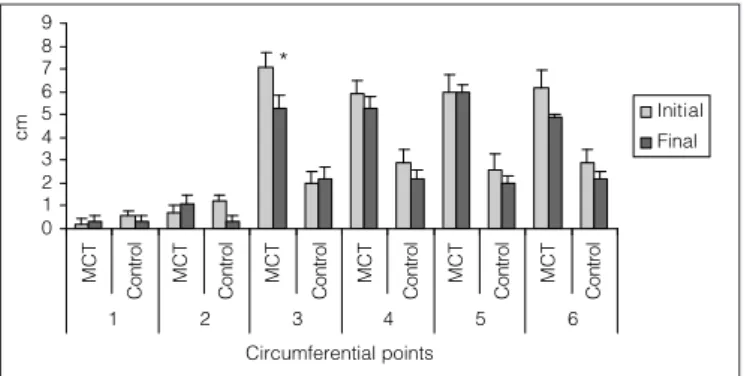
Figure 1 illustrates the mean values for the diferences in circumference measurements between the afected and heal-thy upper limbs of the MCT group, gathered at the beginning and end of the study protocol, in comparison with the mean values for the diferences in these measurements between the afected and healthy limbs of the control group. Statistically diferent values at p<.03 could be seen from analyses of the reduction in the diferences in circumference measurement between the involved and healthy limbs of the MCT group, in relation to the same measurements for the control group. his demonstrated that, in the forearm region, there was a greater reduction (p≤ 0.05) in the MCT group, i.e., more pronounced decreases in lymphedema in the patients of this group.
Figure 2 shows the volumetry for the upper limbs, compa-ring the mean reductions in volume diferences between the two limbs (afected and healthy) and between the groups, at the end of the intervention. From this, it can be seen that there was a greater reduction in the MCT group, at the end of the tre-atment (p≤ 0.05). he mean values for the control group were negative, meaning that there was an increase in the diferences in volume between the afected and healthy limbs of the parti-cipants in this group. Hence, in the end, there was an increase in lymphedema for this group.
Comparisons of the reduction in skinfold thicknesses in the afected upper limb (Figure 3) showed that there were no signiicant diferences in the inal values between the groups.
Figure 1. Comparisons between the mean differences in circumference measurements of the affected and healthy upper limbs of the two groups, before and after the intervention.
0 1 2 3 4 5 6 7 8 9 M C T C o n tr o l C o n tr o l C o n tr o l C o n tr o l C o n tr o l C o n tr o l M C T M C T M C T M C T M C T
1 2 3 4 5 6
Circumferential points
cm
Initial Final
*
Point 1: the metacarpophalangeal joints of the second to fifth fingers; Point 2: an imaginary line going through the metacarpophalangeal joint of the first finger; Point 3: 10 cms below the olecranon; Point 4: 6 cms below the olecranon; Point 5: 6 cms above the olecranon; Point 6: 10 cms above the olecranon.
0 3 6 9 12 15
m
m
MCT Group Control Group
TS BS
TS= triceps skinfold; BS= biceps skinfold.
Figure 3. Comparisons between the mean reductions in skinfolds in the affected upper limbs, at the end of the treatment.
Figure 4. Comparisons between the mean reductions in the quantities of body liquid in the two groups, at the end of the intervention.
0.00 0.50 1.00 1.50 2.00 2.50 3.00 3.50 4.00
Groups
L
it
e
rs
MCT Group
Control Group
Regarding the total body water quantities found by electri-cal bioimpedance, comparisons of the mean reduction in body water between the groups, after the intervention, did not show any signiicant diferences, as seen in Figure 4.
From the patients’ subjective feelings about their lymphe-dema found through the VAS, Figure 5 shows that there was a statistically signiicant decrease (p≤ 0.05) in the degree of discomfort for both groups, comparing the initial and inal va-lues for each group. Regarding feelings of arm heaviness, only the MCT group presented a signiicant decrease (p≤ 0.05) at the end of the intervention. here were no diferences in the feelings evaluated between the two groups.
Discussion
Lymphedema develops from an imbalance between the lymphatic demands and the system’s capacity to drain the lymph. Since high molecular weight proteins that are
extravasated to tissue interstices are solely absorbed by the lymphatic system, if this system loses its drainage capacity because of the destruction or obstructions of the lymph ducts at some point, this causes stagnation of the lymph in the vessel and subsequent extravasation back to the in-terstices22. It also results in a combination of factors such
as obesity. Studies have reported that the patient’s age and whether the surgery was on the dominant or nondominant side were not statistically associated with the development of edema23. However, Freitas et al.24 noted that there were
significant relationships between lymphedema frequency and the weight and age of the patients. In the present study, the patients demonstrated a mean BMI of 26.8 and classified as being pre-obese5, which proved to be an important factor
in establishing lymphatic edema.
he skin is responsible for supericial absorption of the lymph luid and skincare is essential to lymph therapy pro-cedures20. Classical massage was shown to be efective in the
present study, since it assisted in reducing the lymphedema.
-150 -100 -50 0 50 100 150 200 250 300
m
L MCT
Control Group *
Figure 2. Comparisons between the mean reductions in volume difference (ml) in the affected and healthy upper limbs, for the two groups, at the end of the treatment.
*p≤ 0.05.
Figure 5. Comparisons between the degrees of feelings shown by the VAS, before and after the intervention. For discomfort: Degree 1: no discomfort; Degree 2: slight discomfort; Degree 3: moderate discomfort; Degree 4: a lot of discomfort; Degree 5: severe discomfort. For heaviness in the affected limb: Degree 1: no heaviness; Degree 2: slight heaviness; Degree 3: moderate heaviness; Degree 4: heavy; Degree 5: very heavy. For pain in the affected limb: Degree 1: no pain; Degree 2: slight pain; Degree 3: moderate pain; Degree 4: severe pain; Degree 5: unbearable.
0 0.5 1 1.5 2 2.5 3 3.5 4 4.5
D
e
g
re
e
Initial
Final
* * *
Heaviness
Discomfort Pain
MCT Control MCT Control MCT Control
35
Manual lymphatic drainage is used to drain the excess li-quids that bathe the cells and maintain the hydric equilibrium of the interstitial spaces21. he technique used in the present
study enabled reductions in the circumference and volume of the afected limb in the MCT group, thus reinforcing the importance of this procedure for decreasing lymphedema. Manual lymphatic drainage may be indicated together with other treatments, so that it is possible to contain the edema at a comfortable level, reduce the pain and ibrosis and also bring relaxation and provide a feeling of well-being25. In our study,
there was a general improvement in the subjective feelings in the upper limb afected by lymphedema.
Compressive bandaging has been shown to be an efective resource, because it increases the lymphatic absorption and low achieved through prior manual lymphatic drainage26. In
the present study, there were signiicant decreases in the lym-phedema in the patients who had MCT intake in addition to the physical therapy interventions.
With regard to electrical bioimpedance, it is known that its results vary according to the tissue that is being measu-red. It can thus be said that the values obtained were direc-tly proportional to the body fat percentage. In addition to body fat and lean mass evaluations, this method establishes the quantity of body water and is therefore an important follow-up tool for patients with pathological conditions of increased extracellular liquid27. The bioimpedance study by
Cornish et al.28 indicated that there was higher precision
in calculating body water volumes using segmental body impedance, rather than taking the body as a whole. In the present study, this examination was not done segmentally, and therefore it was not possible to obtain definitive con-clusions from evaluating the lymphedema. Nonetheless, it was shown to be important for nutritional evaluation of the patients. Thus, more studies are needed using segmental bioimpedance to evaluate lymphedema.
he biceps and triceps skinfold measurements did not indi-cate any direct relationships with the conventional methods for lymphedema evaluation, since these are traditionally nutritional indicators. According to Shills et al.29, indirect calculation of body
composition by measuring the subcutaneous fat in the skin-folds is the method most used in such investigations. Skinfold measurements have the purpose of estimating total body fat, because there is a relationship between localized fat deposited under the skin and body density30. Measurement of these folds
was used here in an attempt to investigate improvements in skin malleability and the consistency of the lymphedema.
In agreement with the results from Soria et al.13, the use of
MCT by patients with lymphedema was shown to be efective, considering that in the present study there were signiicant reductions in the circumference measurements and volume of the afected limb, in comparison with the group control, thus signifying a decrease in the lymphedema in the patients who used MCT as additional therapy.
MCTs, which are rich in medium-chain fatty acids, are hydrolyzed by pancreatic lipase action and are absorbed in the duodenum more rapidly than are long-chain fatty acids. According to Yokocama and Fagundes31, a low-fat diet that
is rich in MCTs must be used for individuals with intestinal lymphangiectasia and consequent lymphatic insuiciency, in order to decrease accumulations in the lymph ducts and reduce the pressure in these dilated vessels. MCTs are not esteriied or absorbed in the intestinal lymphatic system and chest duct, but enter directly into the portal system: hence the reason for their use. Alcauza and César32 used MCT as
a diet therapy method among women with upper-limb lym-phedema and obtained positive results, with reductions of the clinical symptomatology.
In the present study, it could be seen that the use of MCTs as a means of lymphedema treatment was satisfactory, since the patients who used MCT as a nutritional supplement demonstrated significant improvements compared with the control patients. This also leads us to wonder whether the lipid diet of vegetable oil that was administered in the control group might have constituted a contributory factor for non-regression or even increased lymphedema because that diet was composed of long-chain fatty acids. After absorption of these fatty acids derived from vegetal oil, they leave the intestine in the form of triglycerides through the lymph ducts, incorporated in kilomicrons and transported by the lymphatic system. By increasing the overload in a lym- overload in a lym-phatic system that is already compromised, this may come to negatively influence the involution of the lymphedema. Therefore, the present study draws attention to the fact that ordinary diets that are rich in long-chain fatty acids may not be ideal for people with lymphedema, and sug-r people with lymphedema, and sug-gests that further studies in this area are needed.
References
Guirro ECO, Guirro RRJ. Fisioterapia dermato-funcional – fundamentos, 1.
recurso e patologias. São Paulo: Manole; 2002.
Camargo MC, Marx AG. Reabilitação física no câncer de mama. São 2.
Paulo: Roca; 2000.
Hwang JH, Know JY, Lee KWC. Changes in lymphatic function after 3.
complex physical therapy for lymphedema.Lymphology. 1999;32:15-21.
Petrek AJ, Pressman PI, Smith RA. Lymphedema: current issues in 4.
research and management. Cancer J Clin. 2000;50(5):292-307.
Cuppari L. Guia de nutrição: nutrição clínica no adulto. Escola Paulista de 5.
Medicina. São Paulo: Manole; 2002.
Kissin MW, Querci della Rovere G, Easton D, Westbury G. Risk of lymphedema 6.
following the treatment of breast cancer. Br J Surg. 1986;73:580-4.
Perrin M, Guex JJ.
7. Edema and leg volume: methods of assessment. Angiology. 2000;51(1):9-12.
Staton AWB, Badger C, Sitzia J. Non-Invasive assessment of the 8.
lymphedematous limb. Lymphology. 2000;33:122-35.
Caban ME. Trends in the evaluation of lymphedema. Lymphology. 9.
2002;35:28-38.
Lohman TG, Roche AF, Martorell R. Anthropometric standardization 10.
reference manual. Champaign, Illinois: Human Kinetics; 1991.
Leduc A. Drenagem linfática: teoria e prática. 2ª ed. São Paulo: Manole; 2000. 11.
Herpertz U. Edema e drenagem linfática: diagnóstico e terapia do edema. 12.
São Paulo: Roca; 2006.
Soria P, Cuesta A, Romero H, Martinez F, Sastre A. Dietary treatment of 13.
lymphedema by restriction of long-chain triglycerides. Angiology. 1994;45(8):703-7.
Trauner DA. Medium-chain triglyceride (MCT) diet in intractable seizure 14.
disorder. Neurology. 1985;35:237-8.
Ferreira AMD, Barbosa PEB, Ceddia RB. A influência da suplementação 15.
de triglicerídeos de cadeia média no desempenho em exercícios de ultra-resistência. Rev Bras Med Esporte. 2003;9(6):413-9.
Jeukendrup AE, Saris WHM, Wagenmakers AJM. Fat metabolism during 16.
exercise: a review. Part III: Effects of nutrition interventions. Int J Sports Nutr. 1998;6:121-33.
McKenzie DC, Kalda AL.
17. Effect of upper extremity exercise on secondary lymphedema in breast cancer patients: a pilot study. J Clin Oncol. 2003;21:463-6.
Mamede MV. Reabilitação de mastectomizadas: um novo enfoque 18.
assistencial [teste]. Ribeirão Preto (São Paulo): Escola de Enfermagem da USP; 1991.
Pereira Filho RA, Sevá-Pereira A. Absorção de triglicerídeos de cadeia 19.
média em pacientes com síndrome de alça estagnada. Arq Gastroenterol. 1988;25(2):75-81.
Cassar MP. Manual de massagem terapêutica. São Paulo: Manole; 2001. 20.
Barros MHD. Fisioterapia: drenagem linfática manual. São Paulo: Probel; 2001. 21.
Spence RK, Cahall E. Inelastic versus elastic leg compression in 22.
chronic venous insufficiency: a comparison of limb size and venous hemodynamics. J Vasc Surg. 1996;24(5):783-7.
Segerström K. Bjerle, GS, Nyström A. Factors that influence the incidence 23.
of brachial oedema after treatment of breast cancer. Sacnad JPlast Reconstr Nand. 1992;26:223-7.
Freitas RJ, Ribeiro LF, Taia L, Kajita D, Fernandes MV, Queiroz G S. 24.
Lymphedema in breast cancer patients submitted to modified radical mastectomy. Rev Bras Ginecol Obstet. 2001;23(4):205-8
Kasseroller R. Compendium of vodder’s manual lymph drainage. New 25.
York: Thieme Medical Pub; 1998.
Brentani
26. MM, Coelho FRG. Bases da oncologia. São Paulo: Lemar; 1998.
Correa FHS, Taboada GF, Junior CRMA. Influência da gordura corporal no 27.
controle clínico e metabólico de pacientes com diabetes mellitus tipo 2. Arq Bras Endocrinol Metab. 2003;47(1):62-8.
Cornish BH, Eles PT, Thomas BJ. The effect of electrode placement in 28.
measuring ipsilateral/contralateral segmental bioelectrical impedance. Annals of the New York Academy of Sciences. 2000;904:221-4.
Shills ME, Olson JA, Shike M. Modern nutrition in healph and disease. 29.
Nova York: Lea & Febiger; 1994.
Fett CA, Fett WCR, Marchini JS. Gasto energético de repouso medido vs. 30.
estimado e relação com a composição corporal de mulheres. Arq Bras Endocrinol Metab. 2006;50(6):1050-8.
Yokocama M, Fagundes UN. Linfagiectasia intestinal. The Electronic 31.
Journal of Pediaric Gastroenterology, Nutrition and Liver Dieases. 2003;7(3) [cited 2006 Sep 12]. Available from: http://www.e-gastroped. com.br/sept03/linfangiectasia.htm
César TB, Alcauza MTR, Guirro EC. Oral dietotherapy with medium 32.