IMPACTED CISTERNA MAGNA WITHOUT
SYRINGOMYELIA ASSOCIATED WITH
LANCINATING HEADACHE, THROBBED
NUCHAL PAIN AND PARAPARESIS TREATED
BY POSTERIOR FOSSA DECOMPRESSION
José Alberto Gonçalves da Silva
1, Maria do Desterro Leiros da Costa
2,
Luiz Ricardo Santiago Melo
1, Antônio Fernandes de Araújo
1,
Everardo Bandeira de Almeida
3ABSTRACT - A 29-year-old woman with acute lancinating headache, throbbed nuchal pain and subacute paraparesis underwent brain MRI in supine position that depicted: the absence of the cisterna magna, filled by non herniated cerebellar tonsils and compression of the brain stem and cisternae of the posterior fos-sa, which are aspects of the impacted cisterna magna without syringomyelia and without hydrocephalus. During eight days, pain was constant and resistant to drug treatment. Osteodural-neural decompression of the posterior fossa, performed with the patient in sitting position, revealed: compression of the brainstem, fourth ventricle and foramen of Magendie by herniated cerebellar tonsils, which were aspirated. Immedi-ately after surgery, the headache and nuchal pain remmited. MRI depicted the large created cisterna magna and also that the cerebellar tonsils did not compress the fourth ventricle, the foramen of Magendie and the brainstem, besides the enlargement of posterior fossa cisternae. Four months after surgery, headache, nuchal pain and paraparesis had disappeared but hyperactive patellar and Achilles reflexes remained.
KEY WORDS: headache, nuchal pain, craniovertebral malformation, tight cisterna magna, impacted cisterna magna, Chiari malformation, posterior fossa decompression, paraparesis, herniated tonsils.
Cisterna magna impactada sem siringomielia associada a cefaléia lancinante, dor na nuca terebrante e pa-raparesia tratadas com descompressão da fossa posterior
RESUMO - Uma paciente de 29 anos de idade com quadro agudo de cefaléia lancinante, dor terebrante na nuca e paraparesia subaguda foi submetida a RM do encéfalo, em posição supina, que revelou: ausên-cia da cisterna magna, preenchida por tonsilas cerebelares não herniadas e compressão do tronco encefá-lico e das cisternas da fossa posterior, compatíveis com o diagnóstico de cisterna magna impactada sem si-ringomielia e sem hidrocefalia. Por oito dias a dor foi constante e resistente aos analgésicos. Com a pacien-te em posição sentada, foi realizada descompressão ospacien-teodural-neural da fossa pospacien-terior associada a aspi-ração das tonsilas cerebelares. Os achados perioperatórios foram caracterizados por herniação das tonsilas cerebelares que comprimiam o tronco cerebral, o quarto ventrículo e o forame de Magendie. No pós-ope-ratório imediato houve remissão da cefaléia e da dor na nuca. A RM evidenciou a cisterna magna recém-criada, alargamento do quarto ventrículo e das cisternas do tronco encefálico. Quatro meses depois, a pa-ciente continuava sem cefaléia, sem dor na nuca e sem paraparesia. Entretanto, permaneceu a hiperativi-dade dos reflexos patelares e aquileus.
PALAVRAS-CHAVE: cefaléia, dor na nuca, malformação craniovertebral, cisterna magna impactada, tight cis-terna magna, malformação de Chiari, descompressão da fossa posterior, paraparesia, tonsilas herniadas.
1Neurosurgical Unit of the Hospital Unimed, João Pessoa PB, Brazil; 2Federal University of Paraiba, João Pessoa PB, Brazil; 3 Neuroan-esthesist, João Pessoa PB, Brazil.
Received 3 April 2007, received in fi nal form 17 August 2007. Accepted 17 September 2007.
Many studies on craniovertebral malformations in-dicate that a specifi c, suboccipital headache of vari-able types and duration may affect the patients. In a retrospective study of 249 operated cases of cran-iovertebral malformations, Gonçalves da Silva1 ob-served headache in 149 (59.8%) and nuchal pain in 132 (53%); Milhorat et al.2 studied 364 symptomatic patients with Chiari I malformation and found that the most common symptom was a suboccipital head-ache, experienced by 296 (81%) patients, described as a heavy, crushing, or pressure-like sensation at the back of the head that radiated to the vertex and be-hind the eyes. A distinctive feature of this headache is the tendency to be increased by physical exertion, Valsalva maneuver, head dependency and sudden changes in posture. Their studies also demonstrat-ed in most cases that Chiari-relatdemonstrat-ed headaches could be clearly distinguished from cervicogenic and other headache syndromes. Similar fi ndings were reported by Pascual et al.3 that analyzed the headaches pre-sented by 50 patients with Chiari I malformation. Of these 50, 14 (28%) had suboccipital-occipital head-ache. Headaches correlated to craniovertebral mal-formations affect predominantly the occipital-suboc-cipital region and are usually provoked by cough4,5. Stovner5 studied 34 patients with Chiari I malforma-tion in order to understand the related headache symptoms, particularly the long-lasting attacks or continuous pain. He encountered 20 patients who had or had had headaches and interviewed them by way of a questionnaire. Ten patients had short term cough headaches lasting less than 5 minutes, 14 pa-tients had relatively long-lasting attacks lasting from 3 hours to several days, and 8 patients had continuous headache. Unlike the short term cough headache at-tacks, long-lasting attacks were usually not precipitat-ed by Valsalva-like maneuver and could be differenti-ated from those of migraine and cervicogenic head-ache patients. These characteristics associated with a positive effect of surgical treatment in some patients, suggest a causal relationship between the malforma-tion and the headache. According to this author, this malformation may cause long-lasting headache at-tacks or continuous head pain by compression of the brainstem, central cord degeneration or intracrani-al hypertension.
Hans Chiari6,7 described four types of cerebellar anomalies, which later were named Chiari malfor-mation (CM). Type I was characterized by downward displacement of the cerebellar tonsils and the me-dial portions of the inferior cerebellar lobes, which accompanied the medulla oblongata into the
cervi-cal spinal canal. Type II showed downward displace-ment of portions of the cerebellum, and portions of the inferior vermis, pons, medulla oblongata and, at least, a part of the lengthened fourth ventricle, which reached the disc C4-C5, into the enlarged cervical spi-nal caspi-nal. In type III, the hydrocephalic cerebellum, pons and medulla were inside a cervical meningocele (hydroencephaloceles cerebellaris cervicalis), through a spina bifi da of the fi rst three cervical vertebrae. In type IV, there was hypoplasia of the cerebellum with-out herniation of the cerebellar structures into the spinal canal. Iskandar et al.8 related
fi ve cases of syrin-gomyelia (SM), in which the cisterna magna was fi lled by the cerebellar tonsils. All fi ve cases presented clini-cal improvement after decompression of the posteri-or fossa as well as marked reduction in the size of the syrinx. These authors admitted that this dramatic re-sponse to decompression indicated that this entity has a Chiari-like pathophysiology. Kyoshima et al.9 report-ed four similar cases with an improvement of symp-toms and a reduction in the syrinx size in three pa-tients and a reduction in ventricle size in one. These authors termed the cisterna magna impacted by the cerebellar tonsils “tight cisterna magna” and called the description according to Iskandar et al.8, “Chiari 0 malformation”. Posterior fossa decompression for pa-tients with this malformation is indicated when they present symptoms of compression syndromes of the brainstem and spinal cord, unusual headache types and other uncommon conditions4,5.
This study is based on the rareness of the clinical picture characterized by an excruciating headache, throbbed nuchal pain and paraparesis, associated with impacted cisterna magna without SM.
CASE
Fig 1. Preoperative sagittal MRI shows impacted cisterna magna, diminished fourth ventricle and superior and prepontine cis-ternae.
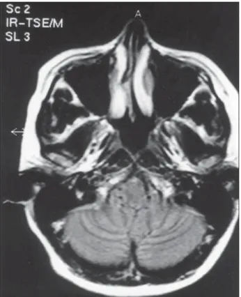
Fig 2. Preoperative axial MRI depicts the absence of the cister-na magcister-na.
Fig 3. Patient in sitting position.
Fig 4. Tonsile herniation observed during the operation in sit-ting position.
Fig 5. Tonsilectomy and large opening of the fourth ventricle.
the fourth ventricle as well as the foramen of Magendie by the cerebellar tonsils which closed this foramen, fi lled the cisterna magna and migrated downwards into the cervical spinal canal (no adherences to the neighboring structures were observed). The left cerebellar tonsil was hypertrophic and reached the C2 level while the right was hypotrophic and situated at C1; the left posterior inferior cerebellar ar-tery (PICA) was enlarged and descended to the C2 level, while the left PICA was hypoplasic. We performed intrapial aspiration of the cerebellar tonsils and made a large open-ing of the fourth ventricle (Fig 5), then sutured the residu-al piresidu-al sac upwards to the dura-mater in craniresidu-al laterresidu-al po-sition and fi nally a dural grafting was made with bovine
ventri-cle as well as of the superior and prepontine cisternae, be-sides the absence of compression of the cerebellar tonsils towards the fourth ventricle and the foramen of Magendie. The headache and nuchal pain disappeared immediately af-ter the operation. Four months laaf-ter, the patient continued without headache, nuchal pain, paraparesis and returned to her job. Nevertheless, the hyperactive patellar and Achilles refl exes remained unchanged.
This study was approved by the Bioethics Research Com-mittee.
DISCUSSION
The pathophysiological mechanisms that underlie headache and nuchal pain in craniovertebral malfor-mations remain unclear. McGirt et al.11 studied 33 cases of Chiari I malformation, presenting with headaches alone, to identify the correlations between headache and CSF fl ow obstruction. The preoperative MRI of the craniovertebral junction was prospectively per-formed in all patients. They observed that regardless of the degree of tonsillar ectopia, occipital headaches were strongly associated with hindbrain CSF fl ow
ab-normalities. Kesler and Mendizaval12 described two cases of diagnosed Chiari I malformation where the primary complaint was headache. These authors ar-gue that CM should be considered in the differen-tial diagnosis of patients who complain of exertional headaches.
To Arnett13, occipital and exertional headaches are associated with tonsillar ectopia encompassing slight descent of the cerebellar tonsils and Chiari I malfor-mation. The headache in this malformation is second-ary to pressure dissociation gradients and perhaps, traction of pain-sensitive tissues. According to this author, the extent of tonsillar descent alone is irrel-evant. The degree of posterior fossa hypoplasia and decrease of the CSF fl ow velocity are more likely the causes of pain symptoms in tonsillar ectopia. On the other hand, Pascual et al.3 and Taylor et al.4admitted that the headache and the nuchal pain are directly related to the degree of tonsilar herniation.
Occipital headache and nuchal pain are frequently observed in cases of craniovertebral malformations like basilar impression (BI), CM and SM1,2,14-16. In a ret-rospective study of 249 operated cases of craniover-tebral malformations, Gonçalves da Silva 1 observed headaches in 149 (59.8%) and nuchal pain in 132 (53%) in the pre-operatory phase. After decompression of the posterior fossa, headache was eliminated in 139 (93.2%) of the 149 cases, while nuchal pain remmited in 121 (91.6%) of the 132 cases.
Gonçalves da Silva et al.17,18described two cases of impacted cisterna magna without tonsilar herniation depicted by MRI in supine position. Both cases pre-sented headache and nuchal pain. During the opera-tion, with the patient in sitting posiopera-tion, cerebellar tonsils herniation was detected. This fi nding indicates that in orthostatic position, cerebellar tonsils hernia-tion may exert compression towards the upper cervi-cal dorsal roots and distortion of the brainstem result-ing in nuchal pain.
This study presents a rare case of impacted cister-na magcister-na associated with lancicister-nating headache and throbbed nuchal pain, at an intensity never identifi ed before in any other patient of our casuistics referred above. The possible pathophysiological mechanisms to explain the severity of the pain is that the herni-ated cerebellar tonsils through the foramen magnum moving up and down could originate compression of the superior cervical spinal cord, cerebellum, medulla and friction of the fi rst cervical roots. This mechanism, along with the described disturbance of CSF due to the compression of the foramen of Magendie, associ-ated with the absence of cisterna magna could have
resulted in headache and nuchal pain.
Iskandar et al.8 were the fi rst to report fi ve cas-es of SM without hindbrain herniation. All cascas-es improved after the decompression of the posterior fossa and the authors suggested that this entity had a Chiari like pathophysiology. Kyoshima et al.9 pub-lished four similar cases with a good recovery of the patients after the decompressive operation of the posterior fossa. These authors named this malfor-mation as “tight cisterna magna” and designated Iskandar’s et al.8 description as “Chiari 0 malforma-tion”. According to Williams19, the cerebellar tonsils herniation may compress brainstem structures and contribute to bulbar and long tracts dysfunctions. In a similar way, the pressing of the cerebellar tonsils into the cisterna magna, without herniation into the cervical spinal canal, causes disturbances to the CSF fl ow at the foramen magnum and can also develop neurological symptomatology by compression of the brainstem. This author also admitted that tonsilar dis-tortion, hindbrain herniation and obstruction of the outlet of the fourth ventricle may cause headache. In some cases, the foramen of Magendie is completely occluded by a membrane and hydrocephalus does not necessarily occur. Probably because of the drainage of the fourth ventricle via the foramen of Lushka.
In this case the foramen of Magendie, the fourth ventricle and the medulla oblongata were com-pressed by the impacted cerebellar tonsils. Probably the compression of the brainstem could explain the paraparesis in this case while the CSF hypertension in the posterior fossa could be responsible for the head-ache and nuchal pain.
Acknowledgements– The authors thank Mr. David Swingler, English Teacher, for the improvement of this
manuscript.
REFERENCES
1. Gonçalves da Silva JA. Malformações occipitocervicais: impressão basi-lar, malformação de Chiari, siringomielia, platibasia. Recife: Editora Uni-versitária/ UFPE, 2003.
2. Milhorat TH, Chou MW, Trinidad EM, et al. Chiari I malformation rede-fi ned: clinical and radiographic fi ndings for 364 symptomatic patients. Neurosurgery 1999;44:1005-1017.
3. Pascual J, Oterino A, Berciano J. Headache in type I Chiari malforma-tion. Neurology 1992;42:1 519-1521.
4. Taylor FR, Larkins MV. Headache and Chiari I malformation: clinical presentation, diagnosis and controversies in management. Curr Pain Headache Rep 2002;6:331-337.
5. Stovner LJ. Headache associated with the Chiari type I malformation. Headache 1993;33:175-181.
6. Chiari H. Über Veränderungen des Kleinhirns infolge von Hydrocepha-lie des Grosshirns. Dtsch med Wschr 1891;17:1172-1175.
7. Chiari H. Über Veränderungen des Kleinhirns, des Pons und der Medul-la Oblongata in Folge von congenitaler Hydrocephalie des Grosshirns. Dtsch Akd Wiss 1895;63:71-85.
8. Iskandar BJ, Hedlund GL, Grabb PA, Oakes WJ. The resolution of syrin-gohydromyelia without hindbrain herniation aĞ er posterior fossa de-compression. J Neurosurg 1998;89:212-216.
9. Kyoshima K, Kuroyanagi T, Oya F, Kamħ o Y, El-Noamany H, Kobaya-si S. Syringomyelia without hindbrain herniation: tight cisterna magna: report of four cases and review of the literature. J Neurosurg (Spine 2)2002;96:239-249.
10. Gonçalves da Silva JA, Holanda MMA. Basilar impression, Chiari mal-formation and syringomyelia: a retrospective study of 53 surgically treated patients. Arq Neuropsiquiatr 2003;61:368-375.
11. McGirt MJ, Nimjee SM, Floyd J, Bulsara KR, George TM. Correlation of cerebrospinalfl uid fl ow dynamics and headache in Chiari I malforma-tion. Neurosurgery 2005;56:716-721.
12. Kesler R, Mendizabal JE. Headache in Chiari malformation: a distinct clinical entity? J Am Osteopath Assoc 1999;:153-156.
13. ArneĴ BC. Tonsillar ectopia and headaches Neurol Clin 2004;22:229-236. 14. Klaus E. Die basiläre Impression. Leipzig: S Hirzel, 1969.
15. Caetano de Barros M. Contribuição ao estudo da impressão basilar as-sociada à malformação de Arnold-Chiari. Tese, Recife, 1959.
16. Arruda JAM. Tratamento da siringomielia associada à malformação de Chiari: análise de 60 casos. Tese, São Paulo, 2001.
17. Gonçalves da Silva JA, Leiros MD, Holanda MMA, Melo LRS, Araújo AF, Viana APB. Impacted cisterna magna without syringomyelia associated with spastic paraparesis: case report. Arq Neuropsiquiatr 2006;64:672-675. 18. Gonçalves da Silva JA, Holanda MMA, Leiros MD, Melo LRS, Araú-jo AF, Almeida EB. Basilar impression associated with impacted cister-na magcister-na, spastic paraparesis and distress of balance: case report. Arq Neuropsiquiatr 2006;64:668-671.