Shizuma Shibata(a) Renata GonDo (a) Élito aRaújo (a)
Carlos Rodrigo de Mello RoeSleR(b) luiz narciso baRatieRi(a)
(a)Department of Dental Sciences, Centre of
Heath Sciences, Univ. Federal de Santa Catarina - UFSC, Florianópolis, SC, Brazil.
(b)Deparment of Mechanical Engineering,
Biomechanics Engineering Laboratory, University Hospital, Univ. Federal de Santa Catarina - UFSC, Florianópolis, SC, Brazil.
influence of surrounding wall thickness
on the fatigue resistance of molars
restored with ceramic inlay
abstract: The purpose of this study was to evaluate the inluence of buccal and lingual wall thickness on the fatigue resistance of molars restored with CAD/CAM ceramic inlays. Forty human third molars were selected and divided into 4 groups, according to the remaining surrounding wall thickness chosen for inlay preparation (n = 10): G1, 2.0 mm; G2, 1.5 mm; G3, 1.0 mm; G4, 0.5 mm. All inlays were made from feldspathic ceramic blocks by a CAD/CAM system, and cemented adhesively. After 1 week stored in distilled water at 37 °C, the speci -mens were subjected to fatigue testing under the following protocol: 5Hz; pre-load of 200 N for 5,000 cycles, followed by increasing loads of 400, 600, 800, 1000, 1200 and 1400 N for 30,000 cycles each. The speci -mens were cycled until failure or completion of 185,000 cycles. The sur -vival rate of the groups was compared using the Kaplan-Meier sur-vival curves (p > 0.05). All specimens withstood the fatigue protocol (185,000
cycles), representing a 100% survival rate. The Kaplan-Meier survival curves showed no difference between groups. It can be concluded that the remaining tooth wall thickness did not inluence the fatigue resis -tance of molars restored with CAD/CAM ceramic inlays.
Keyword: Stress, Mechanical; Ceramics; Inlays.
introduction
Inlay restorations are indirect intracoronary restorations, with no involvement of cusps, and involving a more conservative procedure than crowns.1 While a full crown preparation removes about 67.5% to 75.6% of dental tissue, a typical inlay preparation removes about 20%.2
More-over, inlay restorations have a high clinical success rate in long-term lon -gitudinal studies, namely, a 96% survival rate in 4 years, up to 85.7% in 10 years.3,4 However, a recurrent question regarding inlay restorations relates to cavity design, especially regarding the occlusal isthmus exten -sion, and to when the cusps should be overlapped.5,6,7,8,9 In the literature, recommendations can be found indicating that when the limits of the inlay cavity preparations are closer than 1.5 mm to the functional cusp, or when there is a less than 2 mm remaining thickness, an onlay prepa -ration should be included, with a 2 mm axial reduction.10
This principle of cavity preparation with cuspal coverage or over -lap is based on metal cast restorations that do not adhere to dental substrates, and that are recommended to strengthen a weakened tooth Declaration of interests: The authors
certify that they have no commercial or associative interest that represents a conflict of interest in connection with the manuscript.
Corresponding author: Shizuma Shibata
E-mail: [email protected]
Doi: 10.1590/1807-3107BOR-2014.vol28.0011 Epub Jun 02, 2014
Submitted: Sep 05, 2013
structure.5 However, the advent of adhesive den -tistry, in conjunction with improvements in the mechanical properties of restorative materials and their manufacturing process, has enabled resto -rations to recover all or part of weakened tooth resistance, thus increasing the possibility of more conservative restorative procedures.5,6,7
With the prospects of less invasive preparations and greater preservation of tooth structure, it became necessary to reassess how much of the remaining tooth should be maintained for preparing an indi -rect inlay restoration, without compromising tooth integrity. Therefore, the aim of the study was to eval -uate the inluence of 4 different thicknesses (2.0 mm, 1.5 mm, 1.0 mm and 0.5 mm), at the base of the sur -rounding walls (buccal and lingual), on the fatigue resistance of molar teeth restored with CAD/CAM ceramic inlays. The null hypothesis tested was that the remaining wall thicknesses of cavity prepara -tions would not inluence the fatigue resistance of teeth restored with ceramic inlays.
Methodology
This study was approved by the Human Research Ethics Committee of the Universidade Federal de Santa Catarina (UFSC), Process no. 2054/FR no. 425608.
Forty sound human third molars, without caries or visible cracks, and of similar size (< 5% deviation), were selected. The teeth were cleaned and remained stored in distilled water at 37 ºC throughout the study. All teeth were embedded in acrylic resin (Clássico, São Paulo, Brazil) 3 mm below the cemen -toenamel junction, in a 25 mm diameter PVC cylin -der (Tigre, Joinville, Brazil). A device was developed to standardize the preparation step. When attached to a dental surveyor (Bioart, São Carlos, Brazil), it enabled the high speed handpiece (Kavo, Joinville, Brazil) to be maintained in a stable position, and the long axis of the diamond burs (KG Sorensen, Cotia, Brazil) to be kept parallel to the shaft of the dental surveyor (Bioart, São Carlos, Brazil) and perpendicular to the occlusal surface. The dental preparations were performed with no. 3131 dia -mond burs (KG Sorensen, Cotia, Brazil), followed by no. 3131F (KG Sorensen, Cotia, Brazil) and no. 3131FF diamond burs (KG Sorensen, Cotia, Brazil).
New diamond burs were used after every 5 prepa -rations. The dental surveyor, the high speed hand -piece and the diamond burs were kept in the same position throughout the preparation procedures. The specimens were then moved manually, by the same operator, to perform the dental preparation.
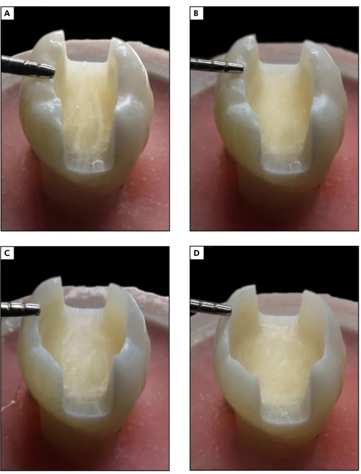
As a last consideration, the characteristics of the cav -ity design were as follows: MOD preparation, without a proximal box for better standardization; divergent internal walls with a 10º to 15º tilt; rounded internal angles; cavosurface angle with no bevel, 4 mm cavity depth from the cusp tips; and buccal and lingual sur -rounding wall thickness according to each group evalu -ated. The tooth wall thicknesses were determined using a digital caliper (Model 727; Starrett, Itu, Brazil), mea -suring the base of each surrounding wall and diving the walls into 4 groups, accordingly (Figure 1A - 1D): G1, 2.0 mm (± 0.1 mm); G2, 1.5 mm (± 0.1 mm); G3, 1.0 mm (± 0.1 mm); G4, 0.5 mm (± 0.1 mm).
The impression, design and milling of the ceramic inlays were performed using an in-ofice CAD/CAM system (Cerec 3D Software V. 3.03; Sirona Dental GmbH, Salzburg, Austria). The design of all the ceramic restorations was determined using the Bio-generic app of the CAD/CAM system. All the resto -rations were made from feldspathic ceramic blocks (Vitablocs Mark II; Vita Zahnfabrik, Bad Säckingen, Germany). After the milling process, the inlays were polished (OptraFine; Ivoclar Vivadent AG, Schaan, Liechtenstein) and their adaptation was checked. All ceramic restorations were cemented adhesively in the dual curing mode. The description of the adhe -sive systems, the cementing agent, and the silane and light-curing unit used in this study is provided in Table 1. After 24 hours stored in distilled water at 37 ºC, the inlay margins were polished (OptraFine; Ivoclar Vivadent AG, Schaan, Liechtenstein), and the inlays were subsequently stored in distilled water at 37 ºC for 7 days before submitting them to the fatigue test.
Figure 1. (A) G1: 2.0 mm. (B) G2: 1.5 mm. (C) G3: 1.0 mm. (D) G4: 0.5 mm.
D b a
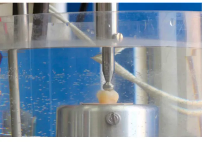
(Certi Foundation, Florianopolis, Brazil). The test was performed by placing the specimen on a metal platform attached to the dynamic testing machine. This platform consisted of a metal base, a resistor (with temperature control) and an acrylic cham -ber ixed to the base, which was illed with dis -tilled water for testing. The water temperature was maintained at 37 ºC to simulate oral temperature.11 After ixing the specimen in the platform irmly, a vertical load was placed directly on the occlu -sal surface, using a 6-mm-diameter metal sphere (Figure 2). Initially, a preload of 200 N for 5000 cycles was applied, using sinusoidal cyclic load -ing at 5 Hz. Physiologically, tooth contact dur-ing the masticatory cycle occurs in 0.25 to 0.33 seconds or 1.57 to 1.58 Hz, on average, taking into account the whole cycle.11,12 Nevertheless, in order to accel -erate the test, a cycle of 5 Hz was used.13 After the pre-load stage, all specimens were subjected to increasing loads of 400, 600, 800, 1000, 1200 and 1400 N for a maximum of 30,000 cycles each. The test was performed by completing the maximum cycles (185,000), or else fracture of the specimen.
The fracture mode was classiied according to the following criteria: Mode I, small fractures in tooth structure or ceramic; Mode II, fracture of one or more cusps, with fracture above the cementoe -namel junction; Mode III, longitudinal fracture compromising the integrity of the tooth or beyond the cementoenamel junction. Mode I and II were considered non-catastrophic failures, and restor -able, whereas Mode III was considered catastrophic and non-restorable.
Statistical analysis
The fatigue resistance of each group was com -pared statistically by the Kaplan-Meier survival curve, which considers the number of specimens that start the test in each group compared to those fractured throughout the test, and which forms a survival probability. The influence of the remain -ing prepared tooth wall thickness was analyzed by comparing the survival curves using a log-rank test at a 5% significance level. Statistical analy -sis was performed with SPSS Statistics 19 (IBM, New York, USA).
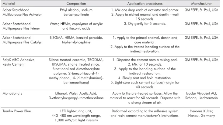
table 1. Adhesive system, resin cement and silane used, together with ingredients and application procedures, accord-ing to the manufacturers.
Material Composition Application procedures Manufacturer
Adper Scotchbond Multipurpose Plus Activator
Ethyl alcohol, sodium benzenesulfinate
1. Mix one drop each of activator and primer. 2. Apply to etched enamel and dentin – wait
15 seconds. 3. Dry gently for 5 seconds.
3M ESPE, St. Paul, USA
Adper Scotchbond Multipurpose Plus Primer
Water, HEMA, copolymer of acrylic and itaconic acids
3M ESPE, St. Paul, USA
Adper Scotchbond Multipurpose Plus Catalyst
BISGMA, HEMA, benzoyl peroxide, triphenylphosphine
1. Apply to the primed enamel, dentin and core material.
2. Apply to the treated bonding surface of the indirect restoration.
3M ESPE, St. Paul, USA
RelyX ARC Adhesive Resin Cement
Silane treated ceramic, TEGDMA, BISGMA, silane treated silica,
functionalized dimethacrylate polymer, 2-benzotriazolyl-4-methylphenol,
4-(dimethylamino)-benzeneethanol
1. Dispense the cement onto a mixing pad. 2. Mix for 10 seconds.
3. Apply to the bonding surface of the indirect restoration. 4. Slowly seat and hold restoration. 5. Light-cure each cement surface/margin for
40 seconds.
3M ESPE, St. Paul, USA
MonoBond S Ethanol, Water, Acetic Acid, 3-ethacryloxypropyl-trimethoxysilane
Apply to the pre-treated surfaces. Allow the material to react for 60 seconds. Disperse with
a strong stream of air.
Ivoclar Vivadent AG, Schaan, Liechtenstein
Tranlux Power Blue LED light-curing unit, 440–480 nm wavelength range,
1,000 mW/cm light intensity
Performed according to the adhesive system and resin cement manufacturer’s instructions.
Results
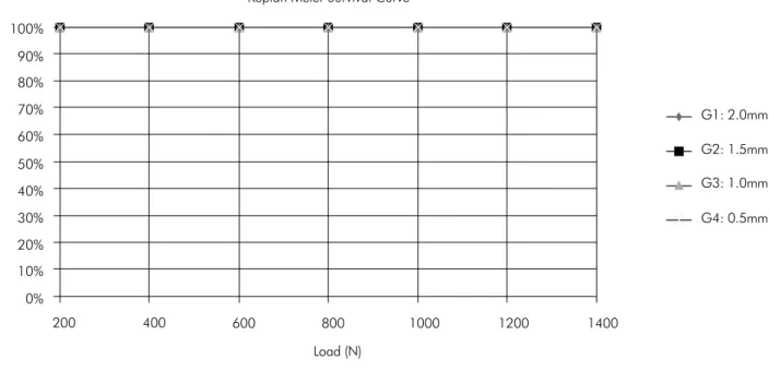
Because all specimens withstood the fatigue pro -tocol, there was no specimen fracture after 185,000 cycles, yielding a 100% survival rate, with no visible signs of failure in the ceramic or the tooth (Table 2). The Kaplan-Meier survival curves showed no differ -ence between the groups, considering that all speci -mens survived the fatigue protocol. It was assumed that all of the specimens were censored,i.e., they
reached the end of study without failing. Therefore, there were no data for post hoc log-rank testing to investigate the inluence of the remaining wall thick -ness on the survival rate (Figure 3).
Discussion
In the present study, no difference was found in the fatigue resistance of molars restored with CAD/ CAM ceramic inlays with 2.0 mm, 1.5 mm, 1.0 mm
or 0.5 mm surrounding wall thicknesses. Therefore, the null hypothesis that surrounding wall thickness does not affect the fatigue resistance of a tooth-res -toration complex was accepted. The current indings are in accordance with the suggestions by Stappert et al.,5 who propose that more studies be performed in
relation to inlay cavity design limits. This suggestion was made after the aforementioned authors assessed the fracture resistance of inlays, onlays and natural teeth, and found no statistical differences in the mean fracture resistance of the tested groups, even after they were submitted to 1.2 million loading cycles.
In the present study, the ceramic inlay thickness and volume varied between groups due to the cavity design – a variable that could inluence tooth strength in each group. However, the inite element analysis showed that the fracture risk of ceramic inlays was not associated as much with ceramic thickness as with ceramic type;
e.g., more rigid ceramics, such as lithium disilicate, have
a lower principal stress, ranging between 20.7 to 22.1 MPa. Conversely, leucite ceramic (a less rigid ceramic) had a greater principal stress, i.e., 27.6 to 29.2 MPa, even
when different thicknesses were tested.14
Compared to the aforementioned ceramics, the feldspar ceramics used in this study had lower values of lexural resistance.15 However, the controlled sin -tering process could account for the high resistance to propagation of the ceramic cracks. In this process, leucite crystals are heated until they become poly -morphic sanidine crystals of feldspar, which have a higher contraction rate upon cooling than the origi -nal crystals.15,16 These characteristics may have led, in part, to the behavior of the restorations observed in this study. Moreover, in ceramic restorations, higher stress concentrations occur on the ceramic restoration surface, as is the case of natural teeth. This characteristic protects the adhesive interface from compression or mastication stresses. Moreover, ceramic inlays can recover tooth structure rigidity better than resin composite restorations.17
Restorations in the oral cavity are predominantly subject to cyclic loading in a wet environment. This is the main cause for the development and growth of cracks, which reduce the strength of restorative materials, or lead to their failure.18,19 The development of a crack can be facilitated by pre-existing faults in table 2. Test groups, number of specimens, number of
fail-ures and specimen survival rate.
Groups Number of
Specimens Failures
Survival Rate
n Percent
G1 – 2.0 mm 10 0 10 100.0%
G2 – 1.5 mm 10 0 10 100.0%
G3 – 1.0 mm 10 0 10 100.0%
G4 – 0.5 mm 10 0 10 100.0%
Total 40 0 40 100.0%
the restoration, occurring during the manufacturing process, or pre-existing cracks in the tooth.20,21 Taking this into account, in-ofice CAD/CAM technology can fashion an indirect restoration in a single session or moment,22 avoiding new cracks during the temporiza
-tion period,20 and can provide restorations made from industrially manufactured ceramic or from composite blocks, which have minor internal laws, as compared with traditional laboratory restorations.21
Results obtained from randomized controlled clinical trials are the best option for comparing or evaluating biomaterials or restorative techniques; however, they present many variables, high costs and ethical issues.11,23 For these reasons, laboratory stud -ies that simulate conditions or situations that may be found clinically, such as fatigue tests using a servo-hydraulic machine, may be a suitable option for the irst and initial evaluation.11 In normal conditions, the chewing cycle varies from 0.2 to 1.5 Hz, and a masticatory load in molar regions is approximately 360 N; in addition, 1.2 million cycles correspond to 5 years of clinical function.5,8 One drawback of fol -lowing the standard guidelines for making labora -tory assessments is the time required to complete the test. Therefore, this study chose to follow a pro -tocol presented by Magne and Knezevic,13 adapted
from an original study by Fennis et al.24 This meth -odology is presented as a balance between the clas -sic fatigue tests with low loads for millions of cycles and the compression tests,25 insofar as the specimens were subjected to loads that were within physiologi -cal limits and that were of extrinsic origin, such as those occurring as a result of trauma.25
Although all of the specimens survived the pro -posed test, the results of this research should be ana -lyzed within its limitations. Krifka et al.9 demonstrated
that preparations with reduced cusps resulted in bet -ter marginal integrity and reduced crack formation than teeth without cusp reduction. Thus, it could be assumed that a higher number of cycles may result in statistically signiicant failures; however, the time required for carrying out the cyclic tests until com -plete fracture is a limitation in fatigue tests. It is a common occurrence to have no failures even after 1.2 million cyclic loads with low load intensity.8 The fatigue protocol performed in this study was chosen precisely for this reason; other studies using the same protocol found fractures in specimens.13,25
Nevertheless, there are other limitations to the present study design. It is possible that the load applied may not have been challenging enough to fracture the tested specimens. In a study performed
Load (N) Keplan Meier Survival Curve
90% 100%
80%
70%
60%
50%
40%
30%
20%
10%
0%
1400 1200
1000 800
600 400
200
Survival rate (%)
G1: 2.0mm
G2: 1.5mm
G3: 1.0mm
G4: 0.5mm
by Saridag et al.,26 the mean fracture strength found in
molar teeth restored by a lithium-disilicate ceramic inlay was 2646.7 (± 360.4) N. Thus, further studies are warranted to determine the inluence of more chal -lenging situations than those applied in the pres -ent study, including the use of higher loads, of test groups with non-restored teeth or inlays cemented with non-adhesive cements, and of test groups with the same wall thickness, but with cusp reduction.
Conclusion
Within the limitations of this study, it may be con -cluded that remaining tooth wall thickness did not
inluence the fatigue resistance of molars restored with CAD/CAM ceramic inlays.
acknowledgements
We would like to thank Dr. Med. Ari Digiacomo Ocampo Moré and technician Daniel João Lúcio for their signiicant contributions to the undertaking and analysis of this study. The authors also wish to acknowledge Laboratório de Engenharia Biomecânica do Hospital Universitário (LEBm - UFSC) for provid -ing the lab facility. The present study was supported in part by the Conselho Nacional de Desenvolvimento
Cientíico (CNPq), grant no. 134164/2011-3.
1. Jackson RD. Indirect resin inlay and onlay restorations: a comprehensive clinical overview. Pract Periodontics Aesthet Dent. 1999 Oct;11(8):891-900; quiz 902.
2. Edelhoff D, Sorensen JA. Tooth structure removal associated with various preparation designs for posterior teeth. Int J Periodontics Restorative Dent. 2002 Jun;22(3):241-9. 3. Krämer N, Ebert J, Petschelt A, Frankenberger R. Ceramic
inlays bonded with two adhesives after 4 years. Dent Mater. 2006 Jan;22(1):13-21.
4. Zimmer S, Göhlich O, Rüttermann S, Lang H, Raab WHM, Barthel CR. Long-term survival of Cerec restorations: a 10-year study. Oper Dent. 2008 Oct;33(5):484-7.
5. Stappert CFJ, Abe P, Kurths V, Gerds T, Strub JR. Masticatory fatigue, fracture resistance, and marginal discrepancy of ceramic partial crowns with and without coverage of com -promised cusps. J Adhes Dent. 2008 Feb;10(1):41-8.
6. Fonseca RB, Fernandes-Neto AJ, Correr-Sobrinho L, Soares CJ. The influence of cavity preparation design on fracture strength and mode of fracture of laboratory-processed composite resin restorations. J Prosthet Dent. 2007 Oct;98(4):277-84.
7. Morimoto S, Vieira GF, Agra CM, Sesma N, Gil C. Fracture strength of teeth restored with ceramic inlays and overlays. Braz Dent J. 2009;20(2):143-8.
8. Stappert CFJ, Att W, Gerds T, Strub JR. Fracture resis -tance of different partial-coverage ceramic molar res -torations: an in vitro investigation. J Am Dent Assoc. 2006 Apr;137(4):514-22.
9. Krifka S, Stangl M, Wiesbauer S, Hiller K-A, Schmalz G, Federlin M. Influence of different cusp coverage methods for the extension of ceramic inlays on marginal integrity and enamel crack formation in vitro. Clin Oral Investig. 2009 Sep;13(3):333-41.
References
10. Banks RG. Conservative posterior ceramic restorations: a literature review. J Prosthet Dent. 1990 Jun;63(6):619-26. 11. DeLong R, Douglas WH. An artificial oral environ
-ment for testing dental materials. IEEE Trans Biomed Eng. 1991 Apr;38(4):339-45.
12. Po JMC, Kieser JA, Gallo LM, Tésenyi AJ, Herbison P, Farella M. Time-frequency analysis of chewing activity in the natu -ral environment. J Dent Res. 2011 Oct;90(10):1206-10. 13. Magne P, Knezevic A. Simulated fatigue resistance of com
-posite resin versus porcelain CAD/CAM overlay restora -tions on endodontically treated molars. Quintessence Int. 2009 Feb;40(2):125-33.
14. 14. Holberg C, Rudzki-Janson I, Wichelhaus A, Winterhalder P. Ceramic inlays: Is the inlay thickness an important factor influencing the fracture risk?. J Dent. 2013 Jul;41(7):628-35. 15. Deany IL. Recent advances in ceramics for dentistry. Crit Rev
Oral Biol Med. 1996;7(2):134-43.
16. Boushell LW, Ritter AV. Ceramic inlays: a case presentation and lessons learned from the literature. J Esthet Restor Dent. 2009;21(2):77-87.
17. Magne P, Belser UC. Porcelain versus composite inlays/ onlays: effects of mechanical loads on stress distribution, adhesion, and crown flexure. Int J Periodontics Restorative Dent. 2003 Dec;23(6):543-55.
18. Kelly JR. Clinically relevant approach to failure testing of all-ceramic restorations. J Prosthet Dent. 1999 Jun;81(6):652-61. 19. Ohyama T, Yoshinari M, Oda Y. Effects of cyclic loading
on the strength of all-ceramic materials. Int J Prosthodont. 1999 Feb;12(1):28-37.
21. Giordano R. Materials for chairside CAD/CAM-produced restorations. J Am Dent Assoc. 2006 Sep;137 Suppl:14S-21S. 22. Mörmann WH. The evolution of the CEREC system. J Am
Dent Assoc. 2006 Sep;137 Suppl:7S-13S.
23. Anusavice KJ, Kakar K, Ferree N. Which mechanical and physical testing methods are relevant for predicting the clini -cal performance of ceramic-based dental prostheses?. Clin Oral Implants Res. 2007 Jun;18 Suppl 3:218-31.
24. Fennis WMM, Kuijs RH, Kreulen CM, Verdonschot N, Creugers NHJ. Fatigue resistance of teeth restored with
cuspal-coverage composite restorations. Int J Prosthodont. 2004 Jun;17(3):313-7.
25. Schlichting LH, Maia HP, Baratieri LN, Magne P. Novel-design ultra-thin CAD/CAM composite resin and ceramic occlusal veneers for the treatment of severe dental erosion. J Prosthet Dent. 2011 Apr;105(4):217-26.