Streptococcus mutans
counts in plaque
adjacent to orthodontic brackets bonded
with resin-modified glass ionomer
cement or resin-based composite
Abstract: This study investigated the number of Streptococcus mutans CFU (colony forming units) in the saliva and plaque adjacent to orth-odontic brackets bonded with a glass ionomer cement – GIC (Fuji Ortho) or a resin-based composite – RC (Concise). Twenty male and female pa-tients, aged 12 to 20 years, participated in the study. Saliva was collected before and after placement of appliances. Plaque was collected from ar-eas adjacent to brackets and saliva was again collected on the 15th, 30th,
and 45th day after placement. On the 30th day, 0.4% stannous luoride gel
was applied for 4 minutes. No signiicant modiication in the number of Streptococcus mutans CFU in saliva was observed after placement of the ixed orthodontic appliances. On the 15th day, the percentage of
Strep-tococcus mutans CFU in plaque was statistically lower in sites adjacent to GIC-bonded brackets (mean = 0.365) than in those adjacent to RC-bonded brackets (mean = 0.935). No evidence was found of a contribu-tion of GIC to the reduccontribu-tion of CFU in plaque after the 15th day. Topical
application of stannous luoride gel on the 30th day reduced the number
of CFU in saliva, but not in plaque. This study suggests that the antimi-crobial activity of GIC occurs only in the initial phase and is not respon-sible for a long-term anticariogenic property.
Descriptors:Streptococcus mutans; Orthodontic appliances; Glass ionomer cements; Tin luorides.
Solange Machado Mota(a)
Carla Enoki(b)
Izabel Yoko Ito(c)
Ana Maria Elias(d)
Mírian Aiko Nakane Matsumoto(e)
(a)Orthodontics Specialist; (b)MSc, Professor
of Orthodontics, Department of Pediatric Dentistry; (e)PhD, Professor of Orthodontics,
Department of Pediatric Dentistry – School of Dentistry of Ribeirão Preto, University of São Paulo.
(c)PhD, Professor of Microbiology, Department
of Clinical Analysis, Toxicology and Bromatology, School of Pharmacy of Ribeirão Preto, University of São Paulo.
(d)PhD, Professor of Statistics, College
of Science and Letters of Araraquara, University of São Paulo.
Corresponding author:
Mírian Aiko Nakane Matsumoto Departamento de Clínica Infantil,
Odontologia Preventiva e Social Faculdade de Odontologia de
Ribeirão Preto - USP Avenida do Café, s/n Ribeirão Preto - SP - Brazil CEP: 14040-904
E-mail: [email protected]
Introduction
Enamel decalciication during orthodontic treat-ment is a serious clinical problem, in particular in patients with poor oral hygiene habits. Recent stud-ies have reported that demineralization of dental surfaces during treatment can be found in 50 to 75% of all patients with ixed orthodontic appliances.1-3
Clinically, white-spot lesions can be seen around brackets. These lesions are incipient carious lesions that can be remineralized by application of luo-ride.4,5 Fluoride-releasing bonding materials and
ce-ments have been used because they reduce the need for patient compliance and potentially inhibit de-mineralization.1-3,6-9
The reduction in decalciication observed when brackets are bonded with luoride-containing mate-rials results from the slow and constant release of luoride, even if in small amounts, in the exact sites where there is higher cariogenic risk. Such reduction may also result from the changes caused on enamel surfaces by the initial higher concentrations of luo-ride.4,10,11
Fluoride-releasing bonding materials also stimu-late the development of a calcium luoride layer on enamel surfaces adjacent to brackets. This layer serves as a potential reserve of luoride, which slow-ly releases luoride ions during the demineralization and remineralization processes. It also acts as a bar-rier against acid challenge.4,12,13
The oral environment of orthodontic patients undergoes changes, such as pH reduction, larger number of sites available for Streptococcus mutans collection, and increased accumulation of food par-ticles, which may lead to an increased number of Streptococcus mutans colony-forming units (CFU) in saliva.14,15 Such changes may contribute to the
de-velopment of the decalciication lesions frequently found at the end of orthodontic treatments.14,16
Several studies have conirmed increases in the number of Streptococcus mutans in saliva during orthodontic treatment. However, these studies have not investigated the use of glass-ionomer cements (GIC) in the placement of orthodontic applianc-es.14,17
Furthermore, scarce data about the use of GIC in bracket bonding have been made available so
far. Studies in the literature report that luoride re-leased from restorations with conventional GIC18-21
and brackets bonded with GIC22-28 may have a
lo-cal cariostatic effect when cariogenic challenge is increased during orthodontic treatment. Therefore, white spots, which are signs of decalciication, may be prevented.
The purpose of this study was to investigate whether luoride released from GIC resulted in low-er Streptococcus mutans counts in plaque adjacent to GIC-bonded brackets than in that adjacent to brackets bonded with resin composites. This study also investigated the effect of a topical application of 0.4% stannous luoride gel.
Material and Methods
Following the approval of the research project by the Ethics Committee of the School of Dentistry of Ribeirão Preto, University of São Paulo (process # 2001.1.827.58.7), 20 male and female patients, aged 12 to 20 years, were selected from the group of pa-tients seen in the Orthodontics Clinic of that institu-tion.
To determine the number of Streptococcus mu-tans CFU, 2.0 ml of non-stimulated saliva were collected from each patient before placement of the appliance. The collected material was placed in prop-erly labeled 15 x 100 mm sterile tubes containing 4 to 5 glass beads. The samples were processed at the Microbiology Laboratory of the School of Pharmacy of Ribeirão Preto, University of São Paulo.
Placement of appliances was performed, and brackets were bonded with a resin-based composite (Concise, 3M-Unitek, Sumaré, São Paulo, Brazil) in one side of the dental arch, and with a GIC (GC Fuji Ortho LC, GC Corporation, Tokyo, Japan) in the other side. The bonding material used in each quadrant was: upper left = Fuji Ortho; upper right = Concise; lower left = Concise; and lower right = Fuji Ortho.
After the placement of appliances was complet-ed, saliva was collectcomplet-ed, and oral hygiene instruc-tions were given to the patient. On the 15th and 30th
maxil-lary lateral incisor, left mandibular central incisor, and right mandibular lateral incisor. Collection was performed continuously with a sterilized probe.
On the 30th day, 0.4% stannous luoride gel was
topically applied for 4 minutes. Saliva and plaque were collected for the last time on the 45th day.
The plaque removed was spread on 15 x 100 mm sterile test tubes containing 4 to 5 glass beads and 2.0 ml of phosphate buffered saline solution (PBS). The test tubes were sent to the laboratory for micro-biological processing.
Saliva and plaque samples were vortexed for two and one minutes, respectively, for dispersion, and submitted to tenfold serial dilutions. After that, 50Pl of each dilution was plated equidistantly on
SB20 agar (tryptone soy yeast agar plus 20% su-crose and 0.2 U/mL Bacitracin; Sigma – St. Louis, MO) and incubated under candle jar system at 37qC
for 2 to 3 days.
Statistical analysis
For statistical analysis, the original data, record-ed as CFU (colony-forming units), were transformrecord-ed into log10(CFU), and all results were expressed as log(CFU). To analyze statistically the materials’ ef-fect on CFU counts in the bioilm, teeth with brack-ets retained with the same material in a same patient were considered as repetitions of measurements. Therefore, log(CFU) was calculated considering the means of CFU counts for brackets bonded with the same material.
The hypothesis of equality of log(CFU) means in saliva in the beginning of the treatment and at the three moments of bioilm collection (i.e., 15, 30 and 45 days after installation of the orthodontic ap-pliances) was tested using the Hotelling’s T2 mul-tivariate test for comparison of means of repeated measurements. To test the hypothesis that the resin-modiied GIC presents greater protection against Streptococcus mutans in the bioilm only in the beginning of the treatment, log(CFU) means in the bioilm adjacent to both materials were compared at each moment, separately. Paired Student’s t-test was used to test the hypothesis of equality between the materials regarding log (CFU) means in the bioilm, at each moment of collection.
Results
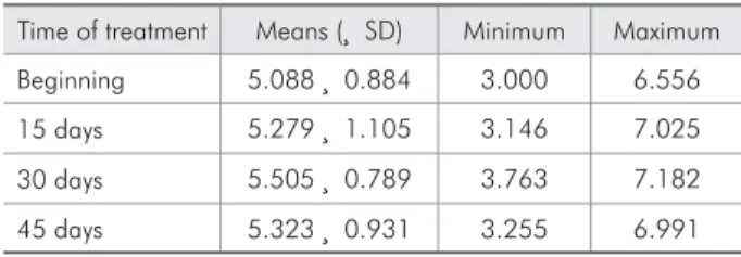
Streptococcus mutans CFU counts in saliva (in the beginning of the treatment and 15, 30 and 45 days after installation of the orthodontic applianc-es) showed that all patients presented moderate to high caries risk throughout the evaluation period (Table 1). Hotelling’s T2 multivariate test did not show statistically signiicant differences among the log(CFU) means in saliva at any of the collection moments (F= 2.298; gl= 3 and 15; p = 0.119), which means that there was no evidence of remarkable al-terations in the patients’ saliva with respect to the CFU counts throughout the study.
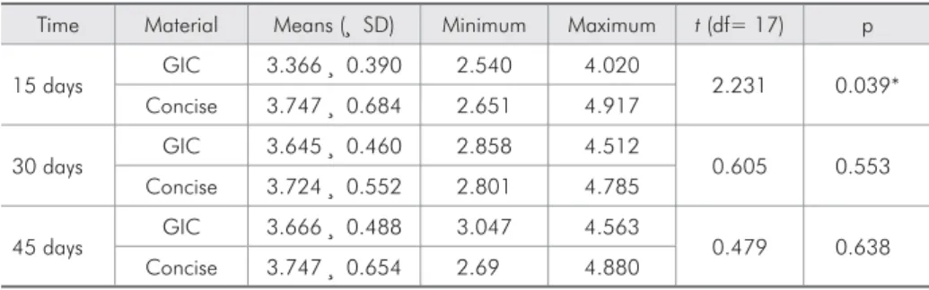
Regarding the effect of the tested materials on CFU formation, it was observed that log(CFU) means in the bioilm adjacent to Fuji Ortho LC 15 days after the beginning of the treatment was sig-niicantly lower than the log(CFU) means in the bio-ilm adjacent to Concise (t= 2.23ª; df= 17; p = 0.039) (Table 2). The results also revealed that there were no statistically signiicant differences among the log(CFU) means in the bioilm of brackets retained with Concise and Fuji Ortho LC, at 30 and 45 days after placement of the appliances. Log(CFU) and the respective means in the bioilm of teeth bonded with Concise and Fuji Ortho LC at the three moments of collection are given in Graph 1.
Discussion
The most important reason to use luoride-con-taining materials is their anticariogenic activity. Such activity can be monitored by means of longi-tudinal studies about luoride release, luoride ef-fects on demineralization, the quality and amount
Table 1 - Means (± SD), minimum and maximum Strepto-coccus mutans log10(CFU) values in saliva, at the beginning of treatment and 15, 30 and 45 days after placement of the appliances.
Time of treatment Means (r SD) Minimum Maximum
Beginning 5.088r 0.884 3.000 6.556
15 days 5.279r 1.105 3.146 7.025
30 days 5.505r 0.789 3.763 7.182
45 days 5.323r 0.931 3.255 6.991
of plaque, or, inally, the occurrence of secondary caries adjacent to bonding material.
Streptococcus mutans is also found on healthy surfaces, and its presence does not always indicate the presence of active caries. However, an increased number of these microorganisms on any surface in-dicates that disease may be present or may develop in the near future.14
Clinical evaluations of GIC have yielded contro-versial results. Some evidence points towards the re-duction of the risk of caries on tooth surfaces adja-cent to glass ionomer restorations.18,19,21,27
After orthodontic treatment and before applianc-es were removed (mean: 9.5 months), the percentage of total Streptococcus mutans CFU found in bacte-rial plaque was lower in areas adjacent to brackets bonded with GIC than in those adjacent to surfaces bonded with a resin composite.27
In our clinical study, statistically signiicant dif-ferences between the two types of material used to bond brackets were found only on the 15th day after
placement of the appliance, when there was a
signif-icant reduction in the number of CFU in the plaque collected from areas adjacent to brackets bonded with resin-modiied GIC. No signiicant differ-ence was found at the other times. Matalon et al.26
(2005) related that reinforced GIC (Fuji Ortho LC) exhibited potent antibacterial activity, which lasted 1 week and diminished over the next 3 weeks.
Fluoride release from GIC was directly associ-ated with its antimicrobial activity, that is, when pH is close to neutral (7.1 – 7.3) and the amount of luo-ride is 140r 25 ppm. Therefore, GIC may be
effec-tive for a short period of time, maybe for only a few days, as shown in our study.28
Fishman, Tinanoff23 (1994) found no association
between the amount of luoride released and antimi-crobial activity of resin-modiied GIC in vitro. On the contrary, the bacterial growth inhibiting effect seemed to be associated with GIC acid release. The reduction in resin-modiied GIC pH and the size of bacterial growth inhibition areas are consistently associated. The largest amount of acid release from resin-modiied GIC and its greatest antimicrobial activity are found immediately after the material is used. As time passes, less acid is released and bacte-rial growth inhibition decreases. A reduced inhibit-ing effect for Streptococcus mutans seems to be as-sociated with the fact that these microorganisms are acid-tolerant.
After application of luoride on old GIC illings, there was no signiicant increase in luoride con-centration on bacterial plaque.24 Our results are in
agreement with the indings of the study cited above – the topical application of stannous luoride gel in our study did not signiicantly reduce the number of Streptococcus mutans CFU in plaque. In contrast,
Time Material Means (r SD) Minimum Maximum t (df= 17) p
15 days GIC 3.366
r 0.390 2.540 4.020
2.231 0.039*
Concise 3.747r 0.684 2.651 4.917
30 days GIC 3.645
r 0.460 2.858 4.512
0.605 0.553
Concise 3.724r 0.552 2.801 4.785
45 days GIC 3.666
r 0.488 3.047 4.563
0.479 0.638
Concise 3.747r 0.654 2.69 4.880
*Significance level: p < 0.05.
Table 2 - Descriptive statistics
of log10(CFU) values in biofilm
for type of material and time of treatment and paired Student’s
t-test for comparison of means
15, 30 and 45 days after placement of the appliances.
0 1 2 3 4 5 6
lo
g(
C
FU
)
15 days 30 days 45 days Concise GIC Concise GIC Concise GIC
means
there were statistically signiicant differences in the number of Streptococcus mutans CFU in the sa-liva of patients after luoride application. However, Corry et al.22 (2003) concluded that luoride release
from Fuji Ortho LC alone fell to minimal values, but with daily addition of extrinsic luoride the lev-els fell initially and then followed an upward trend.
Growth of Streptococcus mutans in plaque was inhibited in vivo in areas adjacent to restorations with conventional GIC and with glass-ionomer silver cement.19,20,29 However, Van Dijken et al.15
(1991) did not ind any reduction in Streptococcus mutans or Lactobacilli in plaque collected from 1-year-old GIC restorations.
Our study found a smaller number of Strepto-coccus mutans CFU in plaque adjacent to brackets bonded with resin-modiied GIC than in plaque ad-jacent to resin composite only on the 15th day after
placement of the appliance. The resin-modiied GIC antimicrobial activity fell along time, and was not reestablished when 0.4% stannous luoride gel was topically applied on the 30th day.
The antimicrobial activity of resin-modiied GIC was only observed in the initial phase of the study, for a short period of time; it does not, thus, seem to be responsible for resin-modiied GIC’s anticario-genic activity in the long run. Gorton, Featherstone10
(2003) found, in an in vivo study, that the resin-modiied GIC’s cariostatic effect around brackets could be observed up to 4 weeks after placement. Maybe resin-modiied GIC activity in the process of caries development is associated with its slow ride release, which results in the presence of luo-ride in enamel or in plaque luid during the soluble phase. Therefore, it may inhibit demineralization
and promote remineralization in sites of highest car-iogenic risk.3
Fluoride activity in remineralization is a result of luoride’s behavior as a catalyst, which is capable of lowering the activation energy required for crystal growth.5 The size and electrostatic charge of
luo-ride permit a more favorable three-dimensional ar-rangement of calcium and phosphate on the crystal surface.
Further in vivo studies about the long-term ef-fect of resin-modiied GIC on plaque should attempt to deine resin-modiied GIC’s actual antimicrobial effect, as well as its magnitude, and how it affects bacterial cells. Although several synergistic and an-tagonistic factors affect clinical studies about the ef-fect of a material on bacterial growth, only clinical studies are able to provide an explanation for the cariostatic potential of a material.
Conclusion
After placement of ixed orthodontic appliances, a signiicant modiication in the number of Strepto-coccus mutans CFU in saliva was not observed. The number of Streptococcus mutans CFU in plaque adjacent to brackets bonded with resin-modiied GIC was smaller than in plaque adjacent to brackets bonded with resin-based composite only on the 15th
day after placement of the appliance. Topical appli-cation of 0.4% stannous luoride gel on the 30th day
did not affect the number of Streptococcus mutans in plaque; the number of microorganisms in saliva, however, was reduced. This study suggests that the antimicrobial activity of resin-modiied GIC occurs only on the initial phase and is not responsible for a long-term cariostatic potential.
References
1. Ögaard B, Arends J, Helseth H, Dijkman G, Van Der Kuijl M. Fluoride level in saliva after bonding orthodontic brackets with a fluoride containing adhesive. Am J Orthod Dentofacial Orthop. 1997;111(2):199-202.
2. Ögaard B, Rölla G, Arends J. Orthodontic appliances and enamel demineralization. Part 1. Lesion development. Am J Orthod. 1988;94(1):68-73.
3. Ögaard B, Rölla G, Arends J, Ten Cate JM. Orthodontic appliances and enamel demineralization. Part 2. Prevention
and treatment of lesions. Am J Orthod Dentofacial Orthop. 1988;94(2):123-8.
4. Basdra EK, Huber H, Komposch G. Fluoride released from orthodontic bonding agents alters the enamel surface and inhibits enamel demineralization in vitro. Am J Orthod Den-tofacial Orthop. 1996;109(5):466-72.
6. Geiger AM, Gorelick L, Gwinnet AJ, Griswold PG. The ef-fect of a fluoride program on white spot formation during orthodontic treatment. Am J Orthod. 1988;93(1):29-37. 7. Papagiannoulis L, Kakaboura A, Eliades G. In vivo vs in vitro
anticariogenic behavior of glass-ionomer and resin composite restorative materials. Dent Mater. 2002;18(8):561-9. 8. Wiltshire WA, Janse Van Rensburg S. Fluoride release from
visible light-cured orthodontic adhesive resins. Am J Orthod Dentofacial Orthop. 1995;108(3):278-83.
9. Zachrisson BU. Fluoride application procedures in orthodon-tic pracorthodon-tice: current concepts. Angle Orthod. 1975;45(1):72-81.
10. Gorton J, Featherstone JD. In vivo inhibition of demineraliza-tion around orthodontic brackets. Am J Orthod Dentofacial Orthop. 2003;123(1):10-4.
11. Pascotto RC, Navarro ME, Capelozza Filho L, Cury JA. In vivo effect of a resin-modified glass ionomer cement on enamel demineralization around orthodontic brackets. Am J Orthod Dentofacial Orthop. 2004;125(1):36-41.
12. Boeckh C, Schumacher E, Podbielski A, Haller B. Antibacte-rial activity of restorative dental biomateAntibacte-rials in vitro. Caries Res. 2002;36(2):101-7.
13. Rix D, Foley TF, Banting D, Mamandras A. A comparison of fluoride release by resin-modified GIC and polyacid-modified composite resin. Am J Orthod Dentofacial Orthop. 2001;120(4):398-405.
14. Rosenbloom RG, Tinanoff N. Salivary Streptococcus mutans levels in patients before, during, and after orthodontic treat-ment. Am J Orthod Dentofacial Orthop. 1991;100(1):35-7. 15. Van Dijken J, Persson S, Sjöström S. Presence of
Streptococ-cus mutans and lactobacilli in saliva and on enamel, glass ionomer cement, and composite resin surfaces. Scand J Dent Res. 1991;99(1):13-9.
16. Stratemann MW, Shannon IL. Control of decalcification in orthodontic patients by daily self-administered application of a water-free 0.4 per cent stannous fluoride gel. Am J Orthod. 1974;66(3):273-9.
17. Vierrou AM, Manwell MA, Zamek RL, Sachdeva R, Tinanoff N. Control of Streptococcus mutans with topical fluorides in patients undergoing orthodontic treatment. J Am Dent Assoc. 1986;113(4):644-6.
18. Qvist V, Laurberg L, Pousen A, Teglers PT. Longevity and cariostatic effects of everyday conventional glass ionomer and amalgam restorations in primary teeth: three-year results. J Dent Res. 1997;76(7):1387-96.
19. Svanberg M. Class II amalgam restorations, glass ionomer tun-nel restorations, and caries development on adjacent tooth sur-faces: a three-year clinical study. Caries Res. 1992;26(4):315-8.
20. Svanberg M, Krasse B, Örnerfeldt HO. Mutans streptococci in interproximal plaque from amalgam and glass ionomer restorations. Caries Res. 1990;24(2):133-6.
21. Tyas MJ. Cariostatic effect of glass ionomer cement: a five-year clinical study. Aust Dent J. 1991;36(3):236-9.
22. Corry A, Millett DT, Creanor SL, Foye RH, Gilmour WH. Effect of fluoride exposure on cariostatic potential of ortho-dontic bonding agents: an in vitro evaluation. J Orthod. 2003;30(4):323-9.
23. Fishman SA, Tinanoff N. The effect of acid and fluoride re-lease on the antimicrobial properties of four glass ionomer cements. Pediatr Dent. 1994;16(5):368-70.
24. Forss H, Jokinen J, Spets-Happonen S, Seppä L, Luoma H. Fluoride and mutans streptococci in plaque grown on glass ionomer and composite. Caries Res. 1991;25(6):454-8. 25. Friedl KH, Schmalz G, Hiller KA, Shams M. Resin-modified
glass ionomer cements: fluoride release and influence on Strep-tococcus mutans growth. Eur J Oral Sci. 1997;105(1):81-5. 26. Matalon S, Slutzky H, Weiss EI. Antibacterial properties of
4 orthodontic cements. Am J Orthod Dentofacial Orthop. 2005;127(1):56-63.
27. Örtendahl T, Thilander B, Svanberg M. Mutans Streptococci and incipient caries adjacent to glass ionomer cement or resin-based composite in orthodontics. Am J Orthod Dentofacial Orthop. 1997;112(3):271-4.
28. Rodriguez JPL, Godoy FG, Lindquist R. Growth inhibition of glass ionomer cements on mutans streptococci. Pediatr Dent. 1994;16(5):346-9.