113 Radiol Bras. 2012 Mar/Abr;45(2):113–117
Imaging evaluation of congenital cystic lesions of the biliary
tract
*
Avaliação por imagem das lesões císticas congênitas das vias biliares
Luis Ronan Marquez Ferreira de Souza1, Flávio Batista Rodrigues2, Lucas Vieira Tostes2, Graziella Borges Barreto2, Mateus Saldanha Cardoso3
Based on the classification of congenital choledochal cysts, images of different cyst types were selected to compose a pictorial essay about this disease, which is not frequently observed in the medical practice, but is included in the differential diagnosis of most common lesions of the biliary tract. Choledochal cysts present nonspecific symptoms and low prevalence. The correct diagnosis based on imaging findings is mandatory to avoid complications, and to aid in the approach and definite treatment.
Keywords: Magnetic resonance imaging; Choledochal cyst; Bile ducts.
Utilizando a classificação dos cistos congênitos de colédoco, imagens de vários tipos de cistos foram selecionadas para compor um ensaio iconográfico sobre esta doença pouco frequente na prática clínica, mas que faz parte do diag-nóstico diferencial das lesões mais comuns da via biliar. Os cistos de colédoco possuem clínica inespecífica e baixa prevalência. O diagnóstico correto por meio dos exames de imagem é uma ferramenta importante para evitar as com-plicações e auxiliar na conduta e tratamento definitivo.
Unitermos: Ressonância magnética; Cisto do colédoco; Ductos biliares.
Abstract
Resumo
* Study developed at the Division of Radiology and Imaging Diagnosis of Faculdade de Medicina da Universidade Federal do Triângulo Mineiro (UFTM), Uberaba, MG, Brazil.
1. PhD, Professor at Universidade Federal do Triângulo Mineiro (UFTM), Uberaba, MG, Brazil.
2. Graduate Students of Medicine, Universidade Federal do Triângulo Mineiro (UFTM), Uberaba, MG, Brazil.
3. MD, Resident of Radiology and Imaging Diagnosis, Univer-sidade Federal do Triângulo Mineiro (UFTM), Uberaba, MG, Bra-zil.
Mailing Address: Dr. Luís Ronan Marquez Ferreira de Souza. Disciplina de Radiologia e Diagnóstico por Imagem – UFTM. Avenida Frei Paulino, 30, Bairro Abadia. Uberaba, MG, Brazil, 38025-180. E-mail: [email protected]
Received July 22, 2011. Accepted after revision January 30, 2012.
Souza LRMF, Rodrigues FB, Tostes LV, Barreto GB, Cardoso MS. Imaging evaluation of congenital cystic lesions of the biliary tract. Radiol Bras. 2012 Mar/Abr;45(2):113–117.
juice reflux with consequential destruction of the mucosa, fibrosis and ductal dilata-tion(1). However, congenital anomalies of
the pancreaticobiliary junction are more frequently found that choledochal cysts. So, most probably, cysts development oc-curs due to an association of multiple fac-tors, and not only because the abnormal development of the ductal system.
DIAGNOSIS
Ultrasonography is the primary method to be utilized in the evaluation of the bil-iary tree, considering its noninvasiveness, swiftness, low cost and wide availability. At ultrasonography, a well defined cystic lesion with variable echogenicity (gener-ally hypoechoic) is observed. The relation-ship between the lesion and the gallblad-der and the biliary tree, as well as the cleav-age planes, must be carefully assessed. Color Doppler can be useful in the differ-entiation of adjacent vascular structures such as the portal vein.
During endoscopic retrograde cholan-giopancreatography (ERCP), the endo-scope is passed and the Vater’s papilla is are most frequently reported, whether in
association or not with previous cholecys-tectomy(3,4). Because of such a clinical
nonspecificity, this condition is included in the differential diagnosis of the most com-mon biliary tract diseases(1).
Imaging methods such as magnetic resonance imaging (MRI) and ultrasonog-raphy (US) are indicated for an early diag-nosis of these cysts and their possible com-plications, besides aiding in a definitive surgical planning.
PATHOGENESIS
Although the pathogenesis of congeni-tal choledochal cysts is discussable, it is considered that most probably such abnor-mality results from abnormal development of the pancreaticobiliary ductal system(5),
as from the fourth gestational week. Such an abnormal development of the pancreaticobiliary ductal system is charac-terized by the extraduodenal junction of the choledochus with the pancreatic duct, out from the influence of the Oddi’s sphincter. The formed duct is longer and is located in a higher position, which causes pancreatic INTRODUCTION
Congenital choledochal cysts are anomalies involving cystic dilatation of any segment of the biliary tract, and are most frequently identified in the common bile duct. The clinical presentation of such con-dition is characterized by a triad of signs and symptoms including abdominal pain, presence of palpable mass and jaundice(1).
An accurate diagnosis is hardly achieved because this triad presents in only one third of patients(2).
characterized. Once the choledochus or pancreatic duct is reached, a contrast agent is injected into the biliary tract and the ra-diographic recording is concomitantly per-formed. Such imaging method was consid-ered as the gold standard until the 70’s decade, but currently it is most frequently utilized immediately before the surgical intervention(5) As ERCP images of patients
with cystic lesions in the biliary tract are analyzed, it is possible to observe relevant alterations in the lesion pathogenesis(6).
One of them is related to the location of the junction between the choledochus and the main pancreatic duct, which is far from the duodenum, creating a long common canal. Typically, such a junction forms an acute angle of 5°–30° but in cases of cystic le-sions this angle is greater and may be > 90°. Because of the necessity of general anes-thesia, ERCP is not indicated for the diag-nosis in children. Additionally, the possi-bility of causing pancreatitis and the limi-tation of the method in the proximal evalu-ation of the biliary tree, which tends to be abnormal, constitute negative points to be taken into consideration.
Magnetic resonance cholangiopan-creatography (MRCP) represents an alter-native for investigating the biliary tree anatomy, because it is noninvasive, does not require contrast injection and presents high accuracy in the identification of ana-tomical variations relevant for surgery(7).
Additionally, MRCP allows the analysis of the proximal biliary tract and the evaluation of the liver and pancreas, facilitating other differential diagnoses, particularly in cases where the study is complemented with MRI of the upper abdomen.
Computed tomography (CT) can also be utilized, particularly with those equipment which allow thinner section thicknesses such as multidetector apparatuses (multi-slice). With this method, the acquisition time is considerably shorter as compared with MRCP, which reduces motion arti-facts. On the other hand, it is associated with inherent risks such as adverse reac-tions to contrast and excessive radiation resulting from repeated exposure at short intervals. At CT, choledochal cysts are seen as fluid collections, with density similar to the gallbladder and peripheral contrast en-hancement of the cystic walls.
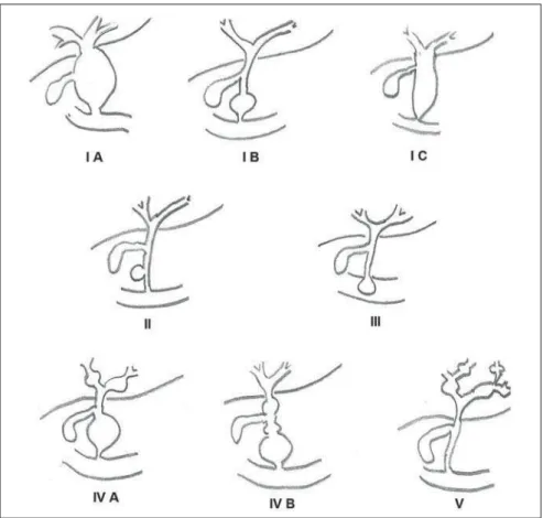
CLASSIFICATION
In the literature, the classification devel-oped by Todani (Figure 1)is the most uti-lized for congenital choledochal cysts and involves the analysis of morphology, loca-tion and number of intra- and extrahepatic cysts by means of cholangiography.
Type I cysts or classical choledochal cysts are restricted to extrahepatic bile
ducts, and are further subdivided into three cyst subtypes as follows: Ia (diffuse) – dif-fuse dilatation of the choledochal duct and expansion through extrahepatic bile ducts (Figures 2 to 5); Ib (focal) – focal dilata-tion of the choledochal duct, with no anomalous pancreaticobiliary junction; Ic (fusiform) – fusiform dilatation of the cho-ledochal duct associated with an anomalous pancreaticobiliary junction(2,8) (Figure 6).
Figure 2. Type Ia cyst. Right hypochondrium ultra-sonography demonstrating cystic lesion with well defined limits containing thin septa and hyperre-fringent echoes adjacent to the right hepatic lobe, in the gallbladder fossa.
Figure 1. Todani’s classification of congenital choledochal cysts.
Figure 3. Type Ia choledochal cyst. Contrast-en-hanced computed tomography demonstrating lobu-lated cystic lesion (arrow) with thin septa within the gallbladder fossa. The lesion is not enhanced after intravenous contrast injection.
The prevalence is higher among children and, among the diagnosed cases, 7% of cysts are detected during pregnancy. Imag-ing findImag-ings, whether at US or other meth-ods, demonstrate choledochus dilatations without any noticeable point of obstruction and with no intrahepatic ducts compro-mise. Pancreaticobiliary junction should be analyzed on thin section RMCP images for differentiation between cysts types Ia and Ic. At US, the cysts content appearance is variable and most frequently a finely het-erogeneous pattern with sparse fine debris is observed.
Type II cysts are supraduodenal diver-ticula which may be found either in the common bile duct or in the common he-patic duct. Generally, these cysts arise from the lateral wall of the duct while the rest of the biliary tract remains normal(2,8) (Figures
7 to 9). At MRI, a small saccular dilatation can be observed near the gallbladder, with well defined limits, hypersignal on T2-weighted images, and communicating with the supraduodenal bile duct.
Type III cysts (or choledochoceles) in-volve a dilatation of the intraduodenal common bile duct segment(2,8) (Figures 10
and 11).Choledochoceles clinically mani-fests, particularly in adults, as abdominal pain and jaundice complicated with cho-langitis and pancreatitis. At MRI, a promi-nence of the major papilla is observed with a signal similar to the one of the biliary tract, projecting towards the wall of the second portion of the duodenum, determin-ing a duodenal filldetermin-ing defect on contrast-enhanced T2-weighted sequences. Figure 4. Type Ia choledochal cyst. Abdominal magnetic resonance imaging – axial TSE T2-weighted sequence demonstrates cystic lesion with regular contours and hypersignal. Same patient on
Figures 2 and 3. Figure 6. Type Ic choledochal cyst. MR-cholang-iography image reconstruction demonstrating fusi-form choledochal dilatation (straight arrow) asso-ciated with anomalous pancreaticobiliary junction (curved arrow). Note the absence of dilatation of intrahepatic bile ducts (arrowheads).
Figure 7. Type II choledochal cyst. MR-cholangiog-raphy image reconstruction demonstrating diver-ticulum (arrow) arising from the lateral wall of com-mon hepatic duct, (Image kindly supplied by Doc-tor Carlos Matsumoto, Unifesp, São Paulo, SP, Bra-zil).
Figure 8. Type II choledochal cyst. Abdominal magnetic resonance imaging, coronal TSE T2-weighted image demonstrates the relation of a small saccular image (arrow) with hepatic duct and gallbladder. (Image kindly supplied by Doctor Carlos Matsumoto, Unifesp, São Paulo, SP, Brazil).
Figure 10. Type III cyst. Cholangiography through Kehr’s drain demonstrates saccular dilatation of dis-tal choledochus (arrow) adjacent to duodenal papilla (choledochoceles). (Image kindly supplied by Doc-tor Eduardo Crema, UFTM, Uberaba, MG, Brazil). Figure 5. Type Ia choledochal cyst. Volume
render-ing image demonstratrender-ing gallbladder (GB) and gi-ant choledochal cyst (arrow).
Type IV cysts are represented by saccu-lar formations in intra- or extrahepatic ducts, and are further subdivided into two subtypes, as follows: IVa – dilatation in-volving both intra- and extrahepatic bile ducts; and IVb – multiple saccular dilata-tions involving only extrahepatic bile ducts(2,8) (Figures 12 and 13). The main
differential on imaging studies is the iden-tification of more than one focus of saccu-lar dilatation, with choledochal compro-mise and dilatation, which differentiates such cyst type from Caroli’s disease.
Type V cysts (or Caroli’s disease) in-volve one or more segmental saccular di-latations of an intrahepatic bile duct which communicate to each other and affect large bile ducts(2,8) (Figures 14 and 15). It is a rare
autosomal recessive disorder which causes varying degrees of inflammation, degen-eration and dilatation of intrahepatic bile ducts resulting from a derangement in the normal embryological development. Caroli’s disease clinically manifests with cholangitis, fever, pain in the hypochon-drium and sometimes jaundice. Associa-tion with intrahepatic biliary calculi, cholangiocarcinoma and hepatic abscess is observed. A quite typical sign of Caroli’s disease is the central dot sign, which cor-responds to enhanced hepatic fibrovascu-lar bundles (hepatic artery and portal vein), protruding into the lumen of dilated intra-hepatic bile ducts.
Figure 14. Type V cyst. MR cholangiography im-age presenting multiple cystic dilatations (arrows) communicating with intrahepatic biliary tree. The common choledochal duct (arrowhead) presents preserved caliber. (Image kindly supplied by Doc-tor Dr. Ricardo Vital, de Uberlândia, MG, Brazil).
Figure 15. Type V cyst. Contrast-enhanced abdomi-nal magnetic resonance imaging, 3D T1-weighted sequence with fat suppression demonstrating seg-mental dilatation of intrahepatic bile ducts (arrow).
Furthermore, extremely rare cases of isolated dilatation of cystic duct are re-ported by Serena Serradel et al.(9) and
Yoon(10), who have suggested that, in the
future, this abnormality should be included in the classification as type VI cyst.
COMPLICATIONS
Most common complications from cho-ledochal cystic lesions include chocho-ledochal
lithiasis, carcinoma of gallbladder and duc-tus choledochus, chronic cholangitis, bil-iary obstruction, esophageal varices, cyst rupture, portal vein thrombosis and hepatic abscess.
Biliary stasis resulting from ductal stenosis, and pancreatic juice reflux as a consequence of the alteration of the junc-tion of the choledochus ductus with the main pancreatic duct, induce chronic irri-tation of the gallbladder epithelium and bile ducts. The progression of the chronic process of epithelial irritation may lead to metaplasia and also to dysplasia(11).
Malignant degeneration is most fre-quently observed in cyst types I, IV and V, Figure 13. Type IVb choledochal cyst. MR-cholan-giography image reconstruction demonstrates mul-tiple saccular formations only in extrahepatic ducts (straight arrow). Note the gallbladder (curved arrow) and the intrahepatic bile duct (arrowhead) with usual appearance. (Image kindly supplied by Doctor Giu-seppe D’Ippolito, Unifesp, São Paulo, SP, Brazil). Figure 12. Type IV, MR-cholangiography image
revealing marked intra- and extrahepatic bile duct dilatation (straight arrows). The intra-peripheral pathway (arrowhead) and gallbladder (curved arrow) are preserved. (Image source: Blasbalg R. Vesícula e vias biliares. In: Caldana RP, D’Ippolito G, editores associados. Gastrointestinal (Série Colégio Brasi-leiro de Radiologia e Diagnóstico por Imagem). 1ª ed. São Paulo, SP: Elsevier; 2011. p. 424). Figure 11. Type III cyst. MR-cholangiography
requiring lifelong follow-up even after complete surgical excision of the cyst(1,3).
Because of the risks and complications, surgical management of the choledochal cyst must be performed as soon as possible. Generally, there are two treatment options, namely, complete cyst resection and cyst drainage. Complete cyst excision is the treatment of choice, in spite of the disad-vantage of requiring bile duct reconstruc-tion.
CONCLUSION
The late diagnosis of congenital chole-dochal cysts results from the low frequency of this condition besides the nonspecificity of clinical signs and symptoms. Therefore, in the suspicion of a possible choledochal cyst, the time to the final diagnosis should be shortened with most appropriate
imag-ing methods such as US and MRCP. The radiologist should classify the cysts, be-sides identifying possible association with other congenital abnormalities and their complications which could eminently make the surgical planning more difficult.
REFERENCES
1. Edil BH, Olino K, Cameron JL. The current man-agement of choledochal cysts. Adv Surg. 2009; 43:221–32.
2. Mortelé KJ, Rocha TC, Streeter JL, et al. Multi-modality imaging of pancreatic and biliary con-genital anomalies. Radiographics. 2006;26:715– 31.
3. Matos C, Nicaise N, Devière J, et al. Choledochal cysts: comparison of findings at MR cholangio-pancreatography and endoscopic retrograde cho-langiopancreatography in eight patients. Radiol-ogy. 1998;209:443–8.
4. Mabrut JY, Bozio G, Hubert C, et al. Management of congenital bile duct cysts. Dig Surg. 2010;27: 12–8.
5. Babbitt DP, Starshak RJ, Clemett AR.
Chole-dochal cyst: a concept of etiology. Am J Roentgenol Radium Ther Nucl Med. 1973;119:57–62. 6. Kim OH, Chung JH, Choi BG. Imaging of the
cho-ledochal cyst. Radiographics. 1995;15:69–88. 7. Lee HK, Park SJ, Yi BH, et al. Imaging features
of adult choledochal cysts: a pictorial review. Korean J Radiol. 2009;10:71–80.
8. Todani T, Watanabe Y, Narusue M, et al. Congeni-tal bile duct cysts: classification, operative pro-cedures, and review of thirty-seven cases includ-ing cancer arisinclud-ing from choledochal cyst. Am J Surg. 1977;134:263–9.
9. Serena Serradel AF, Santamaría Linares E, Herrera Goepfert R. Cystic dilatation of the cys-tic duct: a new type of biliary cyst. Surgery. 1991; 109(3 Pt 1):320–2.
10. Yoon JH. Magnetic resonance cholangiopan-creatography diagnosis of choledochal cyst in-volving the cystic duct: report of three cases. Br J Radiol. 2011;84:e18–22.