Impact of environmental stress on biochemical parameters of bacteria reducing
chromium
Rida Batool
1,2, Kim Yrjälä
2, Shahida Hasnain
1 1Department of Microbiology and Molecular Genetics, University of the Punjab, Quaid-e-Azam Campus, Pakistan.
2
MEM-group, Department of Biosciences, University of Helsinki, Finland.
Submitted: February 18, 2013; Approved: September 9, 2013.
Abstract
Chromium pollution is produced in connection with industrial processes like in tanneries. It has been suggested that bioremediation could be a good option for clean up. The stress effect of variable chromate levels, pHs and growth temperatures on biochemical parameters of two Cr(VI) reducing bacterial strainsPseudomonas aeruginosaRb-1 andOchrobactrum intermediumRb-2 was investi-gated. Transmission electrone microscopy (TEM) was performed to study the intracellular distribu-tion of Cr(VI). It was observed that initial stress of 1000mgmL-1caused significant enhancement of all studied biochemical parameters at pH 7.0 and growth temperature of 37 °C showing great bioremediation potential of the strains. Transmission electron microscopy revealed that the distribu-tion of chromium precipitates was not uniform as they were distributed in the cytoplasm as well as found associated with the periplasm and outer membrane. Fourier transform infrared spectroscopy showed the possible involvement of carboxyl, amino, sulpohonate and hydroxyl groups present on the bacterial cell surface for the binding of Cr(VI) ions. Cr(VI) stress brought about changes in the distridution of these functional groups. It can be concluded that the investigated bacterial strains ad-just well to Cr(VI) stress in terms of biochemical parameters and along that exhibited alteration in morphology.
Key words:Cr(VI), pH, temperature, biochemical parameters, TEM, FTIR spectroscopy.
Introduction
Despite the fact that bacteria are resistant to a variety of compounds, they are at the same time sensitive to even
minute changes in the surrounding environment
(Weilharter et al., 2011). Stressful environmental condi-tions lead to a wide range of responses at the morphologi-cal, physiologimorphologi-cal, cellular and biochemical levels (Gustavs et al., 2009). The ability of bacterial strains to cope with sudden changes in the surrounding environment ensures their ecological dominance under stress conditions. Heavy metal pollution has turned into a major environmental problem and caused severe threats to environmental protec-tion and human health (Järup, 2003). Chromium is among the most hazardous heavy metals (Xuet al., 2009). Due to high solubility, Cr(VI) can easily pass across biological
membranes and exhibit a range of toxic effects evident at cellular and molecular levels (Poljsaket al., 2011). The much less soluble Cr(III) is less toxic. Bacteria adapt differ-ent strategies to combat high level stress along with trans-formation of Cr(VI) to Cr(III) either intra or extracellularlly (Cervantes and Campos-García, 2007). Chromium stress can induce many types of metabolic responses in living or-ganisms such as (a): increased production of metabolites e.g., peroxidase, auxin as a direct response to Cr stress, (b): alterations in the metabolism resulting in the production of new metabolites,e.g., glutathione, proline which may be responsible for resistance or tolerance to chromium stress (Nagajyotiet al., 2010). Heavy metal stress cause severe oxidative damage to biomolecules due to the production of reactive oxygen species (ROS). The high concentrations of ROS led to the disruption of the normal physiological and
Brazilian Journal of Microbiology 45, 2, 573-583 (2014) Copyright © 2014, Sociedade Brasileira de Microbiologia
ISSN 1678-4405 www.sbmicrobiologia.org.br
Send correspondence to R. Batool. Department of Microbiology and Molecular Genetics, University of the Punjab, Quaid-e-Azam Campus, La-hore-54590, Pakistan. E-mail: [email protected].
cellular functioning of the living cells. To combat with such stress, bacteria have developed certain enzymatic systems such as peroxidases (Panda and Choudhury, 2005). One of the major reasons of using the microbes for the control and remediation of metal polluted environment is their bio-chemical versatility, a result of their genetic plasticity and ability to modify physiology as to make them best competi-tor in a constantly changing environment (Murugesan and Maheswari, 2007). To understand the measures adapted by bacteria to cope with stress of hexavalent chromium, differ-ent biochemical parameters of chromium reducing
bacte-rial strains Pseudomonas aeruginosa Rb-1 and
Ochrobacterum intermediumRb-2 were analyzed. Trans-mission electron microscopy (TEM) was performed to lo-calize the distribution of chromium particles intra as well extra-cellularly.
Materials and Methods
Bacterial strains and growth conditions
Pseudomonas aeruginosa Rb-1 (FJ870126) and Ochrobactrum intermediumRb-2 (FJ870125),Gram nega-tive Cr(VI) reducing bacterial strains previously isolated from tannery effluent were obtained from bacterial stock cultures of Department of Microbiology and Molecular Ge-netics, University of the Punjab, Lahore, Pakistan. They were normally grown in Luria Bertani (LB) agar (pH 7.0) at 37 °C.
Biochemical analysis of bacteria reducing hexavalent chromium
Bacterial strains were aerobically grown in Luria Ber-tani (LB) broth supplemented with different hexavalent chromium concentrations (100, 500 and 1000mg mL-1of K2CrO4). Cultures were incubated at variable temperatures
(28, 37 and 40 °C) and pHs (5, 7 and 9) for 24 - 48 hours. Under aseptic conditions, harvesting of cells was done by centrifugation (5,000 x g for 10 min) at 4 °C. All the experi-ments were done in triplicates. Following biochemical pa-rameters of hexavalent chromium reducing bacteria were estimated.
Peroxidase activity and estimation of soluble protein
Peroxidase activity of bacterial strains was deter-mined according to Davy and Murry (1965). Briefly, har-vested bacterial cells were disrupted in cold 0.1 M phos-phate buffer (pH 7.0) by sonication for 5 min (Heilscher Ultrasonic Processors UP 400, S) at 4 °C. The ratio of buffer to bacterial pellet was 4:1 (v/w). The homogenate was centrifuged at 14,000 x g for 10 min. The supernatant was used for the estimation of enzyme peroxidase.
Formula used for peroxidase activity is as follows:
O. D of Test - O. D of Control O. D of Control
Weight
´ of bacterial
pellet (g)
where O.D = Optical density at 470 nm.
For extraction of soluble proteins, samples were pre-pared accordingly (Bhattiet al., 1993) whereas for soluble protein analysis version of Lowry’s method was adopted (Lowryet al., 1951). Amount of soluble proteins was calcu-lated from standard curve obtained by using Bovine serum albumin (BSA) as standard at wavelength of 750 nm on Beckman D-2 spectrophotometer.
Auxin biosynthesis
Auxin production by bacterial strains both in the pres-ence and abspres-ence of K2CrO4 was determined by using
Salkowski colorimetric technique (Glickmann and Des-saux, 1995). Auxin content was estimated by measuring the absorbance at 535 nm with Beckman D-2 spectrophoto-meter. Optical densities of various concentrations of in-dole-3-acetic acid (IAA) (standard) were also measured to construct a standard curve. From the standard curve the ac-tual amount of auxin was measured and calculated as mg gm-1fresh weight of bacteria.
Estimation of proline content
Proline was determined by the modified ninhydrin method (Derminal and Turkan, 2006). Harvested bacterial cells were suspended in 1 mL sterilized distilled water and placed in boiling water bath for 20 min to extract all water soluble compounds in hot water and cooled at room tem-perature. The bacterial suspension was then centrifuged at 13,000 x g for 5 min. The supernatant (200mL) was taken and 150mL distilled water and 1 mL of ninhydrin reagent was added in a test tube and placed in boiling water bath for one hour. The test tubes were cooled on ice to stop the reac-tion. Toluene (6 mL) was added by vigorous shaking and tubes vortexed for 20 seconds. The optical density of result-ing inorganic layer was measured at 520 nm with Beckman D-2 spectrophotometer. The amount of proline produced by bacteria was calculated from standard curve.
Estimation of nitrate reductase activity
Weighed bacterial pellet (1 g) was homogenized with 10 mL ice cold extraction buffer (0.1 M phosphate buf-fer, pH 7.5 containing 0.5 mM EDTA). The extract was fil-tered and stored on ice. 1 mM KNO3 , 0.1 mM NADH,
de-velopment. Optical density was measured at 540 nm with the Beckman D-2 spectrophotometer.
Analysis of nonprotein thiols and estimation of cysteine
Total GSH
(Gamma-L-glutamyl-L-cysteninyl-glycine) and GSSG [Bis (gamma-Glutamyl-L-cysteinyl-glycine) Disulfide] were measured by the GSSG recycling method, with GSSG as the standard (Satohet al., 2002).
Estimation of cysteine and cystine content (mM) of both the strains was done according to Gaitonde (1967) (Gaitonde, 1967).
Fourier Transform infrared spectral analysis
For the FTIR study, bacterial cell pellets were centri-fuged and lyophilized, followed by weighing. Then 20 mg of finely ground biomass was encapsulated in 200 mg of KBr (Sigma) in order to prepare translucent sample disks. The spectra of the lyopholized bacterial cell pellets were obtained by using PerkinElmer spectrum BX FTIR system (Beacon field Buckinghamshire HP9 1QA) equipped with diffuse reflectance accessory with the range of 500-4000 cm-1. All spectra were acquired in transmission mode, by the KBr disc method to get the information specific to the functional groups.
Electron microscopy
The samples for thin-sectioning were prepared as de-scribed (Lounatmaa, 1985). Briefly, the samples were pre-fixed in 2.5% phosphate-buffered glutaraldehyde (pH 7.2) with or without tannic acid for 2 hours at room temperature. The fixed cells were washed three times with phosphate buffer. All samples were post-fixed with phosphate-buf-fered 1% osmium tetra oxide and dehydrated in acetone se-ries and embedded in Taab resin. The thin-sectioned cells were post-stained with uranyl acetate and lead citrate. The samples were viewed using a transmission electron micro-scope (JEM-1200EX), operated at 60 kV.
Statistical analysis
Data was statistically analyzed using SPSS personal computer statistical package (version 16, SPSS Inc, Chi-cago). Analysis of variance (ANOVA) was performed and then means were separated using Duncan’s multiple range test (p = 0.05).
Results
Effect of chromate
There was a significant increase in all the studied bio-chemical parameters (except nitrate reductase) of both the strains with the increase in initial Cr(VI) concentration (Ta-ble 1). At low level of chromate (100 and 500mg mL-1), the soluble protein content ofO. intermediumRb-2 (42.56 and 55.63 mg g-1 cells fresh weight, respectively) was higher
biochemical attributes and Cr(VI) stress 575
thanP. aeruginosaRb-1 (29.2 and 44.37 mg g-1cells fresh weight, respectively) and maximum soluble protein content was also recorded withO. intermediumRb-2 (67.25 mg g-1 cells fresh weight) at 1000mg mL-1of Cr(VI). Hexavalent chromium stress lead to increase in peroxidase activity in both strains. The enzyme activity increased gradually with increasing Cr(VI) concentration for P. aeruginosa Rb-1 that exhibited peroxidase activity of 54.6; 65.64 and 95.34mg g-1cells fresh weight at 100, 500 and 1000 mg mL-1of chromate, respectively compared to control. On the other hand,O. intermediumRb-2 did not show the same pattern of peroxidase activity. It was 50.02; 49.13 and 98.24mg g-1cells fresh weight at increasing concentration of Cr(VI). Auxin content increased with the increasing chromate levels in both strains.O. intermediumRb-2 was more efficient in auxin production than P. aeruginosa Rb-1. At 1000mg mL-1of chromate,O. intermediumRb-2 produced 120.36mg g-1cells fresh weight auxin whereasP. aeruginosaRb-1 produced 84.59mg g-1cells fresh weight. Proline content also increased with higher Cr(VI) concen-tration in both strains relative to control. At 1000mg mL-1 of Cr(VI), P. aeruginosa Rb-1 exhibited a maximum proline content of 988.12mg g-1cells fresh weight andO. intermediumRb-2 revealed proline content of 999.51mg g-1 when compared to respective chromate free control. Nitrate reductase activity of both strains decreased with rising K2CrO4in contrast to the other measured biochemical
pa-rameters.O. intermediumRb-2 had higher nitrate reductase activity than P. aeruginosa Rb-1 at 1000 mg mL-1 of chromate (Table 1).
The content of non-protein thiols (cystine, cysteine, GSH and GSSG) in O. intermediumRb-2 was generally higher than inP. aeruginosaRb-1 under chromate stress with the exception of cystine. At 1000mg mL-1of chromate, O. intermedium Rb-2 exhibited enhanced production of cystine, cysteine, GSH and GSSG content, 4.36 mM, 6.22 mM, 59.18 nM and 70.55 nM, respectively. P. aeruginosaRb-1 also showed enhanced cystine, cysteine, GSH and GSSG content (5.12 mM, 4.53 mM, 22.28 nM and 46.00 nM, respectively) under 1000mg mL-1of K2CrO4
over respective chromate free control (Table 1).
Effect of growth pHs
Generally, pH 7 was optimum for causing increment in biochemical parameters of both strains in chromate sup-plemented conditions compared to chromate free condi-tions. Cr(VI) stress manifested a maximum increase for all the studied biochemical parameters at all growth pHs by both bacterial strains revealed by t- test at p = 0.05. The sol-uble protein content of strainsi.e. P. aeruginosaRb-1 and O. intermedium Rb-2 was highest at pH 7 under both chromate free and chromate supplemented conditions. Sol-uble protein content ofP. aeruginosaRb-1 was found to be 15.51 to 52.82 mg g-1 cells fresh weight whereas O.
intermedium Rb-2 had a protein content of 18.38 and 67.25 mg g-1 cells fresh weight under chromate supple-mented conditions. Peroxidase content of P. aeruginosa Rb-1 andO. intermedium Rb-2 was recorded maximum at pH 7 in Cr(VI) solution exhibiting 95.34mg g-1and 98.24 mg g-1cells fresh weight peroxidase content respectively.
Under Cr(VI) stress, P. aeruginosa Rb-1 and O.
intermedium Rb-2 manifested a maximum increase in auxin content at pH 7 under chromate supplemented condi-tions. Auxin content ofP. aeruginosaRb-1 was in the range of 70.36 to 84.59 mg g-1 cells fresh weight while O. intermediumRb-2 exhibited a higher auxin content, 79.58 to 120.36mg g-1cells fresh weight, at the studied pHs (Ta-ble 2).
Proline content ofO. intermediumRb-2 was found to be higher than proline content ofP. aeruginosaRb-1 at all the studied pHs with maximum at pH 7 under chromate free as well as chromate supplemented conditions. At pH 7, the proline content ofO. intermediumRb-2 was 999.51mg g-1 cells fresh weight whereasP. aeruginosa Rb-1 showed a proline content of 988.12mg g-1 cells fresh weight under Cr(VI) stress. Nitrate reductase activity of both strainsi.e. P. aeruginosaRb-1 andO. intermediumRb-2 were maxi-mum at pH 7 when assayed throughin vitrosystem under chromate free as well as chromate supplemented condi-tions, but chromate lowered the activity. Nitrate reductase activity of O. intermedium Rb-2 was relatively higher (0.58mg g-1fresh weight) thanP. aeruginosaRb-1 (0.36mg g-1fresh weight) at pH 7 under Cr(VI) stress (Table 2).
Non-protein thiols (cystine, cysteine, GSH and GSSG) ofP. aeruginosaRb-1 andO. intermediumRb-2 were maximum at pH 7 under chromate free as well as chromate supplemented conditions. Non-protein thiol con-tent (cysteine, GSH and GSSG) ofO. intermediumRb-2 was significantly higher than forP. aeruginosaRb-1 except in case of cystine content which was higher in P. aeruginosaRb-1 at all studied growth pHs under Cr(VI) stress (Table 2).
Effect of growth temperatures
tempera-biochemical
attributes
and
Cr(VI)
stress
577
Table 2- Effect of variable growth pHs (5, 7 and 9) on biochemical parameters ofP. aeruginosaRb-1 andO. intermediumRb-2 under chromate free and chromate supplemented conditions (1000mg mL-1of K2CrO4).
Strain Parameters Chromate free Chromate supplemented
5 7 9 5 7 9
Rb-1 Soluble protein (mg g-1fresh weight)
5.11±0.06 19.50±0.97 15.44±0.46 15.51±0.17* 52.82±1.47* 23.52±0.38*
Peroxidase (mg g-1fresh weight) 14.78
±0.08 33.62±0.39 21.09±0.26 38.7±1.32* 95.34±1.43* 56.84±0.32*
Auxin (mg g-1fresh weight) 2.73±0.15 13.18±0.59 6.36±0.21 70.36±1.12* 84.59±0.73* 80.12±1.22*
Proline (mg g-1fresh weight) 264.68±0.17 410.52±1.51 337.14±1.20 672.35±0.43* 988.12±0.64* 808.07±0.52*
Nitrate reductase(mg g-1fresh weight) 0.87
±0.02 1.93±0.02 1.03±0.02 0.16±0.03* 0.36±0.02* 0.23±0.02*
Cystine (mM) 0.02±0.00 0.04±0.00 0.03±0.00 1.70±0.12* 5.12±0.05* 2.35±0.01*
Cysteine (mM) 0.48±0.00 0.83±0.01 0.38±0.01 2.5±0.04* 4.53±0.12* 3.18±0.01*
GSH (nM) 0.23±0.01 2.87±0.04 1.90±0.26 11.76±0.56* 22.28±0.54* 5.08±0.20*
GSSG (nM) 0.96±0.02 4.33±0.21 4.03±0.35 10.93±0.27* 46.00±0.85* 12.42±0.30*
Rb-2 Soluble protein (mg g-1fresh weight) 6.83±0.08 19.73±0.50 4.30±0.05 18.38±0.70* 67.25±0.75* 46.83±0.64*
Peroxidase (mg g-1fresh weight) 21.53±0.49 23.35±0.91 23.19±1.03 31.2±0.17* 98.24±0.55* 51.98±0.29*
Auxin (mg g-1fresh weight) 10.09±0.14 16.99±0.66 11.34±0.15 79.58±0.69* 120.36±1.83* 86.00±1.34*
Proline (mg g-1fresh weight) 270.85
±0.18 428.65±1.78 352.49±4.11 707.95±0.46* 999.51±9.88* 815.22±0.53*
Nitrate reductase(mg g-1fresh weight) 0.63
±0.01 2.82±0.05 0.88±0.01 0.13±0.00* 0.58±0.01* 0.18±0.00*
Cystine (mM) 0.03±0.00 0.05±0.00 0.04±0.00 1.38±0.02* 6.22±0.03* 1.73±0.15*
Cysteine (mM) 0.26±0.00 1.00±0.01 0.21±0.00 2.64±0.10* 7.36±0.11* 3.95±0.06*
GSH (nM) 0.53±0.01 3.76±0.13 0.68±0.07 12.07±0.31* 59.18±0.96* 21.75±0.43*
GSSG (nM) 1.53±0.03 6.56±0.05 3.91±0.02 29.02±0.24* 70.55±0.97* 32.68±0.37*
tures whereas P. aeruginosa Rb-1 exhibited 16.74 to 23.35mg g-1cells fresh weight peroxidase activity. Auxin content ofP. aeruginosaRb-1 and O. intermediumRb-2 was recorded highest at growth temperature of 37 °C under chromate free as well as chromate supplemented condi-tions. Relatively higher auxin content (69.33 to 120.36mg g-1cells fresh weight) was shown forO. intermediumRb-2 than forP. aeruginosaRb-1 (65.45 to 84.59mg g-1 cells fresh weight) at the studied growth temperatures under Cr(VI) stress (Table 3).
Proline content ofO. intermedium Rb-2 was higher than forP. aeruginosaRb-1 at all the growth temperatures under chromate free as well as chromate supplemented conditions. Maximum proline content was recorded at 37 °C for both strains,i.e.988.12 and 999.51mg g-1cells fresh weight by Rb-1 and Rb-2, respectively under stress of hexavalent chromium. The growth temperature of 37 °C was found to be conducive for the maximum nitrate reduc-tase activity for both strains when assayed throughin vitro system under chromate free conditions. O. intermedium Rb-2 exhibited relatively higher nitrate reductase activity (0.58mg g-1fresh weight) thanP. aeruginosaRb-1 (0.36mg g-1 fresh weight) under Cr(VI) stress. Non-protein thiols contents ofP. aeruginosaRb-1 andO. intermediumRb-2 was maximum at growth temperature of 37 °C. Cystine, cysteine, GSH and GSSG contents ofO. intermediumRb-2 were found higher than forP. aeruginosaRb-1 at all the studied growth temperatures under chromate supplemented conditions (Table 3).
Transmission electron microscopy
TEM analysis was performed to locate the intra-cellular distribution of Cr(VI). In the thin sections of both strains, cells were having smooth cell surface in the absence of chromium stress (Figure 1 A and C). Upon exposure to Cr(VI), cells of P. aeruginosaRb-1 andO. intermedium Rb-2 showed the increment in size and became irregular in shape. Cr(VI) stress caused lysis of bacterial cells of both bacterial strains (Figure 1 B and D). The thin sections ofP. aeruginosa Rb-1 and O. intermedium Rb-2 showed that precipitates of chromium were distributed in the cytoplasm as well associated with the periplasm and outer membrane (Figure 1 B and D). The cells ofP. aeruginosaRb-1 andO. intermediumRb-2 showed deposition of chromium precipi-tates at the cell periphery, even when grown in the absence of hexavalent chromium (Figure 1 A and C).
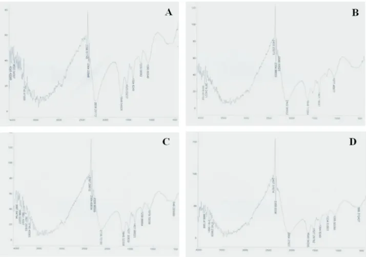
Fourier Transform Infrared (FTIR) spectroscopy
The FTIR spectra of P. aeruginosa Rb-1 and O. intermediumRb-2 grown in L-broth in the presence and ab-sence of 1000mg mL-1of K2CrO4were taken in the range of
500-4500 cm-1 (wave number) in order to determine the role of various functional groups present on the bacterial cell surface and were involved in the uptake of Cr(VI). The
FTIR spectrum pattern of cells ofP. aeruginosaRb-1 and O. intermedium Rb-2 grown in L-broth without Cr(VI) showed the presence of number of functional groups on their cell surface. The prominent absorption peaks in the re-gion of 4000-3500 cm-1were due to OH- symmetric stretch vibration. The absorption peaks in the region of 3500-3200 cm-1were indicative of -OH group and -NH groups; 3000-2500 cm-1showed existence of the carboxylic group; 2600-2500 cm-1exhibited the presence of S-H group and the peaks in the region of 2400-2300 cm-1specified the ex-istence of amines. Absorption peaks at 2260-2100 cm-1 were due to CºC whereas the peak at 1690-1640 cm-1and 1640-1500 cm-1showed the existence of primary and sec-ondary amines and amides, (N-H bending) respectively. Carboxylate ions usually displayed the absorption peaks in the region of 1300-1420 cm-1. Absorption peak in the re-gion of 1239.99 cm-1 was due to presence of sulphonate (SO2O-) groups whereas absorption peaks in the region of
1300-1000 cm-1corresponded to C-O stretching of COOH. Absorption peaks in the region of 750-1000 cm-1showed the existence of S = O, -C-C- and C-Cl functional groups (Figure 2 A and C).
In the FTIR spectra of cells ofP. aeruginosaRb-1 and O. intermediumRb-2 grown in L-broth with 1000mg mL-1 of K2CrO4, shifts were observed in the absorption peaks at
different regions. Major changes were observed in the re-gion of 2500-500 cm-1under Cr(VI) stress shown by both strains (Figure 2 B and D).
Discussion
biochemical
attributes
and
Cr(VI)
stress
579
Table 3- Effect of variable growth temperatures (28, 37 and 42 °C) on biochemical parameters ofP. aeruginosaRb-1 andO. intermediumRb-2 under chromate free and chromate supplemented conditions (1000mg mL-1of K
2CrO4).
Strain Parameters Chromate free Chromate supplemented
28 °C 37 °C 42 °C 28 °C 37 °C 42 °C
Rb-1 Soluble protein (mg g-1fresh weight)
2.34±0.03 19.50±0.97 5.81±0.07 26.40±0.18* 52.82±1.47* 24.55±0.16*
Peroxidase (mg g-1fresh weight) 23.56
±0.75 33.62±0.39 24.87±1.20 31.08±0.83* 92.84±1.41* 83.00±2.32*
Auxin (mg g-1fresh weight) 5.09±0.19 13.18±0.59 8.18±0.23 65.45±1.05* 84.59±0.73* 66.73±1.07*
Proline (mg g-1fresh weight) 362.79±0.36 410.52±1.51 383.01±0.25 820.4±0.59* 988.12±0.64* 911.07±0.59*
Nitrate reductase(mg g-1fresh weight) 1.11
±0.02 1.93±0.02 1.93±0.02 0.24±0.01* 0.36±0.02* 0.26±0.00*
Cystine (mM) 0.03±0.00 0.04±0.00 0.03±0.00 1.4±0.02* 5.12±0.05* 2.36±0.04*
Cysteine (mM) 0.15±0.00 0.83±0.01 0.15±0.00 1.28±0.02* 4.53±0.12* 0.87±0.01*
GSH (nM) 1.76±0.07 2.87±0.07 1.62±0.04 13.07±0.48* 22.28±0.54* 7.50±0.38*
GSSG (nM) 2.92±0.04 4.33±0.21 4.02±0.16 19.69±0.61* 46±0.85* 37.13±0.91*
Rb-2 Soluble protein (mg g-1fresh weight) 5.57±0.06 19.73±0.50 9.09±0.10 31.31±0.35* 67.25±0.75* 62.38±0.70*
Peroxidase (mg g-1fresh weight) 16.74±1.08 23.35±0.91 17.32±0.49 50.74±1.08* 98.24±0.55* 80.63±0.45*
Auxin (mg g-1fresh weight) 6.15±0.11 16.99±0.66 10.45±0.15 69.33±0.45* 120.36±1.83* 75.41±2.79*
Proline (mg g-1fresh weight) 363.44
±3.73 428.65±1.78 410.79±2.60 827.67±0.53* 999.51±9.88* 945.05±5.54*
Nitrate reductase(mg g-1fresh weight) 1.31
±0.01 2.82±0.05 1.81±0.02 0.39±0.03* 0.58±0.01* 0.26±0.01*
Cystine (mM) 0.09±0.00 0.05±0.00 0.05±0.00 3.91±0.03* 6.22±0.03* 3.28±0.05*
Cysteine (mM) 0.27±0.00 1.00±0.01 0.18±0.00 5.18±0.07* 7.36±0.11* 3.83±0.03*
GSH (nM) 1.96±0.05 3.76±0.13 1.53±0.09 10.69±0.42* 59.18±0.96* 13.16±0.24*
GSSG (nM) 4.79±0.02 6.56±0.05 5.34±0.06 17.23±0.26* 70.55±0.97* 23.84±0.39*
et al., 2011). Chromate stress induced in both investigated strains a close to three time higher peroxidase activity at pH7. The higher peroxidase activity under chromate stress can be related to the fact that Cr(VI) causes oxidative dam-age and peroxidases produced by metal resistant bacteria have the ability to protect cellular proteins and DNA from oxidation during stress conditions (Ramírez-Díaz et al., 2008; Pantet al., 2011). Indole acetic acid (IAA) is a com-mon natural auxin and is a comcom-mon secondary metabolite of most of the rhizospheric microorganisms (Yurekliet al., 2003; Khamnaet al., 2010). Growth temperature of 37 °C and pH 7 was found to be optimal for maximum production of auxin by both bacterial strains. Proline is known to be an indicator of stress tolerance and functions as metal chelator. Under stress conditions, intracellular accumulation of pro-line in microbes is a well-documented fact (Köcheret al., 2011). Both strains, i.e. P. aeruginosa Rb-1 and O. intermediumRb-2 exhibited significant enhanced produc-tion of proline under Cr(VI) stress compared to chromate free conditions. Nitrate reductase activity was the only bio-chemical parameter found to be inhibited under Cr(VI) stress treatments in both strains. Inhibition of nitrate reduc-tase activity due to heavy metal stress has previously been reported (Awasthi, 2005; Srivastava and Thakur, 2007).
One of the reasons for the inhibition of nitrate reductase ac-tivity is interference of heavy metal ions with sulphydryl (-SH) groups in enzymes which are involved in determin-ing the secondary and tertiary structure of proteins (Awasthi, 2005). This can lead to lowered enzyme activity.
Intracellular concentration of GSSG increases at the cost of GSH under intense stress conditions (Ackerleyet al., 2006). We observed non-protein thiol production was enhanced under Cr(VI) stress in both studied strains. GSH (Gamma-L-glutamyl-L-cysteninyl glycine) and GSSG [Bis (gamma-Glutamyl-L-cysteinyl glycine) Disulfide] content was peaking at pH 7, 1000mg mL-1of K2CrO4and 37 °C.
GSH concentration was lower than GSSG content. GSH and GSG content ofO. intermediumRb-2 was remarkably higher than forP. aeruginosaRb-1. Cellular exposure to oxidants resulted in reduction in the level of GSH and in-crement in the level of its oxidation product (GSSG). Cysteine and cystine content ofP. aeruginosaRb-1 andO. intermediumRb-2 was highest at pH 7, 1000 mg mL-1of K2CrO4 and 37 °C, but O. intermedium Rb-2 produced
concentration of cysteine in living organisms, it is reported that in several species of eukaryotic algae, cysteine content ranges from 0.6 to 12 mM (Satohet al., 2002). Cysteine
concentrations of both P. aeruginosa Rb-1 and O.
intermediumRb-2 were also within this range. Increase in content of non-protein thiols under Cr(VI) stress suggests their possible involvement in chromate detoxification.
Electron microscopy gives the possibility to study the cell physiology and especially changes in cell structure as a result of exposure to pollutants. Transmission electron mi-croscopic examination of P. aeruginosa Rb-1 and O. intermedium Rb-2 exhibited the distribution of electron dense precipitates intra as well as extra-cellularlly as a re-sult of exposure to chromium. The distribution of precipi-tates was not uniform in case of both bacterial strains and this up take of metals by individual cells within a culture may vary because of physiological reasons. Differential distribution of uranium by the cells ofP. aeruginosaandS. cervisaehas already been reported (Mullenet al., 1989). Intracellular localization of electron dense precipitates in-dicated the intracellular reduction of Cr(VI) as shown in figure 1 (B) and (D). The intracellular reduction pathway
for Shewanella oneidensis was previously reported and Acinetobactersp. strain, PCP3 also showed intracellular lo-calization of electron dense precipitates (Daulton et al., 2007; Srivastava and Thakur, 2007). These precipitates are mainly supposed to be Cr(III) in the form of hydroxyl and carboxyl groups (Bruinset al., 2000; Bencheikh-Latmani et al., 2007). Routinely both bacterial strains were main-tained on Cr(VI) supplemented media, and they accumulate Cr(VI) intracellularly. When the cells were grown in chro-mate free media, they exhibited the deposition of chromium particles at their boundary even in the absence of Cr(VI). This was due to the gradual release of intracellularly accu-mulated Cr(VI) as indicated in figure 1 (A) and (C).
Electron microscopic results showed the distribution of chromium on the bacterial cell surface. Thus, FTIR anal-ysis was performed to investigate the role of functional groups present on the bacterial cell surface in sequestration of chromium. FTIR analysis of the bacterial cells grown with and without Cr(VI) indicated the presence of amino, carboxyl, hydroxyl and sulphonate groups. Cr(VI) stress brought shifts in the absorption peaks. Major shifts in ab-sorption peaks were observed in the region of
4000-biochemical attributes and Cr(VI) stress 581
3500 cm-1, 3500-3200 cm-1, 1300-1450 cm-1 and 1200-1250 cm-1under Cr(VI) stress conditions. These shifts indi-cated binding of the metal ions with certain specific func-tional groups namely; hydroxyl, amino and carboxyl and sulphonate groups, respectively. These functional groups are ionizable and reported to bind with the metal ions (Buenoet al., 2008). Involvement of the carboxyl group in sequestration of chromium with the protein molecules in cyanobacteria under Cr(VI) stress has been described (Pandiet al., 2009). Bacterial cell walls are mainly com-posed of carbohydrates, lipids and proteins thus proposing the possible involvement of above said functional groups in complexation of chromium with the bacterial cell surfaces (Mungasavalliet al., 2007; Lameiraset al., 2008).
Conclusion
It can be concluded that Cr(VI) stress severely alters the bacterial morphology in terms of the shape and size. Cr(VI) stress led to enhancement in production of certain polysaccharides and formation of cell protrusions. These polysaccharides entrapped metal ions present in the sur-rounding environment thus reducing the availability to the bacterial cells. Significant increase of proteins and enzyme activities was exhibited by chromium addition for both Pseudomonas aeruginosa Rb-1 and Ochrobacterum intermedium Rb-2 highlighting their potential for biore-mediation. Ochrobacterum Rb-2 showed a stronger re-sponse in measured biochemical parameters than Rb-1. Variation in the biochemical parameters under Cr(VI) stress may be one of the major reasons of their ecological dominance in metal contaminated environment. Entrap-ment of Cr(VI) by these two strains evident by electron mi-crographs proved them as a good candidates for the remediation of metal contaminated environments.
Acknowledgments
University of the Punjab, Lahore, Pakistan, is ac-knowledged for providing financial assistance for the com-pletion of this study. The Higher Education Commission of Pakistan is also highly acknowledged for providing fund-ing to Rida Batool (IRSIP No. 1-8 /HEC/HRD/ 2009 / 557) to visit the Faculty of Biological and Environmental Sci-ences, General Microbiology, University of Helsinki, Fin-land to perform Electron Microscopy. This research work is the part of Ph.D thesis of Rida Batool.
References
Ackerley DF, Barak Y, Lynch SV, Curtin J, Matin A (2006) Effect
of chromate stress on Escherichia coli K-12. J Bacteriol
188:3371-3381.
Andreoni V, Colombo M, Di-Simine D, Finoli C, Origgi G, Vecchio A, Carzaniga R (1997) Removal of lead from
aque-ous solutions by aBrevibacteriumstrain.In:Rosen, D. (ed).
Modern Agriculture and the Environment, Kluwer Aca-demic Publisher, Dordrecht, Netherland, p. 521-531. Awasthi M (2005) Nitrate reductase activity: A solution to nitrate
problems tested in free and immobilized algal cells in pres-ence of heavy metals. Int. J. Environ Sci Technol 2:201-206. Bencheikh-Latmani R, Obraztsova A, Mackey M, Ellisman M,
Tebo B (2007) Toxicity of Cr(III) toShewanellasp. strain
MR-4 during Cr(VI) reduction. Environ Sci Technol 41:214-220.
Bhatti GA, Qureshi N, Qureshi A, Sultana K (1993) Studies on
heat shock response of wheat seedlings usingE. coliGroEL
antibodies. Pakphyton 5:157-166.
Bruins MR, Kapil S, Oehme W (2000) Microbial resistance to metals in the environment. Ecotox Environ Safe 45:198-207.
Bueno BYM, Torem ML, Molina F, Mesquita LMS (2008)
Bio-sorption of lead(II), chromium(III) and copper(II) by R.
opacus: Equilibrium and kinetic studies. Miner Eng 21:65-75.
Cervantes C, Campos- García, J (2007) Reduction and efflux of chromate by bacteria. Mol Microbiol Heavy Metal 6:407-419.
Daulton TL, Little BJ, Jones-Meehan J, Blom DA, Lawrence F (2007) Microbial reduction of chromium from the hexava-lent to divahexava-lent state. Geochim Cosmochim Acta 71:556-565.
David R, Murry E (1965) Protein synthesis in dark-growth bean leaves. Can J Bot 43:817-824.
Derminal T, Turkan I (2006) Exogenous glycinebetaine affects growth and proline accumulation and retards senescence in two rice cultivars under NaCl stress. Environ Exp Bot 56:72-79.
Gaitonde MK (1967) A spectrophotometric method for the direct determination of cysteine in the presence of other naturally occurring amino acids. J Biochem 104:627-633.
Glickmann E, Dessaux Y (1995) A critical examination of the specificity of the salkowski reagent for indolic compounds produced by phytopathogenic bacteria. Appl Environ Microbiol 61:793-796.
Gustavs L, Eggert A, Michalik D, Karsten U (2009) Physiological and biochemical responses of green microalgae from differ-ent habitats to osmotic and matric stress. Protoplasma 243:3-14.
Järup L (2003) Hazards of heavy metal contamination. Br Med Bull 68:167-182.
Khamna S, Yokota A, Peberdy JF, Lumyong S (2010)
Indole-3-acetic acid production byStreptomycessp. isolated from
some Thai medicinal plant rhizosphere soils. EurAsian J Biosci 4:23-32.
Köcher S, Tausendschön M, Thompson M, Saum SH, Müller V (2011) Proline metabolism in the moderately halophilic
bac-terium Halobacillus halophilus: Differential regulation of
isogenes in proline utilization. Environ Microbiol Rep 3:443-448.
Lameiras S, Quintelas C, Tavares T (2008) Biosorption of Cr(VI) using a bacterial biofilm supported on granular activated carbon and on zeolite. Bioresource Technol 99:801-806. Lounatmaa K (1985) Electron microscopic methods for the study
of bacterial surface structures.In:Korhonen T. K., Dawes,
Anti-gens: Methods for Molecular Characterization, Elsevier Sci-ence Ltd. USA, p. 243-261.
Lowry O, Rosebrough N, Farr A, Randall R (1951) Protein mea-surement with folin phenol reagent. J Biol Chem 193:265-275.
Mullen MD, Wold DC, Ferris FG, Beveridge TJ, Flemming CA, Bailey GW (1989) Bacterial sorption of heavy metals. Appl Environ Microbiol 55:3143-3149.
.Mungasavalli DP, Viraraghavan T, Jin YC (2007) Biosorption of
chromium from aqueous solutions by pre-treated
Aspergillus niger: Batch and column studies. Colloids Surf A: Physicochem Eng Asp 301:214-223.
Murugesan AG, Maheswari S (2007) Uptake of hexavalent chro-mium from aqueous solution employing live, dead and im-mobilized bacterial biomass. J Appl Sci Environ Manag 11:71-75.
Nagajyoti PC, Lee KD, Sreekanth TVM (2010) Heavy metals, oc-currence and toxicity for plants: A review. Environ Chem Lett 8:199-216.
Panda SK, Choudhury S (2005) Changes in nitrate reductase
ac-tivity and oxidative stress response in the mossPolytrichum
communesubjected to chromium, copper and zinc phyto-toxicity. Braz J Plant Physiol 17:191-197.
Pandi M, Shashirekha V, Swamy M (2009) Bioabsorption of chromium from retan chrome liquor by cyanobacteria. Microbiol Res 164:420-428.
Pant PP, Tripathi AK, Dwivedi V (2011) Effect of heavy metals
on some biochemical parameters of Sal (Shorea robusta)
seedling at nursery level, doon valley, India. J Agric Sci 2:45-51.
Poljsak B, Pócsi I, Pesti M (2011) Interference of chromium with
cellular functions.In:Banfalvi, G. (ed). Cellular Effects of
Heavy Metals, Springer. USA, p. 59-86.
Ramírez-Díaz MI, Díaz-Pérez C, Vargas E, Riveros-Rosas H, Campos-García J, Cervantes C (2008) Mechanisms of bac-terial resistance to chromium compounds. Biometals 21:321-332.
Satoh M, Hirachi Y, Yoshioka A, Kobayashi M, Oyama Y. (2002) Determination of cellular levels of non-protein thiols and their correlations with susceptibility to mercury in phyto-plankton. J Phycol 38:983-990.
Srivastava S, Thakur IS (2007) Evaluation of biosorption potency ofAcinetobactersp. for removal of hexavalent chromium from tannery effluent. Biodegr 18:637-646.
Weilharter A, Mitter B, Shin MV, Chain PSG, Nowak J, Sessitsch A (2011) Complete genome sequence of the plant
growth-promoting endophyte Burkholderia phytofirmans strain
PsJN. J Bacteriol 193:3383-3384.
Xu WH, Liu YG, Zeng GM, Li X, Song HX, Peng QQ (2009) Characterization of Cr(VI) resistance and reduction by
Pseudomonas aeruginosa. T Nonferr Metal Soc 19:1336-1341.
Yurekli F, Geckil H, Topcuoglu F (2003) The synthesis of indole-3-acetic acid by the industrially important white-rot
fungus Lentinus sajor-cajuunder different culture
condi-tions. Mycol Res 107:305-309.
All the content of the journal, except where otherwise noted, is licensed under a Creative Commons License CC BY-NC.