Percutaneous pulmonary valve implantation in small conduits:
A multicenter experience
Sebastien Hascoet
a,b,c, José Diogo Martins
d, Haysam Baho
e, Saule Kadirova
f, Fatima Pinto
d, Florent Paoli
g,
Fadi Bitar
h, Abdelfatah abu Haweleh
i, Anselm Uebing
j, Philippe Acar
a, Olivier Ghez
j, Alain Fraisse
j,⁎
aHôpital des enfants, Cardiologie pédiatrique, Centre de Compétence Malformations Congénitales Complexes M3C, CHU Toulouse, 31100 Toulouse, France
b
Hôpital Marie Lannelongue, Pôle de cardiopathies congénitales de l'enfant et de l'adulte, Centre de Référence Malformations Cardiaques Congénitales Complexes M3C- 92350 Le Plessis-Robinson, Faculté de Médecine Paris-Sud, Université Paris Sud, Université Paris-Saclay, France
c
Inserm/UPS UMR 1048 - I2MC, CHU Toulouse, Toulouse, France
dPediatric Cardiology Department, Hospital de Santa Marta, CHLC, Lisboa, Portugal e
King Faycal Specialist Hospital, Jeddah, Saudi Arabia
f
National Research Cardiac Surgery Center, Astana, Kazakhstan
g
La Timone Hospital, Marseille, France
h
American University of Beirut Medical Center, Beirut, Lebanon
i
Queen Alia Heart Institute. Amman, Jordan
jRoyal Brompton and Harefield Hospitals Trust, London, UK
a b s t r a c t
a r t i c l e i n f o
Article history: Received 11 January 2017
Received in revised form 25 September 2017 Accepted 1 December 2017
Available online 5 December 2017
Background: Guidelines allow percutaneous pulmonary valve implantation (PPVI) in conduits above 16 mm diameter. Balloon dilatation of a conduit to a diameterN 110% of the original implant size is also not recommended. We analyzed patients undergoing PPVI in such conditions.
Methods and results: Nine patients (May 2008–July 2016) from 8 institutions underwent PPVI in conduits b16 mm diameter. Five patients with 16–18 mm conduit diameter underwent PPVI after over-expansion of the conduitN 110%. Mean age and weight of the 14 patients was 12.1 (7.7 to 16) years and 44.9 (19 to 83) kg. Median conduit diameter at PPVI was 12 (10 to 17) mm. Median systolic right ventricular pressure was 70 (40 to 94) mm Hg. Procedure was successful in all cases. A confined conduit rupture occurred in 7 patients (50%) and was treated with covered stent in 6. One patient experienced dislocation of 2 pulmonary artery stents that were parked distally. The post-implantation median systolic right ventricular pressure was 36 (28 to 51) mm Hg. Afistula between right-ventricle outflow and aorta was found in one patient, secondary to undiagnosed conduit rupture. This was closed surgically. After a median follow-up of 20.16 (6.95 to 103.61) months, all the patients are asymptomatic with no significant RVOT stenosis.
Conclusions: PPVI is feasible in small conduits but rate of ruptures is high. Although such ruptures remain contained and can be managed with covered stents in our experience, careful selection of patients and high level of expertise are necessary. More studies are needed to better assess the risk of PPVI in this population.
© 2017 Elsevier B.V. All rights reserved.
Keywords:
Percutaneous pulmonary valve implantation Congenital heart defects
Children
1. Introduction
Percutaneous pulmonary valve implantation (PPVI) has emerged as
an alternative to surgery for reconstruction of the right ventricle
out-flow tract (RVOT). Since the first PPVI in 2000
[1]
, multiple studies
have con
firmed its safety and efficacy
[2
–9]
. The Melody valve
(Medtronic Inc., Minneapolis, MN, USA) has been the
first valve inserted
percutaneously in humans and obtained European certi
fication (CE) in
2006 as well as approval for use in the USA in 2010. Mid-term outcome
is good with regards to hemodynamic evolution, functional status and
device durability
[2
–4,7,10]
. Nevertheless guidelines recommend its
use in conduits with nominal diameter equal to or above 16 mm
[7,11]
. Moreover, overdilation of the conduit diameter
N 110% of the
nominal diameter (original implant size) prior to PPVI is not
recom-mended (
www.medtronic.com/safety-info-html
). PPVI in children
with small weight has previously been performed
[8,12,13]
. But little
is known about PPVI ef
ficiency in small and/or over-expanded conduits.
A-priori issues were raised with regards to conduit rupture risk
[14]
and
residual gradient across the valve
[15]
. Thus we analyzed outcome of
patients with off-label PPVI in small and over-expanded conduits.
⁎ Corresponding author at: Royal Brompton & Harefield NHS Foundation Trust, SydneyStreet, London SW3 6NP, UK.
E-mail address:[email protected](A. Fraisse).
https://doi.org/10.1016/j.ijcard.2017.12.003
0167-5273/© 2017 Elsevier B.V. All rights reserved.
Contents lists available at
ScienceDirect
International Journal of Cardiology
2. Methods
2.1. Study design
Between November 2008 and July 2016, 14 patients who underwent off-label PPVI in conduits below 16 mm and/or in conduits below or equal to 18 mm with afinal Melody valve to tube diameter ratio above 110% were retrospectively analyzed. Procedures were performed in 8 centers (Royal Brompton hospital, England, n = 4; CHU La Timone, Marseille, France, n = 3; Lisbon, Portugal, n = 2; CHU Toulouse, France, n = 1; Astana, Kazakhstan, n = 1; Amman, Jordan, n = 1; Jeddah, Saudi Arabia, n = 1; Beirut, Lebanon, n = 1).
Patients were considered for PPVI if they had congenital heart disease requiring pre-vious RVOT surgery, with an indication for pulmonary valve replacement according to cur-rent indications and practices[11]. Patients were discussed and approved for off-label PPVI after a joint cardiac surgical conference. Only patients with“expandable” conduits such as Contegra conduit or homograft or native outflow tract were considered suitable for off-label PPVI. Otherwise, patients with“non-expandable” conduits such as Hancock conduits below 16 mm were not suitable for off-label PPVI and surgical PV replacement was considered. PPVI was contra-indicated in case of active infection or coronary artery compression during balloon testing. Patients with failure attempt of PPVI were not includ-ed in this study. The study protocol conforms to the ethical guidelines of the 1975 decla-ration of Helsinki. Written informed consent for the procedure was obtained from all patients and/or parents before PPVI. The off-label indication of PPVI was explained to the family together with the alternative possibility of surgical valve replacement. The risk of conduit rupture and the potential for emergency surgery were also clearly stated.
2.2. Procedure
Melody valves were used for PPVI. The Melody valve is made of a bovine jugular valved vein (Contegra Pulmonary Valved conduit, Medtronic Inc., Minneapolis, MN, USA) sutured within a Cheatham-Platinium stent (CP stent, NuMED Inc., Hopkinton, NY). The valve was manually crimped and implanted through a dedicated delivery system (Ensemble Transcatheter Delivery System, Medtronic Inc., Minneapolis, MN, USA). It con-sists of a balloon-in-balloon (BiB, NuMED Inc., Hopkinton, NY) catheter delivery system with a retractable sheath that covers the Melody valve once it is crimped over the balloon. The outer balloon is available in 3 sizes: 18, 20 and 22 mm in diameter.
In view of a potential unconfined conduit rupture requiring covered stent implanta-tion in emergency, all the patients had a pre-procedure computerized tomodensitometry to assess proximity of the coronary arteries from the conduit. RVOT calcification was esti-mated on pre-procedure CT scan in each patient, using the following descriptive, semi-quantitative approach: Grade 0 = no calcification, 1 = mild calcification, 2 = moderate calcification that is not circumferential, 3 = moderate calcification that is circumferential or heavy calcification that is not circumferential, and 4 = heavy calcification that is cir-cumferential. All procedures were achieved under general anesthesia. Regarding the risk for conduit rupture during staged balloon dilation of the conduit, surgical back-up was or-ganized on a systematic basis. PPVI was performed by a local specialist assisted by the pri-mary investigator (A.F.) in 13 of the 14 cases. Percutaneous trans-femoral access was used in 13 patients. PPVI was done on a one-stage procedure in all cases. Direct invasive hemo-dynamic measurements and angiographic assessment were made in all patients before and after valve deployment. Minimal internal tube diameter was measured. Progressive balloon dilation of the conduit with high (Mullins, NuMED Inc., Hopkinton, New-York, USA) or ultra-high (Atlas or Atlas Gold, Bard, Tempe, AZ, USA) pressure balloons was per-formed, through a 14 French long Mullins sheath that was advanced immediately below the conduit. A balloon 2 to 4 mm larger than the conduit diameter was initially used. RVOT angiography was performed after every balloon dilation, to rule-out any conduit rupture. In the absence of rupture, step-by step balloon dilation of the conduit was per-formed using 2 mm diameter increment until the diameter of the intended pre-stent was reached. Aortic root angiogram was then performed during this latest balloon dilation to exclude coronary compression in lateral and either right-anterior or left-anterior and cranial projection. In case a confined conduit rupture was diagnosed during progressive balloon dilation of the conduit, a covered stent was immediately crimped on a BIB balloon (NuMED Inc., Hopkinton, New-York, USA) of the same diameter or 2 mm larger than the last balloon used for RVOT dilation. If coronary artery testing was already performed, the covered stent was implanted in the RVOT to treat the conduit rupture. If coronary testing was not yet performed, subsequent balloon dilation of the RVOT was performed with the same or a slightly (2 mm) larger balloon than the last one used for RVOT dilation, with concomitant aortic root angiogram. Covered stent was immediately implanted after coro-nary compression testing. After pre-stenting of the landing zone, Melody valve was im-planted through the Ensemble delivery system using the standardized method[11].
At the time of the procedure, no patient presented ongoing infection on either clinical or biological exams. All patients received antibiotics bolus (25 mg/kg intravenous cefazolin) and anticoagulation (100 UI/kg intravenous heparin) at the beginning of the procedure. Prophylaxis and heparin were repeated if the procedure was prolonged, to maintain the ACTN 250. After PPVI, oral aspirin (100–250 mg once a day) was prescribed life-long. Antibiotic prophylaxis and non-specific prevention measures were recommended following guidelines[16].
2.3. Follow-up
Outcome was assessed in September 2017 in all patients. All local investigators were contacted to obtain clinical and echo data at last visit.
2.4. Statistical analysis
For each patient, we collected demographic characteristics, procedural hemodynamic data, technical details, results and follow-up. Statistical analyses were performed using Stata 11.2 software (Statacorp, Texas, USA). Data are summarized as mean (Standard Deviation), minimal and maximal values. The Shapiro-Wilks test did not reject normality of distribution of continuous variables. Categorical variables were summarized as number and percentages. Pre and post procedural hemodynamic data were compared using a Wilcoxon matched-pairs signed rank test. Fischer Exact and Mann-Whitney tests were used to compare variables of interest among patients with versus without conduit rupture after balloon predilation. Reported P-values are two sided. Values of Pb 0.05 were consid-ered statistically significant.
3. Results (
Tables 1
–3
,
Fig. 1
)
A total of 14 patients were included at a mean age of 12.1 ± 3.0 years
old between November 2008 and July 2016.
Congenital heart defects were cono-truncal defects in 10 cases
(71.4%), congenital aortic valve disease with Ross surgery in 2 cases
(14.3%), transposition of the great arteries in 1 case (7.1%) and
pulmo-nary valve stenosis in 1 case (7.1%). All patients had a right ventricle
to pulmonary artery conduit. A homograft had been used in 10 cases
(71.4%), a Contegra (Contegra Pulmonary Valved conduit, Medtronic
Inc., Minneapolis, MN, USA) in 3 cases (21.4%) and a non valved tube
in 1 case (7.1%). Patient demographics data are presented in
Table 1
.
Procedural data are displayed in
Table 2
. PPVI was performed in a
one-stage procedure in all cases. A trans-femoral approach was used
in 13 cases whereas a left internal jugular vein approach was required
in one patient. Predilation of the conduit was performed in all cases
with simultaneous coronary artery angiogram. Pre-stenting of the
con-duit was performed in 13 cases (92.9%). Only one patient with a 15 mm
homograft had no prestenting at the beginning of the experience. Two
stents were implanted in the RVOT conduit in 4 cases (28.6%). Two
Table 1
Demographics and Diagnostic in patients undergoing percutaneous pulmonary valve implantation in small or overdilated conduits.
Patients (n = 14)
Age, years 12.1 (3.0) 7.7–16
Weight, kg 44.9 (18.3) 19–83
Congenital heart diseases
Commun arterial trunk 5 (35.7%) Tetralogy of fallot with pulmonary atresia 3 (21.4%) Tetralogy of fallot with pulmonary stenosis 1 (7.1%) Pulmonary valve agenesis 1 (7.1%)
Ross procedure 2 (14.3%)
Transposition of the great arteries 1 (7.1%) Pulmonary valve stenosis 1 (7.1%) Number of surgery
1 10 (71.4%)
2 4 (28.6%)
Right ventricle outflow tract
Homograft 10 (71.4%)
Valved conduit (Contegra) 3 (21.4%)
Non valved conduit 1 (7.1%)
Conduit diameter at implantation, mm
12 1 (7.1%) 13 1 (7.1%) 14 4 (28.6%) 15 3 (21.4%) 16 2 (14.3%) 17 2 (14.3%) 18 1 (7.1%)
Conduit diameter at catheterization, mm
10 2 (14.3%) 11 4 (28.6%) 12 2 (14.3%) 13 2 (14.3%) 14 3 (21.4%) 16 1 (7.1%)
Data are presented as frequency (% of total patients in the column) or mean (Standard Deviation) minimum-maximum.
patients had associated lesions of the pulmonary artery bifurcation that
were treated by balloon dilation and stent implantation during the PPVI
procedure.
PPVI was successful in all cases. Mean systolic right ventricle
pressure decreased from 70 to 36 mm Hg (P = 0.002). Mean systolic
right ventricle to systolic aortic pressure ratio decreased from 71% to
33% (P = 0.002). Final systolic right ventricle to systolic aortic pressure
ratio was below 50% in all cases. Seven patients (50.0%) experienced
con
fined conduit rupture after predilation (
Fig. 1
). Covered stenting
was performed and suf
ficient in 6 cases, with no residual leak on post
implantation angiogram. In the last case, a tear at the lower extremity
of the conduit was not diagnosed during the procedure because a
fistula
developed between the RVOT and the aortic root. Then, no contrast
ex-travasation was seen outside of the conduit during the post-dilatation
angiogram in the RVOT. This
fistula was clinically well-tolerated but
be-cause of signi
ficant shunt it was decided to close it. This fistula was
sur-gically closed 14 months after PPVI as the proximity of the aortic end of
the
fistula to the right coronary ostium rendered it unsuitable for
trans-catheter closure. In another patient, bifurcation stenting was initially
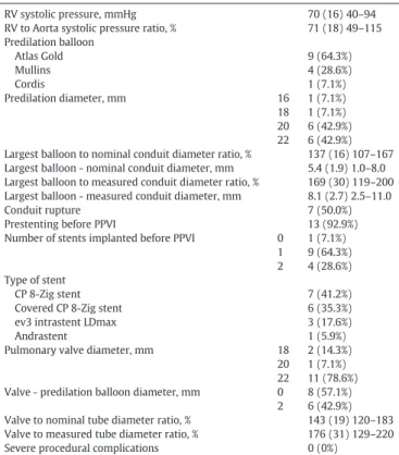
Table 2Procedural details for the 14 patients undergoing PPVI in small or overdilated conduits. RV systolic pressure, mmHg 70 (16) 40–94 RV to Aorta systolic pressure ratio, % 71 (18) 49–115 Predilation balloon Atlas Gold 9 (64.3%) Mullins 4 (28.6%) Cordis 1 (7.1%) Predilation diameter, mm 16 1 (7.1%) 18 1 (7.1%) 20 6 (42.9%) 22 6 (42.9%) Largest balloon to nominal conduit diameter ratio, % 137 (16) 107–167 Largest balloon - nominal conduit diameter, mm 5.4 (1.9) 1.0–8.0 Largest balloon to measured conduit diameter ratio, % 169 (30) 119–200 Largest balloon - measured conduit diameter, mm 8.1 (2.7) 2.5–11.0
Conduit rupture 7 (50.0%)
Prestenting before PPVI 13 (92.9%) Number of stents implanted before PPVI 0 1 (7.1%)
1 9 (64.3%) 2 4 (28.6%) Type of stent
CP 8-Zig stent 7 (41.2%)
Covered CP 8-Zig stent 6 (35.3%) ev3 intrastent LDmax 3 (17.6%)
Andrastent 1 (5.9%)
Pulmonary valve diameter, mm 18 2 (14.3%) 20 1 (7.1%) 22 11 (78.6%) Valve - predilation balloon diameter, mm 0 8 (57.1%)
2 6 (42.9%) Valve to nominal tube diameter ratio, % 143 (19) 120–183 Valve to measured tube diameter ratio, % 176 (31) 129–220 Severe procedural complications 0 (0%)
Data are presented as frequency (% of total patients in the column) or mean (Standard Deviation) minimum-maximum. RV, right ventricle; PPVI, percutaneous pulmonary valve implantation.
Table 3
Comparison of patients with and without conduit rupture during PPVI. Conduit rupture No conduit rupture P-value (n = 7) (n = 7) Age, years 12.2 (3.6) 12.0 (2.6) 0.9 Weight, kg 52.5 (21.4) 37.3 (11.4) 0.2 Type of conduit 0.2 Homograft 6 4 Contegra 0 3
Non valved conduit 1 0
Nominal conduit diameter, mm 15.4 (2.1) 14.6 (1.0) 0.4 Conduit diameter at PPVI, mm 12.6 (2.3) 12.1 (1.5) 0.6 Initial RV systolic pressure, mm Hg 69 (13) 70 (20) 0.8 Initial RV to Aorta systolic pressure ratio,% 77 (20) 66 (17) 0.2 Balloon to nominal conduit diameter ratio, % 135 (15) 140 (18) 0.3 Balloon - nominal conduit diameter, mm 5.1 (1.5) 5.7 (2.4) 0.3 Balloon to measured conduit diameter ratio, % 167 (31) 171 (30) 0.9 Balloon - measured conduit diameter, mm 7.9 (2.7) 8.2 (3.0) 0.8 Data are presented as frequency (% of total patients in the column) or mean (Standard Deviation). RV, right ventricle; PPVI, percutaneous pulmonary valve implantation.
Fig. 1. Lateral angiogram showing a severely stenosed (10 mm diameter whereas initial nominal diameter was 16 mm) homograft in the right ventricle outflow tract (A). After balloon dilation, confined rupture (*) is seen (B). Control angiogram of the right ventricle outflow demonstrates an excellent result after a 22 mm Melody valve implantation (C).
performed because of signi
ficant stenosis. Unfortunately, both stents
embolized during PPVI. They were successfully
“parked” in the distal
pulmonary artery branches and no further intervention was done at
the level of the bifurcation as right ventricular systolic pressure was
35 mm Hg and there was only mild residual stenosis on angiography
at the origin of both pulmonary artery branches. Comparison of
vari-ables among patients with versus without conduit rupture after balloon
dilation is reported in
Table 3
. There was a trend towards more
homo-grafts in patients with conduit rupture. Calci
fication score was not
relat-ed to conduit rupture. It was available in 12 patients including 6 with
rupture and 6 without. Among the 4 cases with a calci
fication score at
4, 2 experienced conduit rupture. The score was 3 or 4 in only 4 of the
patients who experienced conduits rupture whereas all 6 cases with
no conduit rupture had a score of 3 or 4.
Median follow-up duration was 20.16 (6.95 to 103.61) months.
No patient was lost to follow-up. No pulmonary valve replacement,
in-fective endocarditis or death was reported. No pulmonary valve
regur-gitation was observed in 10 patients (71.4%) at last follow-up. A trivial
to mild pulmonary valve regurgitation was reported in 4 patients
(28.6%). Mean peak pulmonary valve gradient at last follow-up was
23.8 mm Hg. All patients were in NYHA 1 functional status.
4. Discussion
In this multicenter cohort study, we observed that off-label PPVI
could be satisfactorily performed in small conduits measuring
b16 mm of nominal diameter and in conduits between 16 and 18 mm
diameter that could be over expanded above 110%. Conduit rupture
oc-curred in 50% of cases but was always con
fined in our experience. Such
conduit ruptures with limited collection did not compromise the
patients and were ef
ficiently treated with covered stents, with the
exception of a
fistula between aortic root and RVOT that was treated
surgically. Finally, before PPVI was performed, most of the patients
(10 out of 14) had only one previous surgical procedure, during
neona-tal period or infancy.
Few data are available with regard to PPVI in small conduits. In a
pre-vious report including 25 patients weighting
b30 kg and undergoing
PPVI, only 4 patients had a conduit below 16 mm diameter. No
compli-cation was reported but only 18 mm Melody valves were implanted
[13]
. The relevance of PPVI in small conduits was recently questioned
by Holzer and Hijazi. They stated that only patients with a RVOT conduit
larger than 16 mm are suitable for PPVI
[15]
. Moreover, in addition to
the risk for conduit rupture, higher residual RVOT gradients following
PPVI in smaller bioprosthesis were observed in another recent study
[17]
. In our patient population, we speculated that there would be
signi
ficant residual RVOT gradient if the conduits were not optimally
expanded. With a 22 mm Melody valve diameter in 76.9% of the
patients, we aimed to implant the largest possible valve. To achieve
this goal,
“aggressive” conduit predilation was performed with a mean
balloon to nominal conduit diameter ratio of 137% (up to 167%) and a
mean balloon to measured conduit diameter ratio of 169% (up to
200%). This predilation step was also crucial to accurately assess the
risk of coronary artery compression with a balloon diameter similar to
the intended Melody valve diameter
[18,19]
. Non-compliant high and
ultra-high pressure balloons were used and best suited for this purpose
[15]
. However in some cases conduit rupture occurred at an early stage
of predilation. Optimal coronary testing using a much higher balloon
diameter after conduit rupture may be life threatening by dramatically
increasing the collection outside of the conduit. In view of this,
preprocedure computerized tomodensitometry was performed on a
systematic basis to potentially exclude any case with risk factor for
cor-onary compression (single origin of the corcor-onary arteries, corcor-onary
ar-tery course close to the RVOT conduit
…)
[18,19]
. Although no patient
was excluded in our study, we would refer to surgery any case at risk
for coronary artery compression on the pre-procedure computerized
tomodensitometry. Following our strategy to favor large landing zones
for PPVI,
final hemodynamic and outcome were nicely improved. All
pa-tients had
final systolic right ventricle to aorta pressure ratio below 50%.
In the present study conduit rupture was observed in 50% of cases,
much higher than the 4 to 9% incidence previously reported in
consec-utive PPVI
[14,20,21]
. They were mainly observed with homografts, as
previously reported
[14]
. However, the risk of death and/or emergency
surgery is not fully clari
fied after conduit rupture. Only one previous
study reported 6 patients undergoing rescue surgeries after PPVI,
in-cluding 2 cases after conduit rupture. Unfortunately, this study collected
patients during early experience with PPVI (2000
–2007) and covered
stent implantation was apparently not attempted in those 2 cases. In a
recent study by Bishnoi et al., 58 cases of conduits tears treated with
covered stent were analyzed. No emergency surgery was needed and
only 4 patients (7%) had uncontained conduit rupture necessitating
pleurocentesis and chest tube placement in 2 of them. Treatment with
covered stent was successful in all cases but one. In this latest case the
uncontained tear was stabilized by covered stent implantation but
be-cause of slow residual leak the patient was eventually operated 3 days
after catheterization
[20]
. Similarly in our patient's population, we
spec-ulated that conduit rupture would be con
fined in the majority of cases
because surgically implanted conduits are surrounded by
fibrosis that
may contain the collection outside of the conduit. In the present study,
conduit ruptures and tears were indeed con
fined and successfully
treated with covered stent implantation, with the exception of the
pa-tient who developed an aorta-to-RVOT
fistula. However, we followed
a rigorous protocol in all our cases. First, we managed to have a covered
stent already crimped on a balloon and ready to be implanted at the
time of conduit predilation. Second, surgical back-up was immediately
available in case of any catastrophic conduit rupture, uncontained and
uncontrolled even after covered stent implantation
[14,20]
. Third,
con-duit predilation was performed through a very progressive and gradual
balloon dilation of the conduit. We always started predilation with a
balloon of no
N125% of the narrowest diameter as this is recommended
[15]
. Further gradual expansion of the conduit was performed with
2 mm increment balloon diameter, until the intended predilation
diameter was achieved to test the risk for coronary compression with
a concomitant aortic root angiogram. Finally, RVOT angiogram was
performed after every balloon dilation. With such protocol no patient
was compromised by conduit rupture and no transfusion or chest tube
drainage was required.
Our study has several limitations. The small number of patients
pre-cluded statistical analysis of risk factors for conduit rupture. There may
also be selection bias and information recall bias which are standard
limitations associated with any retrospective study. Finally, only Melody
valves were used in this study. More recently, other devices such as the
Sapien valve have become available for PPVI and could have also been
used in smaller conduits with a similar approach
[15]
.
5. Conclusion
Off-label PPVI is feasible in small conduits with good hemodynamic
results. However, it is associated with an important risk of conduit
rup-ture. Although we could successfully manage those contained ruptures
with covered stents careful pre-procedure selection of patients and a
high level of expertise are necessary to perform PPVI in this setting.
More studies are needed before any de
finitive conclusion can be made
regarding the risk of PPVI in this patient population.
Funding
None.
Potential con
flict of interest
Alain Fraisse is a consultant and proctor for Medtronic Inc. Other
authors have no con
flict of interest.
References
[1] P. Bonhoeffer, Y. Boudjemline, Z. Saliba, J. Merckx, Y. Aggoun, D. Bonnet, et al., Percutaneous replacement of pulmonary valve in a right-ventricle to pulmonary-artery prosthetic conduit with valve dysfunction, Lancet 356 (2000) 1403–1405.
[2] A. Fraisse, P. Aldebert, S. Malekzadeh-Milani, J.B. Thambo, J.F. Piechaud, P. Aucoururier, et al., Melody (R) transcatheter pulmonary valve implantation: results from a French registry, Arch. Cardiovasc. Dis. 107 (2014) 607–614.
[3] S. Khambadkone, L. Coats, A. Taylor, Y. Boudjemline, G. Derrick, V. Tsang, et al., Percutaneous pulmonary valve implantation in humans: results in 59 consecutive patients, Circulation 112 (2005) 1189–1197.
[4] P. Lurz, L. Coats, S. Khambadkone, J. Nordmeyer, Y. Boudjemline, S. Schievano, et al., Percutaneous pulmonary valve implantation: impact of evolving technology and learning curve on clinical outcome, Circulation 117 (2008) 1964–1972.
[5] E.M. Zahn, W.E. Hellenbrand, J.E. Lock, D.B. McElhinney, Implantation of the melody transcatheter pulmonary valve in patients with a dysfunctional right ventricular outflow tract conduit early results from the U.S. Clinical trial, J. Am. Coll. Cardiol. 54 (2009) 1722–1729.
[6] K. Asoh, M. Walsh, E. Hickey, M. Nagiub, R. Chaturvedi, K.J. Lee, et al., Percutaneous pulmonary valve implantation within bioprosthetic valves, Eur. Heart J. 31 (2010) 1404–1409.
[7] D.B. McElhinney, W.E. Hellenbrand, E.M. Zahn, T.K. Jones, J.P. Cheatham, J.E. Lock, et al., Short- and medium-term outcomes after transcatheter pulmonary valve placement in the expanded multicenter US melody valve trial, Circulation 122 (2010) 507–516.
[8] M. Vezmar, R. Chaturvedi, K.J. Lee, C. Almeida, C. Manlhiot, B.W. McCrindle, et al., Percutaneous pulmonary valve implantation in the young 2-year follow-up, JACC Cardiovasc. Interv. 3 (2010) 439–448.
[9] A. Eicken, P. Ewert, A. Hager, B. Peters, S. Fratz, T. Kuehne, et al., Percutaneous pul-monary valve implantation: two-centre experience with more than 100 patients, Eur. Heart J. 32 (2011) 1260–1265.
[10]P. Lurz, A. Giardini, A.M. Taylor, J. Nordmeyer, V. Muthurangu, D. Odendaal, et al., Effect of altering pathologic right ventricular loading conditions by percutaneous pulmonary valve implantation on exercise capacity, Am. J. Cardiol. 105 (2010) 721–726.
[11]S. Hascoet, P. Acar, Y. Boudjemline, Transcatheter pulmonary valvulation: current indications and available devices, Arch. Cardiovasc. Dis. 107 (2014) 625–634.
[12] K.K. Mallula, D. Kenny, Z.M. Hijazi, Transjugular melody valve placement in a small child with protein losing enteropathy, Catheter. Cardiovasc. Interv. 85 (2015) 267–270.
[13] D.P. Berman, D.B. McElhinney, J.A. Vincent, W.E. Hellenbrand, E.M. Zahn, Feasibility and short-term outcomes of percutaneous transcatheter pulmonary valve replace-ment in small (b30 kg) children with dysfunctional right ventricular outflow tract conduits, Circ. Cardiovasc. Interv. 7 (2014) 142–148.
[14]Y. Boudjemline, S. Malekzadeh-Milani, M. Patel, J.B. Thambo, D. Bonnet, L. Iserin, et al., Predictors and outcomes of right ventricular outflow tract conduit rupture during percutaneous pulmonary valve implantation: a multicentre study, EuroIntervention 11 (2016) 1053–1062.
[15] R.J. Holzer, Z.M. Hijazi, Transcatheter pulmonary valve replacement: state of the art, Catheter. Cardiovasc. Interv. 87 (2016) 117–128.
[16] G. Habib, P. Lancellotti, M.J. Antunes, M.G. Bongiorni, J.P. Casalta, F. Del Zotti, et al., 2015 ESC Guidelines for the management of infective endocarditis: the Task Force for the Management of Infective Endocarditis of the European Society of Cardiology (ESC). Endorsed by: European Association for Cardio-Thoracic Surgery (EACTS), the European Association of Nuclear Medicine (EANM), Eur. Heart J. 36 (2015) 3075–3128.
[17] W. Finch, D.S. Levi, M. Salem, A. Hageman, J. Aboulhosn, Transcatheter melody valve placement in large diameter bioprostheses and conduits: what is the optimal “Landing zone”? Catheter. Cardiovasc. Interv. 86 (2015) E217–23.
[18] A. Fraisse, A. Assaidi, L. Mauri, S. Malekzadeh-Milani, J.B. Thambo, D. Bonnet, et al., Coronary artery compression during intention to treat right ventricle outflow with percutaneous pulmonary valve implantation: incidence, diagnosis, and outcome, Catheter. Cardiovasc. Interv. 83 (2014) E260–8.
[19] B.H. Morray, D.B. McElhinney, J.P. Cheatham, E.M. Zahn, D.P. Berman, P.M. Sullivan, et al., Risk of coronary artery compression among patients referred for transcatheter pulmonary valve implantation: a multicenter experience, Catheter. Cardiovasc. Interv. 6 (2013) 535–542.
[20] R.N. Bishnoi, T.K. Jones, J. Kreutzer, R.E. Ringel, NuMED Covered Cheatham-Platinum Stent for the treatment or prevention of right ventricular outflow tract conduit dis-ruption during transcatheter pulmonary valve replacement, Catheter. Cardiovasc. Interv. 85 (2015) 421–427.
[21] A. Chatterjee, N.S. Bajaj, W.S. McMahon, M.G. Cribbs, J.S. White, A. Mukherjee, et al., Transcatheter pulmonary valve implantation: a comprehensive systematic review and meta-analyses of observational studies, J. Am. Heart Assoc. 6 (2017)https:// doi.org/10.1161/JAHA.117.006432.