Address for correspondence: Dr. Boban Thomas, Cardiac Imaging Section, RM Caselas, Rua Carolina Angelo, 1400-045 Lisbon, Portugal. E-mail: bobantho@gmail.com
INTRODUCTION
The neonatal arterial switch operation (ASO) is now the standard of care for children born with transposition of the great arteries (TGA), with excellent results in most experienced centers. There has always been a concern for the long term outcome of these patients due to the extensive surgical maneuvers employed in the initial
repair. A wide range of long‑term problems such as obstruction of the reconstructed right‑sided tract, the neopulmonary artery (neoPA) and its branches, coronary artery stenosis, neoaortic root enlargement with valvular regurgitation, and recurrent arch obstruction, may occur. In one multicenter study, stenosis of the neoPA and/or its branches was noted in approximately 21% of patients, while others detected increased flow velocity on echocardiography in 24% of patients, followed up over a period of 4.8 ± 3.9 years.[1,2] Abnormal pulmonary blood
flow distribution related to branch pulmonary artery stenosis or hypoplasia has been implicated in reduced exercise capacity and increased ventilatory drive.[3]
Cardiac magnetic resonance imaging (CMRI) is a useful technique to study patients with operated congenital heart disease, especially many years after surgical repair due to inadequate echocardiographic views. We
Stenosis of the branches of the neopulmonary artery after the
arterial switch operation: A cardiac magnetic resonance imaging
study
Boban Thomas, José Diogo Ferreira Martins
1, Nuno Jalles Tavares, Artur Lopes, Fátima F Pinto
1,
José Fragata
2RM Caselas, Lisbon, 1Pediatric Cardiology Service, Hospital Santa Marta, Lisbon, 2Cardiac Surgery, Hospital Santa Marta, Lisbon, Portugal
ABSTRACT
Background : The neonatal arterial switch operation (ASO) is now the standard of care for children
born with transposition of the great arteries. Stenosis of the neopulmonary artery on long‑term follow up is a known complication.
Methods : We performed a retrospective analysis of eleven patients who underwent a cardiac
magnetic resonance imaging (MRI) due to echocardiographic evidence suggestive of stenosis of the neopulmonary artery or its branches (mean estimated Doppler gradient
48 mmHg, min 30 mmHg, max 70 mmHg). A comprehensive evaluation of anatomy and
perfusion was done by cardiac MRI.
Results : The branches of the neopulmonary artery (neo PA) showed decreased caliber in three
patients unilaterally and in two patients, bilaterally. Magnetic resonance (MR) perfusion studies showed concomitant decreased flow, with discrepancy between the two lungs of 35/65% or worse, only in the three patients with unilateral obstruction, by two different
MR perfusion methods.
Conclusions : Cardiac MR can be used as a comprehensive non‑invasive imaging technique to diagnose
stenosis of the branches of the neopulmonary after the ASO, allowing evaluation of anatomy and function of the neoPA, its branches, and the differential perfusion to each lung, thus facilitating clinical decision making.
Keywords : Arterial switch operation, cardiac magnetic resonance imaging, neopulmonary branch
artery stenosis
Access this article online Quick Response Code: Website:
www.annalspc.com
DOI:
report our experience in patients with suspected stenosis of the branches of the neoPA after the ASO, using a CMRI protocol that included morphological analysis, phase‑velocity studies to evaluate flow and angiography using a gadolinium–based contrast.
MATERIALS AND METHODS
The study was conducted over a 24 month period (2009‑2011), during which 60 patients who had undergone the ASO, within 30 days after birth, had routine echocardiographic studies in the pediatric or adult congenital heart disease clinic. Eleven patients showed findings suggestive of stenosis of the neoPA or its branches with a mean estimated Doppler gradient 48 mmHg (min 30 mmHg, max 70 mmHg). These patients then had CMRI exams on a GE 1.5T HDxT system with a 8‑channel cardiac coil, with ECG gating and apnea for some sequences. After an axial scout, a black blood sequence with T1‑weighting was acquired in the axial plane to visualize the neoPA and its branches. Then, axial cine imaging was performed. Subsequent cine imaging of the neoPA in a sagittal orientation was performed to delineate the PA trunk, after which a perpendicular cut was made to perform a phase‑velocity study with the patient breathing freely. After this, a cut was made perpendicular to each branch of the neoPA and a phase‑velocity study was repeated. If flows and morphological appearance confirmed the presence of stenosis anywhere, then an angiographic sequence (either bolus‑tagged or time‑of‑flight) was performed to assess the extent of the stenosis and its morphology. Towards the end of the study, a phantom was placed in the unit and all phase velocity sequences repeated on the phantom while the patient was either seated in the room or placed on a table with continued electrocardiographic monitoring for gating purposes. This sequence was then used for background correction and final analysis in the flow studies.
For all phase contrast (PC)‑MRI studies, the forward flow volume (FFV), backward flow volume (BFV) and the net flow volume (NFV) were calculated with the NFV being the difference between the FFV and the BFV. The perfusion to each lung, designated as differential perfusion, was calculated using two methods – the FFV and the NFV. Differential perfusion of the left lung (for example) through the left branch of the neoPA (LPA) was calculated as follows:
• NFV of LPA/(NFV of LPA + NFV of RPA) × 100% (by the NFV method)
• FFV of LPA/(FFV of LPA + FFV of RPA) × 100% (by the FFV method)
The differential perfusion by each method was correlated as well.
The regurgitant fraction (RF) of each branch was then calculated as BFV/FFV and expressed as a percentage. For practical purposes, mathematically, a larger RF
with decreased NFV on one side would imply a smaller differential perfusion to that lung (when using the NFV method).[4] In the NFV method, a higher regurgitant
fraction, accentuates the difference in perfusion between the two lungs. A flow discrepancy between the two lungs of 35 or 65% or worse has recently been recommended as a threshold for intervention in a BPA.[5] Gradients were not
calculated across sites of stenosis due to technical problems inherent in the phase contrast MR technique. Anatomic diameters were calculated as an indicator of stenosis despite the pulsatility of the neoPA and branches. On the cine sequences, the smallest branch diameter (irrespective of the phase of the cardiac cycle) was used to calculate the Z‑scores of the neoPA and its branches.
RESULTS
The detailed anatomical and flow parameters for each patient we studied are presented in Tables 1‑3. The neoaorta was dilated in nine patients and compressed the trunk of the neoPA in three patients. The branches of the neopulmonary artery showed a decreased caliber in three patients unilaterally and, in two patients, bilaterally. MR perfusion studies showed concomitant decreased flow, with discrepancy between the two lungs of 35 or 65% or worse, only in the three patients with unilateral stenosis, by two different MR perfusion methods. Five patients had stretching of the branches of the neoPA without any difference in the differential perfusion.
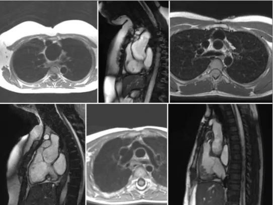
In one patient with bilateral stenosis on anatomic images, the NFV method gave 26% perfusion to the right side while the FFV method gave 41%. In spite of the discrepancy in this case, a strong correlation between the two methods (FFV and NFV) was detected (r = 0.947, P < 0.001). In the other patient with decreased caliber of the branches bilaterally, the cause appeared to be stretching of the branches over a dilated neoaorta. In this patient, despite an anatomic decrease in the caliber bilaterally, no difference in differential perfusion was noted. NeoPA and neoaorticvalvular regurgitation, when detected were clinically insignificant [Figure 1].
The cause of increased Doppler gradient was therefore ascertained to be compression of the trunk of neoPA in three patients, unilateral branch stenosis in three patients, bilateral branch stretching with compression in two patients and stretching of one or both branches without compression in three patients.
DISCUSSION
Stenosis of NeoPA or it's branches remain the commonest cause of late reintervention after ASO. In a comprehensive study of 21 patients, 16 had supravalvular stenosis involving the neoPA alone or along with its branches.[6]
The reported incidence of stenosis at any site in the right ventricular outflow ranging from the subvalvular to the supravalvular region vary between 7‑50%.[7,8] In
one study, the initial incidence was noted to be 52% but dropped to 24% when only gradients above 20 mmHg were considered.[2]
The patients in our series were investigated using CMRI after echocardiography. They showed evidence of significant right‑sided obstruction namely increased velocity at the RV‑PA junction, increase in the velocity of the jet of tricuspid regurgitation or altered interventricular septal motion, indicating high RV pressures. The exact location and quantum of obstruction may be difficult to judge by echo. CMRI confirmed the presence of significant flow difference of greater than 65 or 35% in one lung, compared to the other, using both methods, in only three patients. These three patients had unilateral obstruction predominantly on anatomic images. Flow studies by CMRI, devoid of radiation, obviate the need for perfusion scintigraphy studies to evaluate differential flow to the lungs and have been validated against scintigraphy.[9]
Therefore, scintigraphy was not performed in any of our patients but may be useful in those with discrepant findings by the two methods we used.
Although, we feel that one of the strengths of our study is the use of two methods to calculate the differential perfusion to each lung, some important caveats should be kept in mind. One group has shown that in patients
Table 1: Descriptive anatomical findings in patients
Patient BSA Age Z-AA Z-sinus AR (%) RF (PA) Anatomical findings
1 0.5 1 -2.55 0.19 Mild AR (16) 12.00 Stenosis at origin of the RPA, possible stenosis of LPA 2 1.7 18 -2.40 2.64 Mild AR (8) Dilated neoaortic root, LPA stenosis at origin
3 1.8 18 -1.38 8.52 Dilated neoartic root, BPAs stretched at the bifurcation where the dilated neoaorta then reduces abruptly in diameter
4 0.79 6 0.70 2.64 Mild AR (6),
Qp/Qs 1.5:1 29.00 Dilated neoaortic root directed anteriorly compressing proximal portion of the NPA with mild neopulmonary valve stenosis 5 2 19 0.51 8.82 Mild AR (13) 11.70 Dilated neoaortic root directed anteriorly compressing NPA
6 1.5 10 1.35 4.13 Mild AR (27) 7.30 Dilated neoaortic root directed anteriorly compressing proximal portion of NPA with stretching of the RPA with a decreased calibre compared to the LPA 7 0.6 3 0.27 3.79 Mild AR (14) 11.93 Dilated neoaortic root directed anteriorly compressing proximal portion of NPA
with stretching of the LPA with a decreased calibre compared to the RPA 8 0.65 1 -0.57 0.40 No AR 9.00 Neopulmonary valve stenosis with mild stretching of the LPA
9 1.9 21 -0.10 9.59 Mild AR Dilated neoaortic root with mild reduction in calibre at origin of each BPA 10 0.7 3 -1.05 2.19 No AR 10.00 Dilated neoaortic root with mild kinking at origin of RPA
11 1.7 12 1.00 3.13 Moderate
AR (26) Dilated neoaortic root with reduction in calibre of LPA origin
BSA: Body surface area; RF: Regurgitant fraction; AA: Ascending aorta; AR: Aortic regurgitation; PA: Pulmonary artery. When no value is provided for regurgitant fraction of neoPA or aortic regurgitation, the values were less than 3%
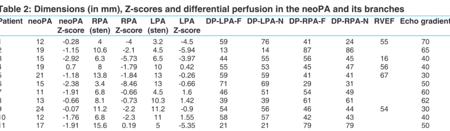
Table 2: Dimensions (in mm), Z-scores and differential perfusion in the neoPA and its branches
Patient neoPA neoPA
Z-score (sten)RPA Z-scoreRPA (sten)LPA Z-scoreLPA DP-LPA-F DP-LPA-N DP-RPA-F DP-RPA-N RVEF Echo gradient
1 12 -0.28 4 -4 3.2 -4.5 59 76 41 24 55 70 2 19 -1.15 10.6 -2.1 4.5 -5.94 13 14 87 86 65 3 15 -2.92 6.3 -5.73 6.5 -3.97 44 55 56 45 16 40 4 19 0.7 8 -1.79 10 0.42 55 53 45 47 56 40 5 21 -1.18 13.8 -1.84 13 -0.26 59 59 41 41 67 30 6 15 -2.38 3.4 -8.46 13 -0.66 71 69 29 31 50 7 11 -1.91 6.8 -0.66 4.5 1.6 46 51 54 49 60 8 13 -0.66 8.1 -0.73 10.3 1.42 39 39 61 61 62 9 24 -0.07 11.2 -2.2 11.2 -0.9 54 56 46 44 54 30 10 12 -1.76 6.8 -2.3 11 1.55 58 57 42 43 40 11 17 -1.91 15.6 0.19 5 -5.35 21 21 79 79 50
DP-LPA-N and DP-LPA-F represent differential perfusion to the LPA using the NFV and FFV methods respectively. RVEF in percentage and echo Doppler gradient in mmHg. Patients 2, 6, and 11 would warrant intervention recommended by findings that are concordant by both methods while patient 1 has equivocal findings as explained in the text. Patients 1 and 3 had bilateral decrease in branch diameters and therefore a clear difference between the two lungs in differential perfusion may not be demonstrable
Table 3: Dimensions, Z-scores and flow in the
neoPA also indexed for BSA
Patient BSA neoPA neoPA/BSA LPA
(FFV) (NFV)LPA (FFV)RPA (NFV)RPA
1 0.5 1020 2040 560 448 390 138 2 1.7 5800 3411 710 655 4800 3813 3 1.8 5856 3253 2791 2571 3504 2126 4 0.79 4088 5174 2344 1979 1872 1760 5 2 7995 3997 4875 4700 3430 3300 6 1.5 5390 3593 3937 3217 1616 1505 7 0.6 1890 3150 779 700 930 678 8 0.65 1900 2923 700 650 1128 1000 9 1.9 9956 4978 5510 4510 4764 3432 10 0.7 2090 2985 1255 1073 895 815 11 1.7 7792 4583 1613 1606 6262 5968
DP: Differential perfusion and NFV and FFV represent net flow volume ml/min and forward flow volume respectively. LPA and RPA are the left and right branches of the neoPA
cases. This is based on our findings on cine imaging where systolic expansion of the neoaorta compresses the branches or the trunk of the neoPA. In some cases, where we observed that the decreased branch diameters were due to the stretching of the branches over the dilated with previously operated tetralogy of Fallot (TOF), the
conventional NFV method underestimated left lung perfusion and they attributed it to a greater regurgitant fraction in the LPA[4] and the FFV method correlated
better with perfusion scintigraphy. In one patient in our series, intervention would have been recommended for the right branch of the neoPA, by the results of one method (NFV) but not by the other (FFV) (patient 1 in Figure 2). This patient had a regurgitant fraction of 64% in the right branch and this may have accounted for the significant difference in both methods.
To the best of our knowledge, there are only two studies that have addressed this problem using CMRI methods.[3,10]
One study[10] reported that the branches of the neoPA
had different diameters during systole (smaller) and diastole as the neoaorta enlarged during systole compressing one or both branches. We also evaluated the diameters of the branches and have noted that a severe narrowing, confirmed by the Z‑score of the stenotic branch, is accompanied by a decrease in perfusion to the lung supplied by that branch. We have also used a phantom correction method to avoid analytical errors as recommended by recent studies.[11,12] All flow studies in
our cohort were performed with the patient breathing freely, which is more physiological than apnea.
In our study, the enlargement of the neoaorta, was a causative factor in the compression of the trunk or stretching of the neoPA or its branches in most of the
Figure 1: From top left to right – dilated neoaorta in a black blood sequence in an axial plane followed by a sagittal plane and a tight stenosis at the origin of the LPA. Bottom left to right – compression of the RVOT and origin of the NPA, kinking of origin of the LPA and mild stenosis at the pulmonary valve
Figure 2: Differential perfusion (DP) to each lung – the left pulmonary artery (LPA) and the right pulmonary artery (RPA) detected by the two methods as described in the text – forward flow volume (FFV) and net flow volume (NFV). In most cases for each branch, either method gives the same value but there is a discrepancy between the two methods for patient 1. The red horizontal line represents the threshold for intervention where one branch has greater than 65% and the other branch with 35% or less. Using the NFV method, but not the FFV method, would recommend intervention in the RPA in patient 1
neoaorta, the Lecompte maneuver may have accentuated this. We cannot rule out the possibility that an acute angle at the bifurcation of the neoPA after the Lecompte maneuver was also a contributing factor to any Doppler gradient detected on echocardiography.
The evolution of the neoaortic and neopulmonary roots after the ASO has been studied. Some authors have reported the neopulmonary valve to be abnormally small or even dysplastic.[13] In contrast, the neoaortic valve
tends to enlarge after the ASO. While the neoaortic root and the annulus is larger after the ASO, compared to normal controls, the diameter at the anastomotic site in the aorta is decreased.[14] The dilatation of the neoaortic
root is progressive while the annulus remains stationary after a while.
Limitations
This was a retrospective analysis of cases in our database where image acquisition had been left to the discretion of the imager. Pulmonary vein flows were not obtained in this study, that could have been useful for internal validation of the flow parameters. RV dimensions and volumes were obtained only in a few patients. The technique is more accurate in unilateral narrowing, if there is bilateral narrowing, flow can be reduced to both lungs while maintaining a differential perfusion that may be equal on both sides.
CONCLUSIONS
A comprehensive CMRI can be used as a non‑invasive imaging technique to diagnose stenosis of the branches of the neoPA after the ASO. Differential perfusion analysis in each lung by the phase velocity method and anatomical findings of narrowing should be the considered in the clinical decision‑making for these patients.
REFERENCES
1. Jatene MB, Jatene IB, Oliveira PM, Moysés RA, Souza LC, Fontes V, et al. Prevalence and surgical approach of supravalvular pulmonary stenosis after Jatene operation for transposition of great arteries. Arq Bras Cardiol 2008;91:17‑24.
2. Bové T, De Muelder F, Vandenplas G, De Groote K, Panzer J, Suys B, et al. Midterm assessment of the reconstructed arteries after the arterial switch operation. Ann Thorac Surg 2008;85:823‑30.
3. Giardini A, Khambadkone S, Taylor A, Derrick G. Effect of abnormal pulmonary flow distribution on ventilatory efficiency and exercise capacity after arterial switch operation for transposition of great arteries. Am J Cardiol 2010;106:1023‑8.
4. Wu MT, Huang YL, Hsieh KS, Huang JT, Peng NJ, Pan JY, et al. Influence of pulmonary regurgitation inequality
on differential perfusion of the lungs in tetralogy of Fallot after repair: A phase‑contrast magnetic resonance imaging and perfusion scintigraphy study. J Am Coll Cardiol 2007;49:1880‑6.
5. Feltes TF, Bacha E, Beekman RH, Cheatham JP, Feinstein JA, Gomes AS, et al. Indications for cardiac catheterization and intervention in pediatric cardiac disease: A scientific statement from the American Heart Association. Circulation 2011;123:2607‑52.
6. Gandhi SK, Pigula FA, Siewers RD. Successful late reintervention after the arterial switch procedure. Ann Thorac Surg 2002;73:88‑93.
7. Lupinetti FM, Bove EL, Minich LL, Snider AR, Callow LB, Meliones JN, et al. Intermediate‑term survival and functional results after arterial repair for transposition of the great arteries. J Thorac Cardiovasc Surg 1992;103:421‑7.
8. Nogi S, McCrindle BW, Boutin C, Williams WG, Freedom RM, Benson LN. Fate of the neopulmonary valve after the arterial switch operation in neonates. J Thorac Cardiovasc Surg 1998;115:557‑62.
9. Sridharan S, Derrick G, Deanfield J, Taylor AM. Assessment of differential branch pulmonary blood flow: A comparative study of the phase contrast magnetic resonance imaging and radionuclide lung perfusion imaging. Heart 2006;92:963‑8.
10. Gutberlet M, Boeckel T, Hogsten N, Vogel M, Kühne T, Oellinger H, et al. Arterial switch procedure for D‑transposition of the great arteries: Quantitative midterm evaluation of hemodynamic changes with cine MR imaging and phase‑shift velocity mapping‑initial experience. Radiology 2000;214:467‑75.
11. Gatehouse PD, Rolf MP, Graves MJ, Hofman MB, Totman J, Werner B, et al. Flow measurement by cardiovascular magnetic resonance: A multi‑centre multi‑vendor study of background phase offset errors that can compromise the accuracy of derived regurgitant or shunt flow measurements. J Cardiovasc Magn Reson 2010;12:5.
12. Holland BJ, Printz BF, Lai WW. Baseline correction of phase‑contrast images in congenital cardiovascular magnetic resonance. J Cardiovasc Magn Reson 2010;12:11.
13. Nakanishi T, Momoi N, Satoh M, Yamada M, Terada M, Nakazawa M, et al. Growth of the neopulmonary valve annulus after the arterial switch operation in transposition of the great arteries. Circulation 1996;94:27‑31
14. Hourihan M, Colan SD, Wernovsky G, Maheshwari U, Mayer JE Jr, Sanders SP. Growth of the aortic anastamosis, annulus, and root after the arterial switch procedure performed in infancy. Circulation 1993;88:615‑20.
How to cite this article: Thomas B, Martins JD, Tavares NJ, Lopes A,
Pinto FF, Fragata J. Stenosis of the branches of the neopulmonary artery after the arterial switch operation: A cardiac magnetic resonance imaging study. Ann Pediatr Card 2013;6:29-33.