Universidade de Aveiro 2016
Departamento de Biologia
MARIA CRISTINA
MONTEIRO
CITOGENOTOXICIDADE DO CRÓMIO E CÁDMIO EM
PLANTAS E EM CÉLULAS HUMANAS
CYTOGENOTOXICITY OF CHROMIUM AND
CADMIUM IN PLANTS AND IN HUMAN CELLS
Universidade de Aveiro 2016
Departamento de Biologia
MARIA CRISTINA
MONTEIRO
CITOGENOTOXICIDADE DO CRÓMIO E CÁDMIO EM
PLANTAS E EM CÉLULAS HUMANAS
CYTOGENOTOXICITY OF CHROMIUM AND
CADMIUM IN PLANTS AND IN HUMAN CELLS
Tese apresentada à Universidade de Aveiro para cumprimento dos requisitos necessários à obtenção do grau de Doutor em Biologia, realizada sob a orientação científica da Professora Doutora Maria da Conceição Vieira Lopes dos Santos, Professora Catedrática da Faculdade de Ciências da Universidade do Porto, e co-orientação da Doutora Helena Cristina Correia de Oliveira, Estagiária de Pós-Doutoramento do Departamento de Biologia da Universidade de Aveiro e do Professor Doutor Francisco Manuel Pereira Peixoto, Professor Associado com Agregação da Universidade de Trás-os-Montes e Alto Douro.
Apoio financeiro da FCT e do FSE no âmbito do III Quadro Comunitário de Apoio (SFRH/BD/48204/2008). FEDER/COMPETE (Projeto
FCT/PTDC/AAC-AMB/112804/2009, BioREM: Integrating multiple
toxicological BIOmarkers in a
phytoREMediation assay of Pb and Cd contaminated sites).
À minha mãe Fernanda Façanha e ao meu namorado Francisco Pinho, por todo o amor e porque sempre desejei a vossa felicidade.
“One, remember to look up at the stars and not down at your feet.
Two, never give up work. Work gives you meaning and purpose and life is empty without it.
Three, if you are lucky enough to find love, remember it is there and don't throw it away.”
In life and science...
“The important thing is to not stop questioning. Curiosity has its own reason for existence. One cannot help but be in awe when he contemplates the mysteries of eternity, of life, of the marvellous structure of reality. It is enough if one tries merely to comprehend a little of this mystery each day.”
Albert Einstein
The mystery will always be present...
“Somewhere, something incredible is waiting to be known.” Carl Sagan
o júri
presidente Prof. Doutor Artur da Rosa Pires
Professor Catedrático, Departamento de Ciências Sociais, Políticas e do Território, Universidade de Aveiro
Prof. Doutor Carlos Manuel Marques Palmeira
Professor Catedrático, Departamento de Ciências da Vida, Faculdade de Ciências e Tecnologia, Universidade de Coimbra
Prof. Doutor Francisco Manuel Pereira Peixoto
Professor Associado com Agregação, Departamento de Biologia e Ambiente, Escola de Ciências da Vida e do Ambiente, Universidade de Trás-os-Montes e Alto Douro (co-orientador)
Prof. Doutora Maria da Conceição Lopes Vieira dos Santos
Professora Catedrática, Departamento de Biologia, Faculdade de Ciências, Universidade do Porto (orientadora)
Prof. Doutor José Joaquim Saraiva Pissarra
Professor Associado, Departamento de Biologia, Faculdade de Ciências, Universidade do Porto
Prof. Doutor Romeu António Videira
Professor Investigador, Departamento de Química, Escola de Ciências da Vida e do Ambiente, Universidade de Trás-os-Montes e Alto Douro
Doutor José Miguel Pimenta de Oliveira
Estagiário de Pós-Doutoramento, Departamento de Biologia, Universidade de Aveiro
Doutora Maria Celeste Pereira Dias
Estagiária de Pós-Doutoramento, Departamento de Ciências da Vida, Faculdade de Ciências e Tecnologia, Universidade de Coimbra
agradecimentos Gostaria de agradecer a todos os que me apoiaram e contribuíram para a realização deste trabalho. Agradeço à FCT pelos financiamentos da bolsa de doutoramento e do projeto BioREM sem os quais este trabalho não seria possível.
Agradeço à minha orientadora principal da tese, Professora Doutora Conceição Santos, por me ter acolhido no seu laboratório, por me ter escolhido para fazer parte da equipa e ter acreditado no meu potencial para desenvolver este trabalho. Agradeço-lhe todo o esforço para que eu seguisse o desenvolvimento de trabalhos numa das minhas áreas de sonho, trabalhar em cultura de células humanas. Toda a orientação com
demonstração de conhecimento, críticas, sugestões e com persistência, grande apoio e compreensão foram essenciais para chegar a esta data com todo este trabalho
desenvolvido. Agradeço à minha orientadora Doutora Helena Oliveira pelo acompanhamento diário no laboratório, pela discussão de ideias e planos que me fizeram crescer e ser capaz, em cada dia, de tomar decisões que conduziram ao seguimento da investigação. Agradeço ao meu orientador Professor Doutor Francisco Peixoto a orientação exemplar e dedicada, a sua capacidade de motivação constante e demonstração de conhecimento, abertura e generosidade. Agradeço-lhe ainda a simpatia e pronta disponibilidade para me acolher na Universidade de Trás-os-Montes e Alto Douro, onde me senti feliz e mais confiante numa nova equipa de investigação, onde também tive a oportunidade de receber conselhos e ajuda de outros professores e colegas de investigação, a todos muito obrigada! Agradeço aos meus orientadores o grande apoio, motivação e compreensão, principalmente na fase de escrita desta tese. Aos meus colegas do Laboratório de Biotecnologia e Citómica agradeço a sua
colaboração, orientação nos trabalhos e o acompanhamento e amizade que se tornou crescente e fundamental no dia-a-dia. À Doutora Glória Pinto e à Doutora Celeste Dias agradeço os ensinamentos de biologia vegetal que me proporcionaram e ao Doutor Miguel Oliveira pela orientação nos ensaios de expressão génica. Agradeço de modo especial à Ana Vasques e Sónia Pinho por me incentivarem e encorajarem sempre, por me acompanharem bem de perto, quer com os pés assentes na terra, quer com o coração. Obrigada pela interajuda, discussão de resultados e conselhos, assim como estou muito grata pelos momentos de conforto, alegria e amizade! Agradeço à Cátia Guerra pela colaboração, enorme força e entusiasmo que irradiava no seu sorriso, bem como à Susana Barros e Andreia Ascenso pela alegria, presença de espírito,
dinamismo e amizade. Aos colegas Fernanda Rosário, Jenny Costa, Bruno Ladeiro, Catarina Remédios, Pedro Pinto, Tiago Pedrosa e Verónica Bastos por contribuírem para um grupo unido e solidário. À Alexandra Fernandes pela simpatia e
companheirismo.
Aos meus amigos Jorge Ferreira, Joana Machado, Helena Silva, Cláudia Machado, Lúcia Noronha, Raquel Ferreira, mana Rosarinho Andrade, Armanda Fernandes e Madalena Conceição agradeço a força que me têm dado ao longo dos anos com a sua presença em diversos momentos, mesmo que às vezes longe geograficamente. Ao Francisco, o meu pilar nesta longa caminhada, por todo o incentivo, presença, paciência, compreensão e por toda a força que me foi dando para que superasse cada obstáculo. Obrigada por acreditares em mim, por seres a pessoa que se preocupa, que me acompanha na vida, por também teres sido um ótimo colega e que partilha a sua sabedoria com entusiasmo. A tua preciosa ajuda no laboratório foi uma enorme motivação para mim, assim como o teu bom humor e simpatia que contagia e alarga fronteiras de amizade. Obrigada pela tua dedicação e amor, que foram essenciais nesta etapa. Agradeço ainda à D. Matilde Pinho toda a compreensão, carinho e ajuda que me deu nesta fase. Agradeço à minha família…à minha mãe Fernanda Façanha, à Florinda Oliveira, à Guida Sousa, e ao Virgílio Silva, pelo amor incondicional, por me acompanharem ao longo da vida e me incentivarem todos os dias neste desafio concreto, para que eu superasse medos, hesitações e dificuldades, contribuindo para a conquista deste objetivo.
palavras-chave Cádmio, crómio, citotoxicidade, genotoxicidade, osteoblastos humanos, Lactuca sativa L.
resumo Os metais têm sido alvo de preocupação devido à sua persistência no
ambiente e potencial toxicidade para os seres vivos, após a atividade humana desmensurada. Nesta tese é dada relevância ao Cr e ao Cd, considerados metais poluentes prioritários. Assim, o objetivo do trabalho foi avaliar e compreender os efeitos cito- e genotóxicos putativamente induzidos por sais de Cr e Cd em plantas e células humanas. O Capítulo 1 expõe as fontes de poluição de Cr e Cd, os seus efeitos tóxicos para os seres vivos e mecanismos de absorção a nível celular em plantas e humanos. Os biomarcadores mais usados na avaliação de exposição e toxicidade de metais foram brevemente discutidos, assim como a possibilidade de realização de ensaios in vivo e in vitro. Dois modelos biológicos foram escolhidos para avaliação da toxicidade do Cr e Cd: a alface (Lactuca sativa L.) e osteoblastos humanos, considerados alvos de acumulação e toxicidade destes metais. Além disso, os efeitos cito- e genotóxicos do Cr e Cd não estavam esclarecidos nos modelos biológicos utilizados. Assim, no Capítulo 2 foram avaliados os efeitos cito- e genotóxicos do Cr e Cd em alface in vivo, enquanto o Capítulo 3 abordou as mesmas questões em osteoblastos humanos in vitro. Cada estudo envolveu a compreensão e integração dos mecanismos de cito- e genotoxicidade em ambos os modelos biológicos em resposta à exposição aos metais. Finalmente, na última secção – Capítulo 4, é apresentada uma conclusão geral, considerando os resultados obtidos para ambos os metais e modelos biológicos e trabalhos futuros relativos aos efeitos e níveis de toxicidade descritos ao longo do trabalho.
keywords Cadmium, chromium, cytotoxicity, genotoxicity, human osteoblasts, Lactuca sativa L.
abstract Metals have been a major concern regarding their persistence in the
environment and potential toxicity to living organisms, after immoderate human activity. In this thesis, it is given relevance to Cr and Cd which are among the priority metal pollutants. Therefore, the aim of this work was to evaluate and understand putative cyto- and genotoxic effects induced by Cr and Cd salts in plants and human cells. In Chapter 1, it is described the source of Cr and Cd pollution, toxic effects to living organisms, and mechanisms of metal uptake at the cellular level, in plants and humans. Briefly, the most used biomarkers in metal exposure and toxicity assessment were discussed as well as in vivo and in vitro testing. Two biological models were chosen for toxicity assessment of Cr and Cd: lettuce (Lactuca sativa L.) and human osteoblasts, both considered targets of these metals accumulation and toxicity. Moreover, the cyto- and genotoxic effects of Cr and Cd had not yet been clarified in both biological models used. Therefore, Chapter 2 begins to address the cyto- and genotoxic effects of Cr and Cd in lettuce in vivo, while Chapter 3 takes over these issues in human osteoblasts in vitro. Each of these studies involved an understanding and integration of the mechanisms of cyto- and genotoxicity in both biological models in response to metal exposure. Finally, over the last section – Chapter 4, a global conclusion is raised considering the results obtained for both metals and biological models, and future work on the effects and levels of toxicity presented along this work are also included in these final remarks.
IX
Index
Abbreviations and acronyms ... XV
CHAPTER 1 – GENERAL INTRODUCTION ...1
Metal environmental pollution ...2
Chromium and cadmium contamination and toxicity – a general approach ...2
Chromium ... 6
Cadmium ... 9
Chromium and cadmium in the trophic chain: some case studies ... 11
Mechanisms of chromium and cadmium uptake in plants and humans ... 14
Chromium ... 14
Cadmium ... 17
Most current biomarkers used in metal exposure and toxicity assessment ... 19
Brief notes on biological models used in metal toxicity assays ... 21
In vivo vs in vitro assays ... 21
Why lettuce and human bone cells as models? ... 22
Objectives ... 25
References ... 26
CHAPTER 2 – CHROMIUM AND CADMIUM CYTO- AND GENOTOXICITY IN PLANTS ... 36
Chapter 2.1 – Chromium-induced cyto- and genotoxicity in lettuce... 37
Abstract ... 38
Keywords ... 38
X
Materials and methods ... 41
Plant culture and growth conditions ... 41
General toxicity and measurement of plant growth ... 41
Total Cr content ... 41
Flow cytometric analysis ... 42
Micronuclei... 42
Mitotic aberrations ... 42
Comet assay ... 43
Antioxidant enzyme activities ... 43
Statistical analysis ... 45
Results and discussion... 45
General toxicity and plant growth ... 45
Total Cr content ... 47
Flow cytometric analysis ... 48
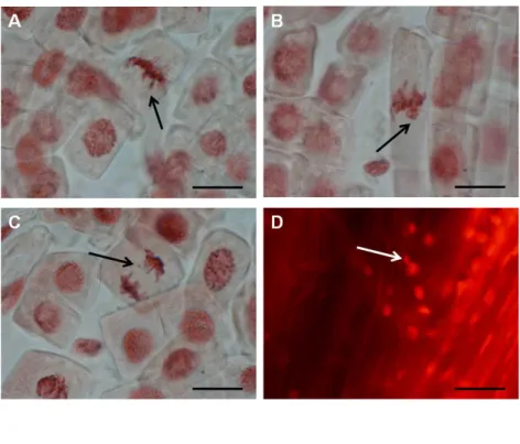
Mitotic aberrations and micronuclei ... 50
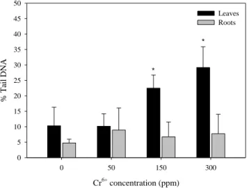
DNA damage ... 51
Antioxidant enzyme activities ... 52
Conclusions ... 55
References ... 56
Chapter 2.2 – Cadmium-induced cyto- and genotoxicity in lettuce ... 60
Abstract ... 61
Keywords ... 61
Introduction ... 62
Materials and methods ... 64
Plant culture and exposure to Cd... 64
Germination rate, plant growth, mortality, IC50, and LC20 estimation ... 64
Cadmium accumulation ... 65
Soluble proteins and antioxidant enzyme activities ... 65
Total antioxidant activity ... 65
Quantification of H2O2 ... 66
XI
Protein oxidation ... 67
Cell membrane permeability ... 67
Comet assay ... 67
Micronuclei... 68
Flow cytometric analysis ... 68
Statistical analysis ... 69
Results ... 70
Germination rate, plant growth, mortality, IC50, and LC20 estimation ... 70
Cd accumulation ... 70
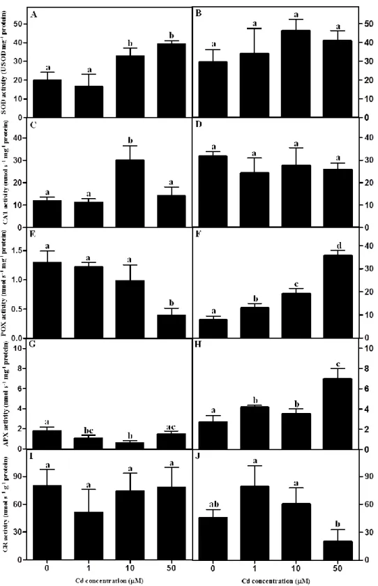
Antioxidant enzyme activities ... 72
Total antioxidant activity and H2O2 content ... 74
Lipid and protein oxidation, and cell membrane permeability ... 75
Comet assay and micronuclei ... 76
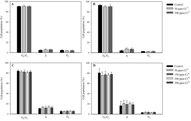
Flow cytometric analysis ... 79
Discussion ... 81
Conclusions ... 86
References ... 88
CHAPTER 3 – CHROMIUM AND CADMIUM CYTO- AND GENOTOXICITY IN HUMAN CELLS ... 94
Chapter 3.1 – Chromium-induced cyto- and genotoxicity in human osteoblasts ... 95
Abstract ... 96
Keywords ... 96
Introduction ... 97
Materials and methods ... 99
Cell culture ... 99
Cell viability ... 99
Cell cycle analysis ... 100
DNA damage ... 101
XII
Statistical analysis ... 102
Results ... 103
Cell viability ... 103
Cell cycle analysis ... 104
DNA damage ... 106
CBMN assay ... 108
Discussion ... 110
Conclusions ... 114
References ... 115
Chapter 3.2 – Cadmium-induced cyto- and genotoxicity in human osteoblasts ... 119
Abstract ... 120
Keywords ... 120
Introduction ... 121
Materials and methods ... 123
Cell culture ... 123
Cell viability ... 123
Cadmium quantification ... 124
Cell cycle analysis ... 124
Gene expression of cell cycle related proteins and DNA damage checkpoints ... 125
Indirect immunofluorescence of microtubules ... 126
DNA damage ... 127
CBMN assay ... 128
Statistical analysis ... 129
Results ... 130
General cell characterization and viability... 130
Cadmium quantification ... 132
Cell cycle analysis ... 133
Expression of genes related to the cell cycle proteins and DNA damage checkpoints ... 134
Cytoskeletal organization ... 136
XIII
CBMN assay ... 138
Discussion ... 140
Conclusions ... 145
References ... 147
Chapter 3.3 – Cadmium-induced mitochondrial dysfunction and oxidative stress in human osteoblasts ... 153
Abstract ... 154
Keywords ... 154
Introduction ... 155
Materials and methods ... 156
Cell culture ... 156
Protein quantification ... 157
Intracellular adenine nucleotides ... 157
Mitochondria isolation ... 158
Mitochondrial respiratory chain enzymes and citrate synthase activities ... 159
Mitochondrial membrane potential ... 160
Fluorescence microscopy of mitochondria ... 160
Intracellular ROS ... 161
Total antioxidant activity ... 161
Protein oxidation ... 162
Lipid peroxidation ... 162
Gene expression of antioxidant enzymes ... 163
Statistical analysis ... 165
Results ... 166
Cell energetic status and activity of mitochondrial respiratory chain enzymes and citrate synthase ... 166
Mitochondrial membrane potential and morphology ... 167
Intracellular ROS and total antioxidant activity ... 168
Protein oxidation and lipid peroxidation... 170
Gene expression of antioxidant enzymes ... 171
XIV
Conclusions ... 174 References ... 175
CHAPTER 4 – GENERAL CONCLUSIONS AND FUTURE PERSPECTIVES .... 179 General conclusions and future perspectives ... 180
XV
Abbreviations and acronyms
ABC – ATP-binding cassette
ABTS – 2,2’-azino-bis(3-ethylbenzthiazoline-6-sulfonic acid)
ADHP – 10-Acetyl-3,7-dihydrophenoxazine
ADP – Adenosine diphosphate
AFLP – Amplified fragment length polymorphism
AMP – Adenosine monophosphate
ANOVA – Analysis of variance
APX – Ascorbate peroxidase
Asc – Ascorbate
ATM – Ataxia telangiectasia mutated
ATM – Ataxia telangiectasia mutated coding gene ATP – Adenosine triphosphate
BER – Base excision repair
BSA – Bovine serum albumin
CAT – Catalase
CAX – Cation exchanger
CBMN – Cytokinesis-block micronucleus
CCNB1 – Cyclin B1
CCNB1 – Cyclin B1 coding gene CCNE1 – Cyclin E1
CCNE1 – Cyclin E1 coding gene
CDC25A – Cell division cycle 25A coding gene CDF – Cation diffusion facilitator
XVI
CDK2 – Cyclin-dependent kinase 2
CDK2 – Cyclin-dependent kinase 2 coding gene CHEK1 – Checkpoint kinase 1
CHEK1 – Checkpoint kinase 1 coding gene CHEK2 – Checkpoint kinase 2
CHEK2 – Checkpoint kinase 2 coding gene CoA – Coenzyme A
Cys – Cysteine
DCF – 2’,7’-Dichlorofluorescin
DCFH2 – 2’,7’-Dichlorodihydrofluorescin
DCFH2-DA – 2’,7’-Dichlorodihydrofluorescin diacetate
DHA – Dehydroascorbate
DHAR – Dehydroascorbate reductase
DMEM – Dubelcco’s modified eagle medium
DMSO – Dimethyl sulfoxide
DMT1 – Divalent metal transporter 1
DNPH – Dinitrophenylhydrazine
DSB – Double strand breaks
DTNB – 5,5'-Dithiobis-(2-nitrobenzoic acid)
DTT – Dithiothreitol
DW – Dry weight
EDTA – Ethylenediaminetetraacetic acid
EGTA – Ethyleneglycoltetraacetic acid
ER – Endoplasmic reticulum
XVII
FBS – Fetal bovine serum
FCM – Flow cytometry
FITC – Fluorescein isothiocyanate
FL – Fluorescence intensity
FPCV – Full peak coefficient of variation
FS – Forward scatter
GAPDH – Glyceraldehyde 3-phosphate dehydrogenase coding gene GPx – Glutathione peroxidase
GR – Glutathione reductase
GSH – Glutathione, reduced form
GSSG – Glutathione, oxidized form
HEPES – 2-(4-(2-hydroxyethyl)piperazin-1-yl)ethanesulfonic acid
HPCV – Half peak coefficient of variation
HPLC – High-performance liquid chromatography
HRP – Horseradish peroxidase
IC – Inhibition concentration
ICP-AES – Inductively coupled plasma atomic emission spectrometry
ICP-OES – Inductively coupled plasma optical emission spectrometry
ISO – International Organization for Standardization
LC – Lethal concentration
LMPA – Low melting point agarose
MDA – Malondialdehyde
MDHA – Monodehydroascorbate
MDHAR – Monodehydroascorbate reductase
XVIII
MFI – Median fluorescence intensity
MMR – Mismatch repair
MMS – Methyl methanosulfonate
MN – Micronucleus/micronuclei
MoM – Metal-on-metal
MT(s) – Metallothionein(s)
MTF1 – Metal-responsive transcription factor 1
MTP – Metal tolerance protein
MTT – 3-(4,5-Dimethylthiazol-2-yl)-2,5-diphenyltetrazolium bromide
NAD(P)H – Reduced nicotinamide adenine dinucleotide (phosphate)
NDI – Nuclear division index
NER – Nucleotide excision repair
NHEJ – Non- homologous end-joining
NMPA – Normal melting point agarose
NPBs – Nucleoplasmic bridges
NRAMP – Natural resistance associated macrophage protein
1O
2 – Singlet oxygen
O2• - – Superoxide anion
•OH – Hydroxyl radical
8-OHdG – 8-oxo-2’-deoxyguanosine
PBS – Phosphate buffer saline
PC(s) – Phytochelatin(s)
PCS – Phytochelatin synthases
PCR – Polymerase chain reaction
XIX
PI – Propidium iodide
PMSF – Phenylmethylsulfonyl fluoride
POX – Guaiacol peroxidase
PRI – “Post-R point” index
PVP – Polyvinylpyrrolidone
qPCR – Quantitative polymerase chain reaction
RBCs – Red blood cells
RER – Rough endoplasmic reticulum
Rho123 – Rhodamine 123
RNase – Ribonuclease
RNS – Reactive nitrogen species
ROS – Reactive oxygen species
RT – Room temperature
SD –Standard deviation
SDS – Sodium dodecyl sulphate
SE – Standard error
SNP – Single nucleotide polymorphism
SOD – Superoxide dismutase
SS – Side scatter
SSB – Single stranded breaks
TAA – Total antioxidant activity
TBA – Thiobarbituric acid
TBARS – Thiobarbituric acid reactive substances
TCA – Tricarboxilic acid
XX
TRITC – Tetramethylrhodamine
Tris-HCl – Tris(hydroxymethyl)aminomethane hydrochloride
UV – Ultra violet
WPB – Woody plant buffer
WST1 – 2-(4-Iodophenyl)-3-(4-nitrophenyl)-5-(2,4-disulfophenyl)-2H-tetrazolium, monosodium salt
ZIP – Zinc-related iron-related protein
ΔΨm – Mitochondrial membrane potential ε – Extinction coefficient
1
2
Metal environmental pollution
Metals occur naturally in Earth’s crust but episodes of environmental metal contamination are increasing mostly due to anthropogenic activities including agriculture (e.g., liming, sewage sludge, irrigation waters, pesticides and fertilizers) or municipal/industrial and mining effluents and wastes (Nagajyoti et al. 2010). Despite most dramatic increases of environmental contamination with metals had been occurring since the industrial revolution (18th-19th centuries), some examples of metal contamination and/or intoxication were already reported in ancient Roman and Greece (Gilbert 2012). Also, the increasing use of metals over the last centuries “has significantly altered the natural distribution of metals in the environment” (Gilbert 2012). These contaminants are, therefore, spread through the soils, water and air, where they become available and may be accumulated in living organisms (Nagajyoti et al. 2010).
Metals have been classified as essential metals or micronutrients (e.g., Cu, Zn, Fe, Mn), and non-essential metals (e.g., Cd, As, Pb, Hg) to living organisms. Metals from both groups may be toxic to plants or animals (Nagajyoti et al. 2010). The form/association of metals may differ in the environment, which alters their bioavailability and consequently their level of toxicity. This evidence is particularly significant in studies of (eco)toxicology and of environmental fate of metals, and their transport/accumulation through the food chain (Gilbert 2012). Thus, metals’ pollution and their persistence in the environment, as well as their toxicity to living organisms have been considered by international/governments agencies, like the Environmental Protection Agency (2013), the Agency for Toxic Substances and Disease Registry (2014), and European Comission (2001). For example, the Agency for Toxic Substances and Disease Registry has launched a substance priority list (of 275 substances) ranking Cd in the 7th place, and Cr6+, Cr6+ trioxide, and Cr in the 17th, 66th and 78th positions, respectively (Agency for Toxic Substances and Disease Registry 2014).
Chromium and cadmium contamination and toxicity – a general approach
Data published by the British Geologic Survey (2012) showed that from 2008-2012 China has been the major producer of Cd (and metals in general), and that the world’s total production of this metal over these years had remained overall constant. On the other hand, South Africa was the major producer of Cr (44% of world total Cr production). Also the
3
demand for Cr has dramatically increased these last decades, as Cr use increased from less than 2 million (in the early 20th century) to around 25 million tonnes in 2012 (British Geologic Survey 2012).
Supporting the scientific and public concern on metals’ pollution, it should also be highlighted that among different types of contaminants (e.g., chlorinated hydrocarbons, mineral oil, polycyclic aromatic hydrocarbons) affecting Europe, metals are the main category, with average values of contamination reaching 34.8% and 30.8% in soil and groundwater, respectively (Panagos et al. 2013).
Contaminations with Cr and Cd have been found in several Portuguese areas mostly associated to mining and industrial activities. The map below (Figure 1) summarizes the existing sources of mineral extraction (metallic and energetic), where some of the reported Cr and Cd-contaminated regions were identified. For example, in an active mine located in Coval da Mó high contamination levels with Pb, Zn and Cd were reported (Ferreira da Silva et al. 2009). Abandoned mines are still sources of metal contamination, like the Lousal mine where water and sediments contain high concentrations of Cd and other metals (Luís et al. 2011). In sampling sites of Ave River, water and sediments presented contamination with Cd and other metals, with the highest levels being found in areas near industries and highways (Pinto et al. 2011).
Since 1999 the Portuguese Environment Agency has been performing regular analyses of hazardous substances in monitoring stations of water sources (interior, estuary and coastal waters). The most recent report (Portuguese Environment Agency 2012) shows that Cd is among the most detected metals in sediments of these waters in the period of 1999-2004. The highest levels of Cd were found in monitoring stations near mines (e.g., Dornelas do Zêzere near Panasqueira mine (the major W mine of Europe) and in areas with a great urban and industrial impact (e.g., Lisbon and Setubal areas) (Figure 1).
4
Figure 1. Reserves of metallic and radioactive minerals in Portugal, and several regions contaminated with Cd, Cr or both metals. In general, in contaminated sites that are not located near mineral reserves, the main source of Cd and/or Cd presence/contamination is due to industrial and urban activity (adapted from Direcção Geral de Energia e Geologia (2013)).
In addition to the detection of contaminating metals in water, aquatic organisms such as barbel (Barbus sp.), harvested in the rivers Vouga, Douro, Mondego, Tejo, Sado, and Guadiana, showed contamination with Cd in their liver, with the highest levels observed in fishes from Sado River. Moreover, in all monitoring stations of estuary or coastal waters Cd was detected being the highest values found in Viana do Castelo, Castelo do Queijo, and
5
Lagos. Also in all stations of estuary or coastal waters Cd was found to accumulate in mussels and plankton (Portuguese Environment Agency 2012). The same report showed that Cr was also detected in some monitoring stations (e.g., Ribeira de Pernes) in water, and in the liver of fishes captured in Sado River (Portuguese Environment Agency 2012).
Environmental contamination with Cr in Portugal is mostly linked to industrial activities, and the Portuguese Environment Agency (2012) also identified the main and most recent national areas having Cr soil contamination (excluding mining areas), or where Cr and/or Cd represent a concern (including other contaminants). The national agency has approved investments for environmental recovery of soils contaminated with Cr and/or Cd. For example, investments were approved to recover soils of a former steel industry in Seixal (Cr and Cd contamination), and to recover the treatment plant of the tannery industry of Alcanena contaminated with Cr (Branco et al. 2005) (Figure 1).
More watercourses in other regions of the country had sediments contaminated with Cr (among other metals). Particularly relevant are the Aveiro canals, near former industries of ceramic and metallurgy, and the Murtosa canal that receives wastewater discharges from the Chemical Complex of Estarreja (Martins et al. 2013) (Figure 1).
In addition to environmental contamination, occupational exposure has also been identified as a main way of human contamination. As an example of these two ways of contamination, individuals working in Panasqueira mine, and populations living nearby, had higher concentrations of As, Cr, Mg, Mn, Mo, Ni, Pb, S, Se, and Zn compared to individuals living in non-contaminated areas (Coelho et al. 2014). Curiously, the populations environmentally exposed (and mainly women compared to men) showed higher levels of these metals (Coelho et al. 2014) and DNA damage (Coelho et al. 2013) than a similar population that was occupationally exposed. The higher frequency of MN (only in females) and chromosome aberrations was proposed by the authors to be related to environmental exposure (Coelho et al. 2013). Also, in a U mine area (in Cunha Baixa, Mangualde) contaminated with several metals (including Cd), local wild animals (mice and earthworms) accumulated high levels of Cd (Lourenço et al. 2012, 2013).
6
Chromium
Plants contamination by Cr: environmental contamination with Cr and its toxicity
depend on multiple conditions (e.g., Cr oxidation state, soil/water pH and model organism) (Peralta-Videa et al. 2009). Particularly, the oxidation state (Cr, Cr2+, Cr3+, Cr4+, Cr5+, and Cr6+) is crucial, being Cr3+ and Cr6+ the most stable forms (Zayed and Terry 2003), and Cr6+ the valence most widely studied concerning toxicity. The oxidative state of Cr affects Cr solubility and bioavailability in the environment, as well as its absorption, accumulation and toxicity in living organisms (Panda and Choudhury 2005). Contamination of crops with Cr may occur through contaminated irrigation water, soil and air/aerosols.
Contrarily to Cr6+, the effects of Cr3+ in plants are rather unknown (e.g., Song et al. 2014), and it has been widely assumed that Cr3+ is not necessary to plants and presents low toxicity or even may stimulate growth at low doses. For example, in bean plants, Cr3+ at low concentrations (0.25-1 µM) enhanced plant growth (Bonet et al. 1991). In another study, the response of seedlings of sorghum exposed to 50 and 100 µM Cr6+ or Cr3+ for 10 days was compared (Shanker and Pathmanabhan 2004). The results showed that Cr3+ led to lower accumulation of Cr in plant organs than Cr6+, and in both conditions the highest level of Cr was found in roots, followed by leaves and stems. Furthermore, 50 µM Cr3+ did not induce plant growth inhibition, neither increased ROS content in leaves, while Cr6+ reduced plant growth and was more cytotoxic (Shanker and Pathmanabhan 2004). Compared to the effects of Cr3+, Cr6+ induced higher levels of ROS, and lipid peroxidation, and stimulated antioxidant enzymes (SOD, DHAR, GR, CAT, and APX), but decreased GSH. Therefore, at the cellular level, Cr6+ induced an unbalance between ROS production and antioxidant defenses, i.e. Cr6+ induced oxidative stress (Shanker and Pathmanabhan 2004). More recently, Song et al. (2014) demonstrated in barley (Hordeum vulgare) that the Cr3+ toxicity, measured by the metal effects on root elongation, decreased with increasing activity of Ca2+ and Mg2+ (but not with K+ or Na+). The effect of pH was also explained by the H+ competition with Cr3+ and the concomitant toxicity of CrOH2+ in solution.
Induction of growth inhibition by Cr6+ was reported in maize (Sharma et al. 2003), citrillus (Dube et al. 2003), and alfalfa (in particular, root and shoot growth inhibition) (Peralta et al. 2001). In these studies, maize also presented chlorosis together with decrease of chlorophyll a and b contents and CAT inhibition (Sharma et al. 2003); citrillus showed
7
necrotic leaves and alterations in minerals content (Dube et al. 2003); and alfalfa had decreased seed germination (Peralta et al. 2001).
Our laboratory has extensively studied the toxic effects of Cr6+ in crops, particularly in Phaseolus vulgaris: in addition to root growth inhibition (Rodriguez et al. 2011), our group showed that Cr6+ negatively affected photosynthesis in pea plants after 28 days of exposure, e.g., by changing the morphology of chloroplasts, decreasing photosynthetic pigments content, the rate of CO2 assimilation, as well as Rubisco activity (Rodriguez et al. 2012). Furthermore, genotoxic effects of Cr6+ were also observed in these plants, i.e., Cr6+ induced DNA damage, polyploidization, and mutations, associated with cell cycle arrest at G2 phase (Rodriguez et al. 2011, 2013).
Human contamination by Cr: Despite the consensus that Cr6+ is toxic for both plants and animals (O’Brien et al. 2013; see Chapters 2.1 and 3.1), Cr 3+ is essential to animal diet, contributing to the normal metabolism of proteins, carbohydrates and lipids (Peralta-Videa et al. 2009). Some in vivo (mouse, rat and fruit fly) and in vitro (mouse and human cell lines) studies showed that Cr3+ used in nutritional supplements was not significantly toxic, while other studies showed that Cr3+ induced DNA damage, mutations, decrease of cell viability, or increase of cancer rates in offspring in mice (Levina and Lay 2008). Cr3+ supplements are freely available in the market, e.g., Cr3+ picolinate that was approved and considered safe by Food and Drug Administration (USA) and by Food Standards Agency (UK). But both agencies and the Agency for Toxic Substances and Disease Registry have been following published scientific data about toxicity of Cr3+ compounds used as dietary supplements. Considering this followed data the Agency for Toxic Substances and Disease Registry alerts for supplement consumption that should be taken with care, mainly at excessive doses (Wilbur et al. 2012).
Human/animal contamination with Cr may occur by several ways, including ingestion of contaminated water or food (via the food chain), occupational exposure (Keegan et al. 2008), and use of Cr-containing medical devices (e.g., dental (Eliasson et al. 2007), hip or other joint orthopedic prostheses (Sampson and Hart 2012)) often made of stainless steel or Co-Cr alloys (Gunaratnam and Grant 2008; Sampson and Hart 2012). During the use of some of these Cr-containing prostheses metal debris are released in the form of particles or ions (Cr6+ (Eiselstein et al. 2007)) and spread to many tissues (Gunaratnam and Grant 2008;
8
Campbell and Estey 2013) where they can lead to cyto- and genotoxicity (see more details in Chapter 3.1).
An experimental study demonstrated that mice exposed to Cr6+ suffered from oxidative stress leading to an increase of lipid and protein oxidation (Ben Hamida et al. 2013). The authors also demonstrated that the levels of nonenzymatic antioxidants (GSH, nonprotein thiol, vitamin C) as well as antioxidant enzyme activities (GPx and SOD) decreased despite of an increase of CAT activity. Also several biomarkers of liver injury (e.g., aspartate transaminase, alanine transaminase and lactate dehydrogenase activities, bilirubin, and albumin levels) increased (Ben Hamida et al. 2013).
The toxic effects of Cr6+ in chrome electroplating workers were evaluated in a research. For that, blood Cr levels and biomarkers of oxidative stress such as lipid peroxidation, thiol groups and antioxidant capacity of plasma were analyzed (Zendehdel et al. 2014), demonstrating Cr6+ induced biochemical toxicity. Another study demonstrated that chronic exposure to Cr6+ could disrupt blood element homeostasis (Song et al. (2012) vide Song et al. (2014)). These authors demonstrated in Sprague-Dawley rats that among blood, serum, and RBCs, the latest is the most sensitive to Cr6+ exposure. Song et al. (2014) also found dose-response relationships among rats exposed to Cr6+: Ca, Mg, and Mn in blood, Fe, Mg, and Se in serum, and Mg and Zn in lung tissue decreased with exposure. However, Ca, Co, Cr, Mg, Mn, and Se in RBCs, and Ca, Co and Mo increased in lung after Cr6+ exposure (Song et al. 2014).
In general, it has been demonstrated that absorbed Cr in mammalians distributes to nearly all tissues and it mostly accumulates in kidney and liver. Moreover, bone is also a major target, thus contributing to long-term persistence of Cr in the organism (Wilbur et al. 2012).
9
Cadmium
Crops contamination by Cd: As stated above Cd is used in industry (e.g., mines,
production of batteries, coatings and plating, pigments, plastic stabilizers, and also alloys (Faroon et al. 2012)). The improper disposal of wastes in the environment has led to Cd spread and contamination, accumulating in plants that are grown in contaminated soils. For example, high levels of Cd accumulated in edible parts of 20 plant species (e.g., lettuce, tomato, bean, carrot, and spinach plants) harvested from several sites around a Zn plant in Huludao, in China (Zheng et al. 2007). Also other wild organisms like mushrooms living in contaminated areas accumulate Cd (Petkovšek and Pokorny 2013).
Humans are generally exposed to Cd by ingestion of contaminated food (e.g., crops, mollusks, and crustaceans). In particular, cereals such as rice and wheat, green leafy vegetables, potato, carrot and celeriac may contain higher Cd levels than other edible plants and even more than fish and meat (Järup and Akesson 2009). Many urban gardens, like some from the urban area of Porto, have been found to contain soil contaminated with Cd, other metals and other toxic compounds at levels exceeding maximum values allowed by governmental laws, and at higher levels than those quantified in rural areas (Rodrigues et al. 2013). Therefore, plants harvested in urban areas are at risk of being contaminated with metals. This occured in harvested plants from home gardens of the municipality of Celje in Slovenia. Among these plants root and leafy vegetables accumulated more Cd compared to grain vegetables (Bešter et al. 2013).
Concerning Cd accumulation and its effect on plant growth, a research study showed that four plant species, Zea mays (sweet corn), Triticum aestivum (wheat), Cucumis sativus (cucumber), and Sorghum bicolor (sorghum) that were grown for 5 days in an artificial soil contaminated with increasing Cd concentrations, accumulated this metal in their tissues, mostly in roots (An 2004). Although seed germination was insignificantly affected by Cd, the growth of seedlings decreased in a dose-dependent manner. Moreover, between root and shoot growth evaluation, root growth was the most sensitive endpoint of Cd toxicity in these plant species. In particular, sorghum was the species accumulating the highest levels of Cd and the most sensitive to Cd, followed by cucumber, wheat, and sweet corn (An 2004). Similarly to Cr6+, in most species, Cd is also able to decrease the levels of chlorophyll pigments by substituting Mg2+ (Mysliwa-Kurdziel and Strzałka 2002), thereby inducing
10
chlorosis (Sanità di Toppi and Gabbrielli 1999). Cd may cause necrotic lesions in plant organs (Sanità di Toppi and Gabbrielli 1999) and may affect photosynthesis (e.g. in lettuce (Monteiro et al. 2009b; Dias et al. 2012)). Cd also induces oxidative stress by increasing ROS levels and changing the activity of antioxidant enzymes e.g. in lettuce (Monteiro et al. 2009b; Chapter 2.2). Cd is genotoxic to plants, as this metal induced DNA damage detected by comet assay in species like Allium cepa, and Nicotiana tabacum (Bandyopadhyay et al. 2011), or MN formation in Vicia faba (Souguir et al. 2010) (), and microsatellite mutations in lettuce, which were dependent on plant age and exposure conditions (Monteiro et al. 2007, 2009a).
Tobacco plants accumulate high levels of Cd in their leaves, and because of that, inhalation of tobacco smoke is an important source of Cd exposure in humans (Järup and Akesson 2009).
Human contamination by Cd: Cd has a long biological half-life in humans/animals,
particularly in kidneys and liver. Chronic ingestion of Cd might lead to different pathologies, like neurological diseases, infertility, diabetes, cancer, or renal and bone injuries like osteoporosis and osteomalacia (Oliveira et al. 2009; Nair et al. 2013).
Cadmium is known to cause a disease called “Itai Itai disease”, characterized by loss of bone tissue and failure of the kidneys. This disease was first mentioned in the early 20th century, when the population living near the Jinzu river basin in Toyama, Japan, was exposed to Cd by drinking contaminated water and by ingesting contaminated rice grown in that area (Nordberg 2009). More recently, a study involving a population living in an industrial complex in Korea showed high levels of Cd in urine associated with osteopenia and osteoporosis in adults, being the older people and female the groups presenting lower bone mineral density (Shin et al. 2011).
Cadmium is classified as carcinogenic to humans based on animal studies and occupational exposure to workers (IARC 1993). For example, wild mice living in a Cd-contaminated mine in Cunha Baixa, showed accumulation of Cd in bones, kidney and liver. These mice also showed altered gene expression of tumor suppressing genes (Lourenço et al. 2013). In addition, these mice and earthworms showed DNA damage (Lourenço et al. 2012, 2013).
11
Chromium and cadmium in the trophic chain: some case studies
As reported above, a way of human contamination by Cr and Cd includes ingestion of contaminated water and food (including contaminated crops). Some ecotoxicological studies have been done to evaluate the potential of metal transfer/biomagnification through different levels of the trophic chain. But compared with toxicological studies in isolated organisms, the consequences and fate of metals from plants/producers through terrestrial food chain has been less studied. Most research involved studies of metal transfer in the trophic chain in aquatic environments. Some authors (Wallace and Luoma 2003; Seebaugh and Wallace 2004) suggested that trophically available metal [i.e., metal associated with heat shock proteins such as MT, high density proteins (such as enzymes), and organelles] could support the bioenhancement of Cd transfer along aquatic food chains.
Some of the biological models used to assess metal bioaccumulation in situ or in lab conditions include crops such as pea (Rodriguez et al. 2011) and lettuce (Monteiro et al. 2009b), and other species (Zheng et al. 2007) including the hyperaccumulator plant species like Thlaspi spp. (Monteiro et al. 2010). Regarding animals/consumers, models as earthworms (e.g., Eisenia fetida (Alonso et al. 2009)), aquatic organisms like isopods (e.g., Porcellio dilatatus (Monteiro et al. 2008)), mollusks (e.g., oyster (Kurochkin et al. 2011)), crustaceans (e.g., Daphnia magna (Regaldo et al. 2009), crayfish (Kuklina et al. 2014)) and foraminiferal species (Martins et al. 2013); snails (e.g., Scheifler et al. 2002; Ebenso et al. 2013); rodents (Lourenço et al. 2012, 2013; Tête et al. 2014) and birds (Coeurdassier et al. 2012) were used. Also, microalgae (e.g., Chlorella vulgaris (Regaldo et al. 2009), diatoms (Ferreira da Silva et al. 2009)), bacteria (e.g., Megharaj et al. 2003; Khan et al. 2010; Kumar et al. 2012), and fungi as edible mushrooms (e.g., Kalač 2010) were also used. These organisms are considered bioindicators of metal contamination in ecosystems because they can be used for monitoring purposes in contaminated sites by evaluating metal bioaccumulation and toxicity in these living organisms.
The availability of a metal does not only depend on the total metal concentration in soil/water by itself, but it also depends on the pH of soil/water, metal form, organic matter content, chelating agents, as well as the species of living organism and the route of exposure (Rogival et al. 2007; Peralta-Videa et al. 2009). Thus, the bioconcentration factor (ratio between the metal concentration accumulated in the organism and the metal concentration
12
in the environment) is consequently different (Peralta-Videa et al. 2009). In a study of Marchese et al. (2008) the bioconcentration factor of Cr was calculated in several aquatic organisms from different levels of the food chain following 28 days of exposure to Cr-contaminated sediments. Plants (Ceratophyllum demersum) presented the highest value, 718.66 ± 272.91, followed by worm (Limnodrillus udekemianus) with 172.55 ± 80.8, the crab (Zilchiopsis collastinensis) with 67.72 ± 35.4, and the fish (Cnesterodon decemmaculatus) with 23.11 ± 12.82 (Marchese et al. 2008). Moreover, through the food chain, metals can be assimilated at different rates. For example, Calhôa et al. (2006) showed that Cd was more efficiently assimilated by isopods fed with lettuce contaminated superficially with 100 µM Cd(NO3)2 than by isopods fed with biologically contaminated lettuce (i.e., lettuce grown and exposed to 100 µM Cd for 7 days). This fact was related to the form of Cd present in lettuce: in biologically exposed lettuce, Cd was bound to thiol groups forming complexes of e.g., Cd-GSH, or Cd-Cys, reducing Cd availability; on the other hand, in lettuce superficially exposed to Cd, the metal was more available, leading to a high level of accumulation in isopods (Calhôa et al. 2006). Another study showed that Cd bioavailability to isopods was dependent on the subcellular partitioning of the metal in lettuce and two species of Thlaspi spp. (T. caerulescens, a hyperaccumulator species, and T. arvense, a nonaccumulator species) (Monteiro et al. 2008). Also Ding et al. (2013) used Amaranthus hypochondriacus L. and the insect Prodenia litura to characterize Cd allocation along these trophic levels and corresponding detoxification strategies. When exposed to contaminated soil, A. hypochondriacus leaves accumulated high levels of Cd and the concentration in P. litura larvae increased with increasing Cd concentrations in the leaves used as food supply. The authors found high Cd transfer coefficients from soil to leaf and from larvae to feces. A. hypochondriacus leaves showed the highest contents of Cd in pectates and in protein-integrated forms (Ding et al. 2013). On the other hand, cell fractions of P. litura larva showed that the type of proteins dominant for metal-binding compartmenting Cd were those obtained in the heat-stable protein fraction. These data supported that the way Cd is retained/accumulated at subcellular level is crucial for the mechanisms of Cd sequestration and excretion by P. litura larva feeding (Ding et al. 2013). Also in the worm species Limnodrilu hoffmeisteri, Cd showed different levels of bioavailability depending on its association with molecules or organelles. Cd associated with high density proteins and heat shock proteins was fully available to the consumer
13
Palaemonetes pugio, while Cd bound to organelles was less (70%) trophically available, and Cd bound to metal-rich granules was unavailable (Wallace and Lopez 1997).
Many ecotoxicological assays involve monitoring of contaminated areas, and organisms living there to assess metal bioaccumulation. For example, in several metal-contaminated sites near an active non-ferrous metallurgic factory in south of Antwerp, in Belgium, the metals As, Cd, Cu, Pb and Zn were found in soil (Rogival et al. 2007). In that study, two foods of the diet of wood mice – acorns (seeds of Quercus robur) and earthworms – were collected. Regarding Cd, the metal concentration in soil was positively correlated with Cd concentration in acorns and earthworms. Moreover, wood mice also living near the factory accumulated Cd in liver and kidneys, in a dose dependent manner related to the Cd concentration present in mouse diet (Rogival et al. 2007), supporting Cd transfer and bioaccumulation.
Juvenile snails (Helix aspersa), fed for 4 weeks with rape plants previously contaminated with Cd, showed Cd accumulation (Scheifler et al. 2002). In another study, other snail species (Limicolaria aurora) was fed for 4 weeks with edible mushrooms that were harvested in three contaminated (Cd, Cr, and other metals) farm sites in Niger Delta (Nigeria) (Ebenso et al. 2013). However, the metal uptake by the snails was low and bioaccumulation was not enough to consider the snails improper for human consumption (< 1 mg Kg-1 Cr or Cd, (FAO/WHO 2001 vide Ebenso et al. 2013)).
Compared to Cd, less ecotoxicological assays have been done regarding Cr contamination of the environment and its transfer through the trophic chain. Some studies show the possibility of Cr accumulation and toxicity in organisms. For example, Kuykendall et al. (2006) exposed two species of fish, fathead minnows and largemouth bass (Micropterus salmoides) (predator), to 2 ppm Cr6+ in water for 4 days. Cr6+ led to DNA-protein crosslink formation in erythrocytes of both species (higher levels in fathead minnows). When largemouth bass individuals were fed with fathead exposed to Cr6+ DNA-protein crosslinks also increased. Thus, both water and diet intake with Cr6+ led to DNA-protein crosslink formation in a predator fish (Kuykendall et al. 2006).
In Nord-Pas-de-Calais, France, where a former Pb and Zn smelter is located, some vegetables produced in agricultural soils and home gardens contained high concentrations
14
of Cd and Pb at levels higher than permissible values for human consumption (Douay et al. 2013). In other polluted areas, in Zhejiang Province, China, there are agricultural fields located near local industries (e.g., metal smelting and battery making) where high levels of Pb, Cd, Cr, Hg, and As were found (Liu et al. 2013). Almost 300 vegetable species were sampled from these soils and depending on the plants species metals accumulated at different levels: e.g., celery accumulated the highest Cr concentration and tomato the lowest, and asparagus and lettuce plants accumulated higher Cd and Cr concentrations compared to rape. According to the model of risk assessment applied in this study, Cd is the metal causing the greatest cancer risk (Liu et al. 2013).
Mechanisms of chromium and cadmium uptake in plants and humans
Chromium
Uptake of Cr by plants and translocation from roots to shoots depends on the metal oxidation state, which in turn depends on soil pH, organic matter, chelating compounds, and microorganisms present in the rhizosphere (Manara 2012). In particular, in acidic soils Cr6+ compounds are more mobile and also more easily reduced to Cr3+. Moreover, the organic matter, Fe2+, and sulfites can readily reduce them to Cr 3+, whereas manganese oxides can oxidize Cr3+ to Cr6+ (Becquer et al. 2003). In addition, some compounds from root exudates, like organic acids may reduce Cr6+ and/or form complexes, increasing the solubility and mobility of Cr through the root xylem (Bluskov et al. 2005).
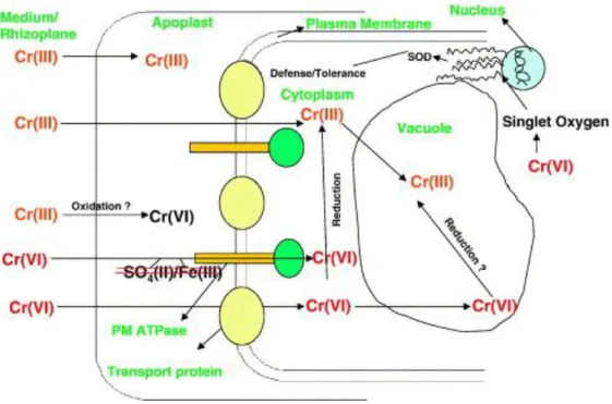
In the current proposed model of Cr uptake and toxicity in plant roots (Figure 2), both Cr6+ and Cr3+ are taken up via symplast (Shanker et al. 2005) and plants may be capable of reducing Cr6+ to Cr5+, Cr4+ and Cr3+, contributing to detoxification of Cr6+ (Santos and Rodriguez 2012). Uptake of Cr6+ by root cells occurs by active transport through carriers of sulphate, phosphate and iron ions, while Cr3+ is taken up by passive transport (Shanker et al. 2005).
Figure 2 also shows that Cr6+ may be immobilized in vacuoles of root cells and possibly reduced to Cr3+ as a way of detoxification and prevention of Cr6+ translocation from roots to leaves (Shanker et al. 2005). In fact, Cr content is frequently very much higher in roots than in other parts of the plant. For example, it was evaluated the uptake of Cr3+ and Cr6+ in water
15
hyacinth, and it was found that both Cr states accumulated preferably in the root and that Cr6+ was more translocated (Paiva et al. 2009). In tumbleweed, the same pattern of Cr3+ and Cr6+ uptake was observed (Gardea-Torresdey et al. 2005). Unlike other metals (e.g., Cd), Cr is unable to induce PCs in plants (Shanker et al. 2005), and it is thought that Cr may induce the synthesis of MT, although the mechanisms involved are not yet clear (Shanker and Pathmanabhan 2004).
16
In animal cells Cr6+ (e.g., CrO
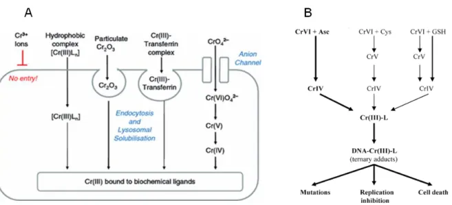
42-) uptake (Figure 3A) occurs via anion channels (sulphate and phosphate) (Beyersmann and Hartwig 2008), like in plant root cells. In the extracellular environment Cr6+ may be reduced to Cr3+ as a way of prevention of toxicity, and the existent Cr3+ ions enter slowly into the cell by diffusion or phagocytosis (Collins et al. 2010; Zhitkovich 2011). According to Beyersmann and Hartwig (2008) and as shown in Figure 3A, Cr3+ can form complexes with hydrophobic ligands (e.g., 1,10-phenanthroline, 2,2’-bipyridine, picolinic acid) that permeate by diffusion through plasma membrane. Moreover, Cr2O3 particles can be taken up by phagocytosis, and then solubilized in lysosomes and Cr3+ is released. Cr3+ can also bind to transferrin (competing with Fe2+) and enter the cell by endocytosis (Figure 3A).
Figure 3B shows that once in cells, Cr6+ is rapidly reduced to Cr5+, Cr4+ and Cr3+ by reducing enzymes (e.g., NADPH cytochrome c reductase, GR) and non-enzymatic reducers like Asc, GSH, and Cys (Afolaranmi et al. 2008). As Cr6+ is being reduced, ROS are generated in Fenton or Haber-Weiss reactions (Raghunathan et al. 2009). These ROS and intermediate forms of Cr can induce genetic damages, including Cr-DNA adducts, Cr-DNA crosslinks, Cr-DNA-protein crosslinks, DNA interstrand crosslinks, oxidative DNA damage, DNA DSB/SSB and abasic sites (O’Brien et al. 2003).
Figure 3. (A) Model of Cr uptake in animal cells, proposed by Beyersmann and Hartwig (2008). (B) Reduction
of Cr6+ by Asc, Cys, and GSH, and formation of Cr-DNA adducts that may lead to cyto- and genotoxic effects. L – ligand (Asc, GSH, Cys, or histidine) (Adapted from Zhitkovich (2005)).
17
Cadmium
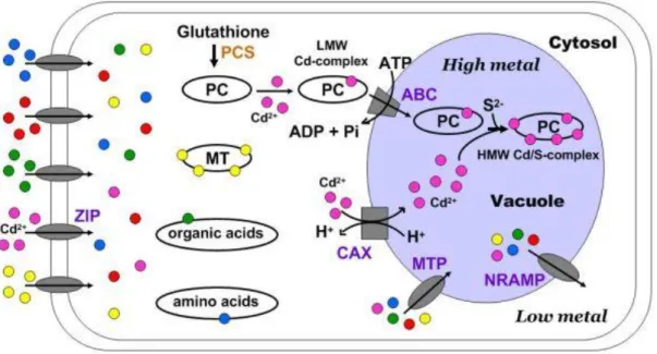
Uptake of Cr in plants depends on the rhizosphere factors, as previously mentioned for Cr. When Cd ions come in contact with root cells, they can be retained in the cell wall containing pectic sites, hystidyl groups, and extracellular carbohydrates (e.g., callose and mucilage) (Manara 2012). Yang and Chu (2011) presented the mechanism of Cd uptake in plant root cells that is shown in Figure 4.
Figure 4. Cd uptake and sequestration in vacuoles of plant cells (Yang and Chu 2011).
Similarly to Cr, Cd enters the root cell via symplast (Clemens et al. 2002). The cell membrane of a root cell has several families of transporters (also observed in other living organisms) involved in metal uptake and homeostasis, and some of them like the ZIP family (carrier of e.g., Fe2+, Mn2+, Zn2+), has been associated with Cd2+ carriage into the cytoplasm (Yang and Chu 2011). In addition, Cd uptake can be reduced by membrane efflux transporters (CDF family) that pump Cd ions outside the cell, and these transporters can also carry Cd into the xylem (Manara 2012). The increase of Cd ions in cytoplasm increases the generation of Cys-rich metal binding peptides – PCs, which were identified in plants, fungi and diatoms, and apparently may also exist in some animals (Clemens et al. 2001). These are synthesized from GSH by PCS. Cd is then complexed with PC (due to thiol and carboxyl groups), originating a low molecular weight complex that is sequestered into the vacuole through an ABC transporter. Other Cd ions that enter into the vacuole via CAX transporters
18
(Ca2+/H+ antiports) and sulfide bind to low molecular weight complexes to produce the high molecular weight complex (Yang and Chu 2011). The vacuole is, therefore, the main storage compartment of Cd (Manara 2012), but when necessary, MTP and NRAMP transporters regulate the flux of metal ions (Yang and Chu 2011). Also in response to high Cd levels in cytoplasm, MTs (Cys-rich metal binding polypeptides (Cobbett and Goldsbrough 2002)), organic acids (e.g., malate, citrate and oxalate), amino acids (e.g., hystidine, and proline), GSH, and phytate (a main storage of phosphorous in plants) are able to chelate Cd (Yang and Chu 2011; Manara 2012), decreasing Cd availability to induce ROS formation and cause cellular damages. Besides being a chelating agent, MT is able to eliminate •O2- and •OH by reaction of Cys residues, thus it can reduce the levels of ROS induced by Cd in cells (Yang and Chu 2011). Cd is translocated from roots to shoots as complexes and via xylem(Hossain et al. 2012).
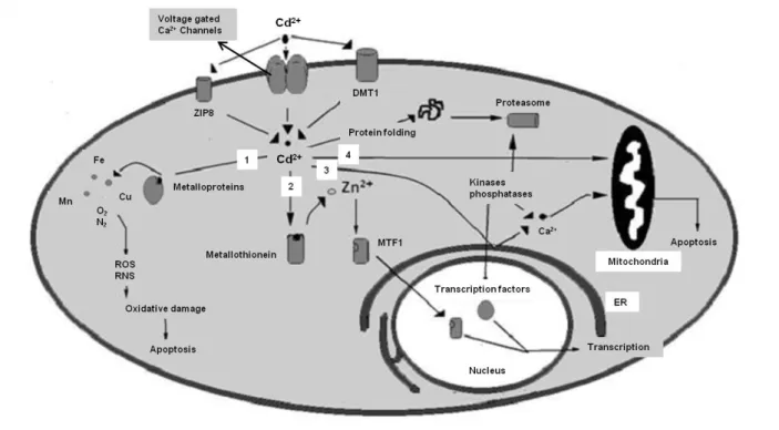
Uptake of Cd in animal cells may occur by different ways. Complexes of Cd with MT, albumin or other proteins enter the cell by endocytosis. Moreover, when Cd is free (Cd2+) or complexed with Cys, GSH, it can bind to active sites of carrier proteins and channels of substrates as amino acids, oligopeptides, organic anions or organic cations (e.g., the organic anion transporters Oat-1, Oat-3) to enter the cell (Bridges and Zalups 2005; Sabolić et al. 2010). Cd ions can also be transported by carrier proteins and channels (mainly DMT1, voltage gated Ca2+ channels and ZIP8) involved in the uptake of divalent cations like Ca2+, Fe2+, and Zn2+. Figure 5 shows Cd entering the animal cell (according to Sarkar et al. (2013)) by these ion transporters and, when inside the cell Cd may take different pathways: (1) Cd competes with Fe2+, Mn2+, Cu2+, Zn2+, for active sites of MT either leading to misfolding of the protein or production of ROS/RNS that contribute to oxidative damage and, ultimately, to cell death; (2) Cd can also bind to MT displacing Zn2+ which in turn binds to MTF1 transcription factor that encodes a transcription factor that induces the expression of MT and other genes involved in metal homeostasis; (3) Cd ions can also bind to ER membrane leading to release of Ca2+, which in turn can lead to apoptosis by caspases activation, or Ca2+ can activate some kinases and phosphatases resulting in transcription of cell cycle genes (e.g., related with cell cycle progression, therefore influencing cell proliferation); binding of Ca2+ may also lead to proteasomal degradation of proteins. When in the cell, (4) Cd also induces apoptosis by mitochondrial pathway (Sarkar et al. 2013).
19
Figure 5. Cd uptake and some cytotoxic effects in animal cells (Adapted from Sarkar et al. (2013)).
Chronic exposure to low Cd doses is highly correlated with Cd accumulation and cellular damages in kidneys, and Cd concentration in urine has been used as biomarker of long-term Cd exposure. However in acute exposure with high Cd doses, Cd mainly accumulates in liver, and Cd concentration in blood has been used as biomarker of a recent exposure to Cd (Godt et al. 2006). Besides bones, liver and kidneys, Cd can also accumulate in testis, spleen, heart, lungs, thymus, salivary glands, epididymis, prostate, pancreas, and central nervous system (Sarkar et al. 2013).
Most current biomarkers used in metal exposure and toxicity assessment
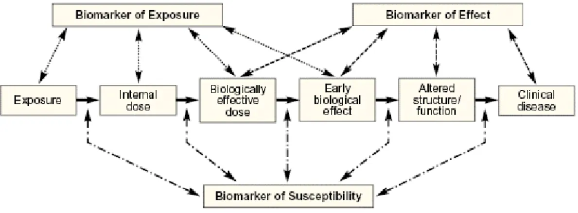
In the assessment of metal exposure and toxicity, different biomarkers have been used at all levels of biological organization (from molecules to cells, individuals, populations, communities and ecosystems) (Mussali-Galante et al. 2013). Figure 6 explains how biomarkers can be classified. The use of biomarkers allows the assessment of exposure level – biomarkers of exposure – and the extent of toxic response, including changes in the structure, function/performance of the cell/tissue/organism – biomarkers of effect (Bearer
20
2001; Mussali-Galante et al. 2013). For example, concerning biomarkers of exposure, one can consider the measurement of a contaminant or related metabolites (e.g., Cd, Cd-MT, Cr, Cr-GSH) in target organs/tissues or in body fluids. The set of biomarkers (of exposure and effect) are considered biomarkers of susceptibility allowing prediction of individual’s susceptibility to a particular compound/xenobiotic (Mussali-Galante et al. 2013). Biomarkers of susceptibility are therefore valuable tools for diagnostics providing insight into the mechanisms of action and the pathways and targets of the compound/xenobiotic. A set of biomarkers can also be used in biomonitoring, and risk assessment involving predictive evaluations in non-intentionally exposed organisms, populations, or ecosystems.
Figure 6. Biomarkers of exposure, effect and susceptibility in an individual under xenobiotic exposure (Bearer
2001).
Therefore, the ideal combination of biomarkers should combine different types of biomarkers, and consider all levels of biological organization, including molecules, cell, organism, population, community and ecosystem (Mussali-Galante et al. 2013). Moreover, this ideal combination of biomarkers should allow a distinction between reversible and irreversible effects, by identifying the most sensitive changes occurring within the shortest period of time, and using minimally invasive techniques to the living being under study.
At cellular level (in both plants and animals), metal biomarkers are frequently classified according to the cell’s ability for metal uptake and cytotoxic effects (Aziz et al. 2014). Cytotoxicity is usually evaluated by measuring changes in cell viability, oxidative stress, cell cycle progression, mitochondrial dysfunction, and cell death. In particular, these
21
biomarkers have been largely used in human cell lines (AshaRani et al. 2009; Aziz et al. 2014). Other biomarkers are focused on measuring genotoxic effects. We highlight here DNA damage measured by comet assay – i.e., DNA DSB/SSB and alkali-labile sites; presence of MN in e.g., Allium sepa L. (Seth et al. 2008) and human cell lines (AshaRani et al. 2009); DNA-ploidy changes, which were identified, for example, by FCM in Pisum sativum (Rodriguez et al. 2011); and microsatellite instability, which was found in Lactuca sativa L. (Monteiro et al. 2009a) and in P. sativum (Rodriguez et al. 2013), exposed to Cd and Pb, respectively. These data support that at the cellular level one should combine cytotoxic with genotoxic biomarkers.
The combination of multiple biomarkers of cytotoxicity and genotoxicity has been exhaustively investigated in animals and less in plants. Some of the cytotoxic and genotoxic biomarkers used to assess metal toxicity are commonly used in animals and plant models (e.g., oxidative stress, loss of membrane integrity, DNA damage, mutations, MN) (Monteiro et al. 2007, 2009b; Rodríguez-Serrano et al. 2009; Wu et al. 2010; Rodriguez et al. 2011; Patnaik et al. 2013).
Brief notes on biological models used in metal toxicity assays
In vivo vs in vitro assays
Toxicological research usually involves in vivo assays with exposure of animals to chemicals, or in vitro assays using cell/tissue culture, or isolated organs. In vivo assays are closer to real exposure conditions because the response of an organism to a chemical is influenced by the function of organs, circulatory and lymphatic systems in superior animals, or vascular system in plants. Moreover, assays with living organisms imply different perspectives of exposure, i.e. acute exposure, or chronic exposure throughout their life cycle, where effects on reproduction may be observed. While in vivo assays using plants do not raise ethical issues (however it requires some logistic requirements), in vivo assays using animals have, nowadays, many restrictions. Regarding the value of animal welfare (without suffer, pain and discomfort) it is considered that an “animal is any vertebrate animal non human and cephalopods (as it was proved they suffer pain), including autonomous larval and/or reproduction forms” (European Commission 2010). In general, in vivo assays follow guidelines of good practices when using animals, following the principle of 3Rs firstly
22
advocated by William Russell and Rex Burch (Russell and Burch 1959), and actually into force by the European Commission (2010). This principle incites for Reduction of the number of animals used per treatment condition, Refinement of protocol methodologies providing the best possible welfare conditions, and Replacement of in vivo assays by in vitro, or in silico assays, or the use of lower organisms and/or at early stages of development (i.e. with or without incomplete development of neuronal system) (European Commission 2010)). In the European directive it is stated that “An experiment shall not be performed on an animal, if another scientifically satisfactory method of obtaining the result…, is reasonably and practicably available” (European Commission 2010).
Why lettuce and human bone cells as models?
Toxicity assays on the effects of metals in humans cannot be done directly (only biomonitoring, risk assessment, and in silico evaluations) clearly due to ethical issues, thus in vivo assays using e.g., rats, mice, pigs, or in vitro assays have been performed. But using animal models not always reflects the human response to the same chemical. Therefore, in vitro assays using human cells are promising alternatives, providing preliminary data that may help in planning future trials in vivo. Other alternatives are the reduction of the number of animals and analyses of the necessary biomarkers only to avoid or at least reduce animal suffering.
In this thesis, metal (Cr and Cd) toxicity is assessed in two biological models, plants in vivo (Chapters 2.1 and 2.2.), and human cells in vitro (Chapters 3.1-3.3.). Some plant species, including crops like lettuce (L. sativa L.) are recommended by OECD for standard ISO toxicity tests (OECD 2006). Lettuce was also chosen because it is a widely commercialized crop and able to accumulate metals (e.g., accumulation of Cd, Cr, Ni, Pb, among others, in edible parts (Peris et al. 2007)), and several toxicity effects have previously been observed in lettuce exposed to Cd (Monteiro et al. 2007, 2009a, 2009b, 2010). However, most of these studies provided non systematized data (e.g., focusing only some particular aspects of plant cellular response to Cd), being mostly focused on e.g., oxidative stress (e.g., Monteiro et al. 2009b). Besides, data provided by Monteiro et al. (2009b) only used one dose, 100 µM, currently acknowledged as too high. Therefore, to decipher the mechanisms underlying Cd cyto- and genotoxicity and the cell responses, it is crucial to study, in a more systematic and