Universidade de Trás-os-Montes e Alto Douro
Pilot study on Bovine Fasciolosis in Praia
slaughterhouse in Santiago – Cape Verde
Dissertação de Mestrado em Medicina Veterinária
Sara Machado da Silva Levy Cruz
Orientador: Professora Doutora Maria Madalena Vieira-Pinto
Universidade de Trás-os-Montes e Alto Douro
Pilot study on Bovine Fasciolosis in Praia
slaughterhouse in Santiago – Cape Verde
Dissertação de Mestrado em Medicina Veterinária
Sara Machado da Silva Levy Cruz
Orientador: Professora Doutora Maria Madalena Vieira-Pinto
For my mother My father Bárbara Fernando And Carlos.
“It's a dangerous business, Frodo, going out your door. You step onto the road, and if you don't keep your feet, there's no knowing where you might be
Acknowledgments
Durante este trabalho, houve muitas pessoas que me ajudaram e apoiaram de variadíssimas maneiras. Umas menos, outras mais. As suas palavras ou ações não passaram despercebidas nem tampouco foram esquecidas. A elas sou grata:
Em primeiro lugar, à minha orientadora, Professora Doutora Maria Madalena Vieira-Pinto, que entrou no meu barco, apoiou o projeto, que só esteve confirmado mesmo à altura da hora, quando já estava a entrar no avião. Tenho de agradecer todos os “puxões de orelha”, o interesse interminável, os momentos “aha” elucidativos e todas as oportunidades que me proporcionou.
À Professora Doutora Manuela Calado, por tudo o que me ensinou, com tanta paciência, e também por todo o carinho e interesse. Agradeço também todo o apoio que recebi por parte do IHMT, ao António, à Cátia e ao Tiago, por todas os pequenos conselhos no laboratório.
À Professora Doutora Isabel Pires, pela ajuda constante e carinho, não só neste projeto, mas durante todo o MIMV. À Professora Doutora Justina Prada, estou grata pelo corte para as lâminas histopatológicas deste estudo. À Professora Doutora Paula Rodrigues por todo o trabalho que envolveu as fotografias, foi incansável e estou muito agradecida.
À Eng.ª Teresa Coutinho do Laboratório de Parasitologia da UTAD, pelo incansável trabalho com os meus parasitas.
À Dr.ª Teresa Mateus por todo o trabalho que teve, fico eternamente agradecida.
Ao Dr. Pedro Roquette, por ser sempre um marco e um apoio. Por ser sempre o meu “Pedro Roquel”.
Tenho a agradecer o apoio incondicional por parte do pessoal da DGASP, MDR. Ao Coordenador da Pecuária, Eng.º João Fonseca pela oportunidade, pelo valor que sempre me deu, por nunca me ter fechado a porta e por me ter sempre incluído e feito sentir que fazia parte da equipa. À Eng.ª Lígia Matos, por todos os conselhos, por todos os esclarecimentos sem hora, por
todo o saber partilhado, por me ter dado gosto pelo trabalho e me ter ensinado a trabalhar em equipa. Ao Dr. Afonso Semedo por me ter recebido, ajudado a coordenar o meu estágio e pelo apoio. Ao Dr. José Luís pelo rigor e profissionalismo. À Eng.ª Helena Gomes por toda a companhia, amizade e companheirismo.
A todo o pessoal do Porto da Praia, Ziza, Jaime, César, Miqueias, Luis, Cesarina, Alberto e Belmira, por todos os ensinamentos, apoio e companheirismo.
A todos os funcionários do Matadouro da Praia, principalmente ao Miguel, ao Nelson e ao Gabi. Estou grata pelo que me ensinaram, por me terem recebido tão bem, por nunca me recusarem nada e pela humildade.
À Dr.ª Margarida Correia, porque sem ela, o meu estágio não existiria. Obrigada pela insistência, pela comunicação e pela ajuda. Igualmente ao Dr. Edson Santos por toda a ajuda preciosa que gentilmente cedeu.
A uma das pessoas mais importantes neste estágio, o meu tio Abraão Levy, pela hospitalidade, pelos eternos ensinamentos, por toda a disponibilidade, por todo o amor, por me ter feito sentir em casa, por eu sentir, neste momento, que Cabo Verde é a minha segunda casa. Por tanta coisa que nunca conseguirei descrever. À Amália pela companhia, pelas conversas, pela hospitalidade. À minha prima Paula Levy, por me ter mostrado tanta coisa e me ter recebido tão bem. Ao meu primo Yannick Levy Monteiro, pela companhia e amizade.
Queria agradecer a todos os meus amigos. Aos BIGS, por todo o companheirismo e amizade. Aos meus amigos Daniela e Fernando por todo o apoio, tempo e amor. Ao André por significar tanto e, embora não me tenha acompanhado nesta aventura, esteve sempre presente. Obrigada por me ensinar tanto e por aceitar o que lhe transmito, sempre. À Diana, Luiza e Sofia pelo companheirismo e momentos. Ao Karol, grande companheiro e amigo.
Queria agradecer à Elisabete, que trabalha em minha casa desde os meus 8 anos, que me educou juntamente com os meus pais. Estou eternamente agradecida por todo o apoio, conselhos e companhia ao longo de todo o meu percurso, académico e de vida.
Aos meus padrinhos, ambos falecidos durante a execução deste trabalho, por todo o apoio ao longo de toda a minha vida. Sempre presentes e nunca esquecidos.
À minha prima Isabel Matos por ter gosto em me ensinar e por me ter sempre apoiado. À Maluda e à Meena.
À minha irmã Bárbara, por todo o apoio, pelo amor fraternal e por toda a ajuda e conselhos. Ao meu irmão Fernando pela companhia, pelo amor e por me tentar sempre agradar em todas as situações, através de palavras ou ações. Por nunca me dizer que não a absolutamente nada.
Ao Carlos, pelo companheirismo, pelo respeito, pelo ensinamento mútuo, por partilhar um plano comigo, pelo empenho desmedido em nós.
Ao meu pai por todo o apoio.
À minha mãe por todo o apoio, pelo seu sacrifício, por me incentivar sempre a dar o meu máximo e acreditar que sou capaz, mesmo quando eu não sou. Por apostar em todas as minhas aventuras e me dizer para ir em frente e não olhar para trás.
Abstract
Fasciolosis is a disease caused by a parasitic trematode, usually Fasciola hepatica or
Fasciola gigantica, which affects animals and man. This disease has socio-economically
devastating effects.
In the present study, the occurrence level of Fasciolosis in Praia slaughterhouse, in Santiago, Cape Verde, was calculated. From 131 bovines slaughtered, during the study period, 12 (9%) had Fasciolosis compatible lesions.
The parasite’s genetic characterization was made, through Restriction Fragment Length Polymorphism Analysis of PCR-Amplified Fragments (PCR-RFLP), in order to reliably confirm the presence of Fasciola gigantica instead of Fasciola hepatica. 11 faecal samples, from the cases with FCL (Fasciolosis compatible lesions), were analyzed through simple sedimentation techniques. From the 12 samples, 7 contained Fasciola spp. eggs.
A sample of liver and lymph node were collected for histopathological analysis. The liver had findings compatible with chronic hepatitis with microvacuolar hepatic degeneration, extensive areas of localized fibrosis, portal inflammatory infiltrate and hyperplasia of the bile ducts whereas the lymph node revealed follicular and diffuse reactivity, accumulation of a dark brown to black pigment in the macrophages’ cytoplasm localized in the medullary sinuses and discreet inflammatory infiltrate constituted by eosinophils.
Resumo
A Fasciolose é uma doença causada por um parasita da classe Trematoda, do género
Fasciola, sendo normalmente Fasciola hepatica ou Fasciola gigantica, que afecta animais e o
homem. Esta doença tem efeitos socioecónomicos devastadores. Estima-se que perto de 600 milhões de animais e 2.4 milhões de pessoas, no mundo inteiro, sofrem de Fasciolose.
No presente estudo, a taxa de ocorrência de Fasciolose no Matadouro da Praia, em Santigo, Cabo Verde, foi calculada. Dos 131 bovinos abatidos, durante o período de estudo, 12 (9%) tinham lesões compatíveis com Fasciolose.
Foi feita a caracterização genética do parasita foi feita, através de Restriction Fragment Length Polymorphism Analysis of PCR-Amplified Fragments (PCR-RFLP), para confirmar, com certeza, que a infecção era causada por Fasciola gigantica e não Fasciola hepatica.
11 amostra de fezes, dos casos com FCL (lesões compatíveis com Fasciolose), foram analisados através da técnica de sedimentação simples. Das 11 amostras, 7 continham ovos de
Fasciola.
Duas amostras, uma de fígado e outra de gânglio linfático, foram recolhidas para se proceder a análise histopatológica. O fígado apresentava lesões compatíveis com hepatite crónica com degenerescência hepatocitária microvacuolar, áreas extensas de fibrose localizada, infiltrado inflamatório portal e hiperplasia dos ductos biliares. O gânglio linfático possuía reatividade folicular e difusa, acumulação de pigmento castanho escuro a negro, no citoplasma de macrófagos localizados nos seios medulares e possuía também discreto infiltrado inflamatório por eosinófilos.
Index
Introduction…….………1
Objectives……….2
Literature Review……….………...3
1. Cape Verde – Food Safety Related Services………...………...3
2. Fasciola spp……….………..5
2.1. Taxonomy...………..…….………..5
2.2. Morphology………..………...6
2.2.1. Differentiation between species…………...………...8
2.3. World distribution…..………..9
2.4. Life cycle………..………..10
3. Fasciolosis………..………...12
3.1. Historical background………...………..12
3.2. Fasciolosis transmission and dissemination………...………….13
3.2.1. Larvar development………...………14
3.2.2. Normal definitive hosts and adaptation to new definitive hosts………...15
3.3. Fasciolosis development…………...15
3.4. Clinical and lesional presentation………17
3.5. Diagnosis………...………..19
3.5.1. Differential diagnosis between Fasciola hepatica and Fasciola gigantica…...20
3.6. Prevention and Control………....21
3.7. Economic losses related with Fasciolosis………22
3.8. Fasciolosis in Cape Verde – Human and Animal……...……….23
4. Importance of slaughterhouse and sanitary inspection…...……….26
Material and Methods………...………...29
1. Slaughterhouse characterization...………..29
2. Slaughter volume register and causes for meat condemnation…...………...33
3. Evaluation of fasciolosis as a cause of meat condemnation…...………...33
3.2. Sample collection………34
4. Laboratory analysis………...35
4.1. Molecular analysis……….………...………...35
4.1.1. Total genomic DNA extraction……….………35
4.1.2. Polymerase Chain Reaction Amplification (PCR)…..……….36
4.1.3. Restriction Fragment Length Polymorphism (RFLP)…...………37
4.2. Faecal Analysis………...………...38
4.3. Histopathological Analysis………...……….39
Results and Discussion………...………..40
1. Slaughter volume register and causes of meat condemnation………..40
2. Evaluation of fasciolosis as a cause of meat condemnation………...………...40
2.1. Lesion register and classification………...………42
3. Laboratory analysis………...………...50
3.1. Molecular analysis………...………..50
3.1.1. Total genomic DNA extraction………...………51
3.1.2. Polymerase chain reaction amplification (PCR)………...……….51
3.1.3. Restriction fragment length polymorphism (RFLP)………...………...52
3.2. Faecal analysis………...………53
Conclusions………55
Index of figures
Figure 1- Map of Cape Verde………...3
Figure 2- Anatomy of Fasciola hepatica……….……….7
Figure 3- Fasciola gigantica (A) with detailed cephalic cone (B), uterus carrying eggs (C), ovaries, vitelline duct, ootype (D), testes (E) and vitellaria (F)……….7
Figure 4- Fasciola tegument with spines (black arrows) directed backwards………..…….8
Figure 5- Fasciola gigantica (A) and Fasciola hepatica (B)………..………...8
Figure 6- Fasciola egg………..………10
Figure 7- Life cycle of Fasciola hepatica…...………..12
Figure 8- Affected gall bladder from a bovine with Fasciolosis...………16
Figure 9 – Bovine liver with bile ducts’ thickening, on the left lobe…………...……….18
Figure 10- Bovine liver with hyperplasia of the bile ducts…….………..19
Figure 11- Calcium deposit (black arrow) and a Fasciola gigantica fluke (white arrow)…...….19
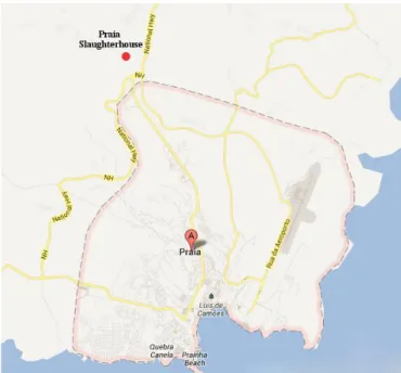
Figure 12- Map with the location of Praia slaughterhouse...………….…………...………29
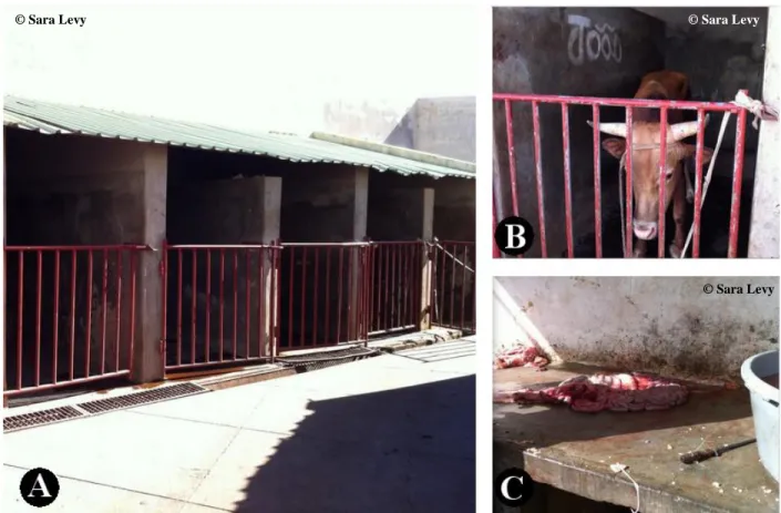
Figure 13- Slaughterhouse’s outside area composed by the stables (A), with a stabled animal before slaughter (B) and a stone table for preparation of the digestive organs (C)………...30
Figure 14- Carcass hanged for evisceration………...31
Figure 15- Parts of the slaughter procedure: digestive organs being cleaned (A), carcass cut to be weighted (B) and removal of the brain (C)………..………..31
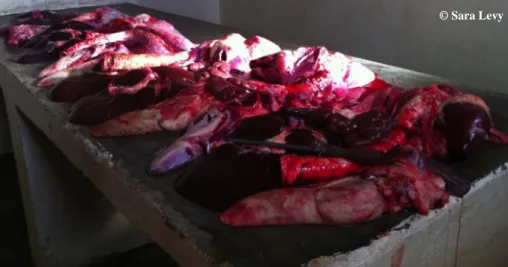
Figure 16- Organs on a stone table, after evisceration of the carcass, and before being inspected……….……….………..32
Figure 17- Fasciola inside the hypertrophied bile duct (A). Fasciola moving on the surface cut (B)……….………33
Figure 18- Sample. Microtube containing the fluke in ethanol at 98%...34
Figure 19- Rectum containing faeces……….……….35
Figure 20- 18 S, 5.8 S, 28 S tracts and internal transcribed spacers of the ribosomal DNA gene segment. ……….………..37
Figure 22- Level 1 of lesion score: liver exterior with slight evidence of bile ducts’ hypertrophy
(A) and, after the parenchyma’s cut, appearance of bile ducts’ hypertrophy (B)………….…….43
Figure 23- Level 2 of lesion score: liver exterior with evidence of bile ducts’ hypertrophy (A)
and, after the parenchyma’s cut, appearance of bile ducts’ hypertrophy (B).………43
Figure 24- Calcium deposit inside the bile duct (A) that was later removed (B)………….…….44 Figure 25- Level 3 of lesion score: liver exterior with evidence of bile ducts’ hypertrophy (A),
after the parenchyma’s cut, appearance of bile ducts’ hypertrophy (B) and internal calcification (B, C)………..……44
Figure 26- Level 4 of lesion score: liver exterior with evidence of bile ducts’ hypertrophy (A),
after the parenchyma’s cut, appearance of bile ducts’ hypertrophy with internal calcification (B) and presence of purulent material (C)…………..……….….45
Figure 27- Hepatic lymph node from a liver with FCL (A) and respective histopathological slide
(B)………..……….…46
Figure 28- Histopathological slide of a liver with extensive areas of localized fibrosis (A) and
portal inflammatory infiltrate (B)……….…..46
Figure 29- Bovine faeces near a watercourse (A and B) in S. Domingos, Santiago………….…49 Figure 30- Lymnaea spp. near a watercourse in S. Domingos, Santiago………..49 Figure 31- Water being pumped from a watercourse (A) into a horticultural production (B), in S.
Domingos, Santiago………50
Figure 32- Agarose gel electrophoresis of extracted DNA from the 19 samples, seen under
ultraviolet light on a transilluminator (Alphamanager™HP, Alpha Innotech)…...………..51
Figure 33- Agarose gel electrophoresis of the amplified ITS 1 region from the 19 samples, seen
under ultraviolet light on a transilluminator (Alphamanager™HP, Alpha Innotech)…………...52
Figure 34- Agarose gel electrophoresis of the amplified ITS 1 region from the 19 samples, after
digestion with TasI (FastDigest® Tsp509I, Fermentas)……….52
Index of tables
Table 1- Fasciolosis Compatible Lesion score………..34 Table 2- Nucleotide sequence of the primers used in the PCR………..37
Table 3- FCL cases for each FCL score level. ………...42 Table 4- Fasciolosis cases’ record. Level of liver rejection and respective FCL score level….47 Table 5- Record of Fasciola spp. eggs in faeces and respective FCL score level………….….54
Abreviations
ARFA Agency of Supervision and Regulation of Food and Pharmaceutical Products
bp Base pairs
C. novyi Clostridium novyi
cox1 Cytochrome c oxidase subunit 1
CT Computerized Tomography
CTAB Hexadecyltrimethylammonium Bromide
DNA Deoxyribonucleic Acid
ECV Cape Verdean Escudo
EDTA Ethylenediamine Tetraacetic Acid
ELISA Enzyme-Linked Immunosorbent Assay
ENSA National Food Safety Strategy
ETB Ethiopian Birr
EtBr Ethidium bromide
F. gigantica Fasciola gigantica
F. hepatica Fasciola hepatica
FAO Food and Agriculture Organization
HACCP Hazard Analysis and Critical Control Points
IGAE Inspection of Economic Activities
INE National Statistics Institute
ISO International Organization for Standardization
ITS Internal Transcribed Spacers
L. truncatula Lymnaea truncatula
MDR Ministry of Rural Development
NaCl Sodium chloride
nad1 NADH dehydrogenase subunit 1
NADH Nicotinamide Adenine Dinucleotide Hydride
NCBI National Center for Biotechnology Information
pH Negative logarithm for hydrogen ion
PNSA National Food Strategy Program
PVC Polyvinyl chloride
rDNA Ribosomal DNA
RLFP Restriction Fragment Length Polymorphism
Rpm Rotations per minute
TAE Tris-acetate + EDTA buffer
TE Tris-HCL + EDTA buffer
US United States
Introduction
This work was based in two internships: the first was in Praia, Cape Verde, and the second was at Instituto de Higiene e Medicina Tropical in Lisbon, Portugal.
The first had the duration of 6 months and started on September 2012. During the first 4 months (from September to December 2012), several veterinary activities were performed, such as harbour and airport inspection, large animals’ clinic and meat inspection at Praia slaughterhouse. This last activity, started in November, highlighted the importance of Fasciolosis’ cases observed during bovine inspection. Fasciolosis is a disease caused by members of the genera Fasciola, usually by Fasciola hepatica or Fasciola gigantica (Gracey et al., 1999). The first is widely distributed in Europe, North America, South America, Asia, Australia, New Zealand and some North African countries, Kenya and South Africa, whereas the second is present in southern Europe, South and Southeast Asia, South America and wide-spread in most of Africa (Norbury, 2008). This disease exists in three forms, acute, subacute and chronic, being the last the most common, and usually affects the bile ducts and liver parenchyma of ungulates (cattle, sheep, goats, buffaloes, deer, elk, moose and equidae) and also man (Gracey et al., 1999).
Since hepatic lesions related to fasciolosis can be seen during sanitary inspection at slaughterhouse level, this procedure could be used to monitor the disease. At slaughterhouse level, the economic losses associated with Fasciolosis are mainly related to meat condemnation. During animal production, the economic losses includes reduced production and quality of wool, reduced lambing percentages, poor lambs’ growth rate, increased costs for replacement stock, reduced production and quality of milk, lower growth rates and lower feed conversion rates in fattening cattle, expensive anthelmintic treatment (Boray, 2007).
In some countries, both species of Fasciola (Fasciola hepatica and Fasciola gigantica) are present (Huang et al., 2004 and Amer et al., 2011). Genetic characterization is the only reliable method of differentiation between the two species and can be achieved via PCR- Restriction Fragment Length Polymorphism (RFLP).
Due to the importance of Fasciolosis in slaughtered bovines, a study was carried out, from January to February 2013, to investigate the Fasciolosis prevalence during that period as well as to analyse the related lesions and its influence in meat condemnation. Additionally, Fasciola
species identification was carried out by means of genetic analysis in Instituto de Higiene e Medicina Tropical.
The faeces of the animals with FCL (Fasciolosis compatible lesions), were analysed in order to confirm the presence of Fasciola spp. eggs. Since bovines graze near watercourses, they can contaminate the water with faeces containing eggs this way infecting the snails. These waters are exhaustly used to water horticultural productions, possibly contaminating them and potentially infecting humans.
Objectives
Considering the importance of Fasciolosis in slaughtered bovines for human consumption, several aims were outlined for this study:
Monitoring of the sanitary inspection activities.
Determine Fasciolosis occurrence level in Praia slaughterhouse.
Investigate the Fasciolosis influence as a meat condemnation in bovines slaughtered for consumption.
Characterization of the hepatic lesion level associated with Fasciola infection.
Genetic characterization of Fasciola collected from bile ducts of infected livers, in order to confirm the presence of Fasciola gigantica rather than Fasciola hepatica and to discard the presence of Fasciola hybrids.
Determination of Fasciola’s eggs elimination by animals with hepatic lesions compatible with Fasciolosis.
Literature Review
1. Cape Verde – Food Safety Related Services
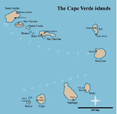
Cape Verde is an African country constituted by ten islands on the Atlantic Ocean (Figure 1), nine of which are habited, while one, Santa Luzia, is desert. The eastern island is located 460 km from Senegal’s coast and 1500 km south of the Canary Islands, whereas the western island is located 830 km from Senegal. The islands are situated in the Macaronesian region and can be divided into two groups: Barlavento, that is constituted by the northern islands: Santo Antão, São Vicente, Santa Luzia, São Nicolau, Sal and Boavista; and Sotavento, that is composed by the southern islands: Maio, Santiago, Fogo and Brava. The biggest island is Santiago, with 990 km2, and is where the capital of the country, Praia, is located (Gomes, 2006).
Cape Verde has a population of, approximately, 500 000 inhabitants according to the last census, made in 2010. The majority of the population is female (50,5%) and lives in urban areas (61,8%). The most populated island is Santiago with 273 919 (55,7%) inhabitants (INE, 2010).
The climate is dry and the average temperature in Cape Verde is, approximately, 25ºC being September the hottest month (average 27ºC) while February the coldest month (average 22ºC). There are two seasons, the rainy season, from August to October, and the dry season, from December to June; July is considered a transition period. The rain is concentrated, irregular and intense. Dry years may occur when it doesn’t rain in the country, highly affecting the agricultural and animal production. The trade winds as well as the high relative humidity also influence the climate (Amaral, 1964).
The land in Cape Verde is arid so the natural vegetation is scarce and the long dry period stimulates the lack of vegetation. There are around 755 vegetal species in this country, where 83 are considered endemic (Gomes, 2006). Even though the agricultural conditions aren’t the most desirable in Cape Verde, 45,2% of the Cape Verdean population still dedicate themselves to agricultural production (Ministério do Desenvolvimento Rural, 2004).
Animal production is an important way of means in Cape Verde. The last census, made in 2004, showed that there were 9877 bovine holdings, 25498 caprine holdings, 2982 ovine holdings and 29937 swine holdings. According to the same census, Santiago was the island with the highest number of holdings per specie. The island includes 73,4%, 54,3%, 86,9% and 58,3% of the country’s total bovine, caprine, ovine and swine holdings, respectively (Ministério do Desenvolvimento Rural, 2004).
Historically, Cape Verde always had enormous difficulties managing the lack of food resources due to the hard climate conditions. There are even reports of deaths by malnutrition and hunger in the 1940s, in this country. The situation propagated until the mid-seventies, when Cape Verde gained independence. After 1975 the government decided to implement policies to try and improve the food situation in the country. But even today, Cape Verde imports around 80% of its food (Mascarenhas, 2013).
Poverty can influence food safety and Cape Verde has a significant percentage of population living below the poverty line. Approximately 27% of the population lives in this situation, mainly in rural areas, where lack of basic sanitation, electricity and water sources is a well-set reality. Due to this matter, in 2004, the Ministry of Environment and Agriculture of Cape Verde approved, with FAO’s (Food and Agricultural Organization) technical support, a strategy and a sequential program to fight this issue: National Food Safety Strategy (ENSA) and National
Food Strategy Program (PNSA), respectively. ENSA and PNSA had several aims including to guarantee food safety and quality in order to protect the health of consumers (Pinto, 2011).
Several organizations are in charge of food safety and hygiene, in Cape Verde. The Agency of Supervision and Regulation of Food and Pharmaceutical Products (ARFA) is an independent organization tutored by the Ministry of Tourism, Industry and Energy. Its function is to establish hygiene and safety technical standards regarding the food and pharmaceutical chain, to activate surveillance and alert systems on risk situations, to regulate and apply hygiene and safety standards like ISO and/or HAACP, to economically regulate the market, monitoring the price/quality relationship (ARFA, 2013).
The Inspection of Economic Activities (IGAE) is the national inspecting authority, specialized in prevention and repression of anti-economic infractions and infractions against the public health. This authority is a part of the Criminal Police and belongs to the Ministry of Tourism, Industry and Energy of Cape Verde. Its function is to work in the field, supervising and triggering actions (punitive or not) to enforce the law compliance regarding the food and non-food sectors. IGAE stands on a credibility, scientific, transparency and confidentiality base (IGAE, 2011).
The Agricultural and Livestock Services work as a branch of the Ministry of Rural Development (MDR). Regarding sanitary inspection, they are responsible for the safety of food of animal origin, supervising the production and importation. Therefore, they’re the authority responsible for inspection of imported food as well as inspection of the animals slaughtered for human consumption, in each slaughterhouse (MDR, 2013).
2. Fasciola spp.
2.1. Taxonomy
Regarding taxonomy and according to the National Center for Biotechnology Information (NCBI) (2013), Fasciola has the following taxonomic classification:
Phylum: Platyhelminthes Subphylum: Neodermata Class: Trematoda
Subclass: Digenea Superorder: Anepitheliocystida Order: Echinostomida Suborder: Echiostomata Superfamily: Echinostomatoidea Family: Fasciolidae Genus: Fasciola
Species: Fasciola hepatica (Linnaeus, 1758), Fasciola gigantica (Cobbold, 1856),
Fasciolopsis buski (Lankester, 1857), Fasciola jacksoni (Cobbold, 1869), Fascioloides magna (Bassi, 1875), Protofasciola robusta (Lorenz, 1881)
2.2. Morphology
Fasciola hepatica is one of the biggest trematodes in the world. It can reach a length of 30
mm and a width of 13 mm and has a leaf-like appearance. The fluke has a pronounced widening, giving the appearance of having shoulders (Roberts & Janovy, 2009). Its color is pale brown and its form and shape is flat and oval, respectively (Gracey et al., 1999).
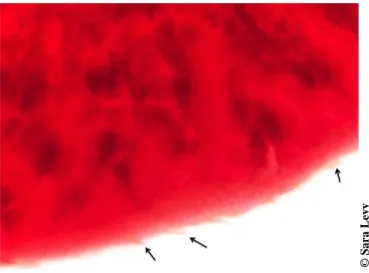
Fasciola species are bilaterally symmetrical and have a tegument with spines (Figure 4),
directed backwards, that are greatly responsible for liver cirrhosis (Norbury, 2008).
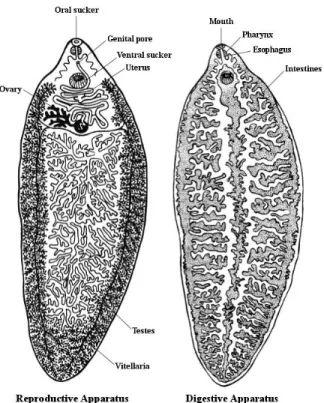
All Fasciola species possess two suckers, one ventral and one oral, with similar size, located on the cephalic cone (Figure 3B) (Norbury, 2008). The digestive system is composed by pharynx that is connected to the esophagus, followed by branched intestines. A pair of anterior ganglia and three pairs of nerve cords constitute the nervous system of this parasite (Gracey et
al., 1999).
Regarding the reproductive apparatus (Figure 2), the fluke’s testes (Figure 3E) are highly branched and occupy almost the entire posterior part of the parasite. The ovary (Figure 3D) is located above the testes and opens in a genital pore above the ventral sucker. The vitellaria (Figure 3F) are highly developed and are branched in the lateral and posterior region of the body (Norbury, 2008). The uterus (Figure 3C) is short and has a spiral shape, between the ovary and the cirrus shape (Gracey et al., 1999).
Figure 2- Anatomy of Fasciola hepatica. Adapted from Vignau et al. (2005).
Figure 3- Fasciola gigantica (A) with detailed cephalic cone (B), uterus carrying eggs (C),
ovaries, vitelline duct, ootype (D), testes (E) and vitellaria (F). Bar=1 mm (A). Original magnification: 20 X (B, C, D, E, F). © S a ra L ev y
Figure 4- Fasciola tegument with spines (black arrows). Original magnification: 50 X. 2.2.1. Differentiation between species
Fasciola species are polymorphic and can morphologically vary, depending on the
definitive host. Due to this fact, phenotypic and morphologic analysis can’t always be used to distinguish the species (Stunkard, 1957; Lee & Zimmerman, 1993 and Lofty et al., 2003).
Fasciola gigantica is, usually, morphologically distinct of Fasciola hepatica (Figure 3).
The cephalic cone of Fasciola gigantica is shorter in proportion (Norbury, 2008) and this specie also possesses less defined shoulder appearance than Fasciola hepatica (Gracey et al., 1999).
Figure 5- Fasciola gigantica (A) and Fasciola hepatica (B).
© S a ra L ev y © S a ra L ev y
Fasciola hybrids have been found in several countries like Japan (Hashimoto et al.,
1997), Korea (Agatsuma et al., 2000), China (Huang et al., 2004), Vietnam (Le et al., 2008) and Egypt (Amer et al., 2011).
To resolve the identification problem, as well as the hybridization phenomenon, several techniques have been conducted, in order to sequence different DNA regions for both Fasciola
hepatica and Fasciola gigantica, from several locations. The DNA regions that were amplified
were cytochrome c-oxidase subunit 1 (cox1) and the NADH dehydrogenase subunit 1 (nad1), from mitochondrial DNA; and internal-transcribed spacer 1 (ITS1) and 2 (ITS2), and 28S rDNA (28S) from nuclear DNA (Hashimoto et al., 1997; Agatsuma et al., 2000; Marcilla et al., 2002; Huang et al., 2004; Le et al., 2008; Ai et al., 2011; Amer et al., 2011 and Rokni et al., 2010).
2.3. World distribution
Fasciola species need specific conditions, as acceptable moisture and temperature, in
order to survive. Both parasite and intermediary host depend on a set of suitable environmental conditions (Norbury, 2008).
F. hepatica is more predominant and has a cosmopolitan distribution; it is usually located
at higher altitudes, because the temperature is lower. F. gigantica is confined to the tropics (Norbury, 2008).
F. hepatica is widely distributed in Europe, North America, South America, Asia,
Australia, New Zealand, some North African countries, Kenya and South Africa. F. gigantica is present in southern Europe, South and Southeast Asia, South America and wide-spread in most of Africa (Norbury, 2008).
Some countries have both species, like China (Huang et al., 2004) and Egypt (Amer et al., 2011). Hybrids have been discovered around the world: Japan (Hashimoto et al., 1997), Korea (Agatsuma, 2000), China (Huang et al., 2004), Vietnam (Le et al., 2008) and Egypt (Amer et
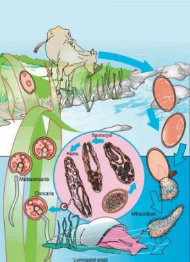
2.4. Life Cycle
Fasciola species are hermaphrodites (Gracey et al., 1999).
Sexual reproduction usually occurs, however, self-fertilization is also a possibility (Fletcher et al., 2004, cited by Norbury, 2008).
The adult Fasciola hepatica lives in the definitive host’s bile ducts. The eggs are carried, firstly to the bowel lumen with the bile, and then to the exterior with faeces (Bowman, 2009). These eggs are oval, yellow, measure 120 mm to 150 mm and have an operculum (Figure 6) (Vignau et al., 2005).
Figure 6- Fasciola spp. egg.
The next phase of the cycle, the development of the miracidia, will only happen if the eggs fall into water. If this occurs, the eggs will complete their development into miracidia and hatch within 9 to 10 days, if the weather is warm. Cold water tends to delay their development (Roberts & Janovy, 2009).
The miracidium is completely covered with cilia and has a conical papilla at its anterior end for boring into the snail. It escapes from the egg by pushing the operculum aside and when it’s free, it swims in search of a specific snail. The miracidium has to find a suitable snail (usually
Lymnaea truncatula) within 24 hours, otherwise, it dies (Bowman, 2009).
The miracidium finds the snail by chemical stimulation (Vignau et al., 2005). When it pierces inside the later, it loses it cilia and migrates to the gonad or the digestive gland of the snail and forms a sporocyst (Bowman, 2009).
© T er es a M a te u s
germinal cell, will become a germinal ball, through growth and repeated divisions, and will later develop into a redia (Bowman, 2009). Each sporocyst has 8 to 12 rediae (Vignau et al., 2005).
The rediae grow until they burst the sporocyst and migrate through the snail, eating its tissue on the way. The redia has germinal balls that will develop into a second generation of rediae. Each germinal ball of the second-generation rediae will develop into a cercaria (Bowman, 2009).
Cercariae are rounded (Vignau et al., 2005) and have certain adult organs: oral and ventral sucker, mouth, pharynx, forked intestine and primitive reproductive organs, and a tail. When fully developed, the cercariae leave the redia through a birth pore and make their way out of the snail into the water (Bowman, 2009). If the water, in which the snail is located, dries, the mollusc penetrates the mud in order to survive, carrying the cercariae with it. When water is present again, the snail emerges from the mud and sheds the cercariae (Roberts & Janovy, 2009).
Once in water, cercariae will quickly attach to any object, lose their tail and develop a thick, transparent cyst around themselves. They can encyst freely in water if they do not find an object within a short time (Roberts & Janovy, 2009). In this phase, the cercariae become metacercariae, the infective forms to the definitive host that includes cattle, sheep, and man (Bowman, 2009).
When ingested, in water or vegetation, the juvenile flukes excyst in the small intestine and cross the peritoneal space to the liver, where they penetrate its parenchyma and wander around for two months (Roberts & Janovy, 2009). After that, they enter the bile ducts, mature into adult flukes, and begin laying eggs after, approximately, a month and a half (Bowman, 2009).
The life cycle of Fasciola gigantica is similar to that of Fasciola hepatica (Figure 7). The big difference is that each phase takes more time, about 3 or 4 weeks longer than Fasciola
hepatica. The intermediary hosts are also snails of the genus Lymnaea, but the species are more
Figure 7- Life cycle of Fasciola hepatica. Adapted from Bowman, D. (2009).
3. Fasciolosis
Fasciolosis is an acute, subacute or chronic disease caused by members of the genera
Fasciola. It usually affects the bile ducts and liver parenchyma of ungulates (cattle, sheep, goats,
buffaloes, deer, elk, moose and equidae) and man (Gracey et al., 1999).
Usually, Fasciolosis is considered an important veterinary disease due to the economic losses caused in livestock and human Fasciolosis is viewed as a secondary disease (Mas-Coma et
al., 1999a).
3.1. Historical background
As stated by Bouchet (2003), Fasciola hepatica was present in the wetland pre-historical sites in Switzerland and France. Fasciola eggs were found in sediments and coprolites. This same author refers that in Middle Ages, these trematode infections accompanied humans and their domestic animals, since several parasite samples were found in excavations of archaeological sites.
across Fasciolosis. He described the disease as “sheep liver rot” (Mufti, 2011).
Francisco Redi made the first drawing of the adult Fasciola hepatica in 1668. Govard Bidloo followed, in 1698, by writing and illustrating a book about Fasciolosis in sheep (Mufti, 2011).
In 1737, Jan Swammerdam described the cercaria and redia and some years later, in 1760, Pallas discovered the disease in a man. Professor C. L. Nitzsch recognized the similarity between the cercaria and the adult Fasciola hepatica, in 1816, and as a consolidation of these studies, Johannes Steenstrup published a book, in 1844, where he referred that trematodes had two generations (Roberts & Janovy, 2009).
In the mid-1800s there were circumstantial evidences that indicated that molluscs were involved in Fasciola hepatica transmission. In 1880, George Rolleston, an Oxford teacher, thought that the common slug was Fasciola hepatica’s intermediary host. So, Rolleston invited A. P. Thomas to determine Fasciola hepatica’s lifecycle. Thomas discovered one specimen of L.
truncatula infected with redia and cercaria and found similarities between them and the adult Fasciola hepatica. He, then, proceeded to infect the snail with the miracidium and followed the
development of the parasite (Roberts & Janovy, 2009).
At the same time, Rudolph Leuckart was able to trace the development of F. hepatica to
L. truncatula, as well, and published the results 10 days before Thomas, but credit was given to
both investigators. Although they explained a big part of the cycle, how the definitive host got infected was still left undiscovered. It was only between 1892 and 1893 that a Brazilian researcher in Hawaii, named Adolph Lutz, was able to demonstrate that the ruminants got infected by eating the juvenile cysts that were encysted in the vegetation. Although he was working with Fasciola gigantica, the principle was the same since the life cycle of both species was considered equal (Roberts & Janovy, 2009).
3.2. Fasciolosis transmission and dissemination
Domestic and wild animals can act as definitive hosts of Fasciola spp., this depending on the geographical location. Sheep, cattle and goats are the usual definitive hosts (Mas-coma et al., 2005). However, studies have shown that pigs and donkeys can be infected by Fasciola species, as well (Mas-Coma et al., 1999b ).
Human are also definitive hosts and play a significant part in the transmission of Fasciolosis, especially in human hyperendemic zones (Mas-Coma et al., 1999b).
The liver fluke development is highly influenced by environmental conditions because the infection of both intermediary and definitive hosts depends on an association with fresh water, and larval development occurs within species of freshwater snails that, in its turn, are very dependent upon environmental factors. This transmission is greatly influenced by human activities, with the main definitive host species being domestic animals (Mas-Coma, 2005).
The spread of Fasciolosis is usually caused by the important Fasciola hepatica capacity in colonize and adapt to new environments, even in extreme conditions (Mas-Coma, 2005).
One of the biggest reasons for the successful geographical expanding of Fasciola
hepatica was the exportation of European livestock, to the five continents. The fluke has adapted
to other mammal species like camelids and marsupials (Mas-Coma, 2005).
As above-stated, new hosts can occur. This event highly contributes to the spread of the disease. Fasciola hepatica can adapt to a broad range of Lymnaea species and these species have a wide distribution and spread (Mas-Coma, 2005).
3.2.1. Larvar development
Fasciola species have a big capacity of expansion, related with their ability to adapt and
colonize new environments. Researches made in the Northern Bolivian Altiplano, compared the development of Fasciola hepatica in this area and Europe. Molluscs in this high altitude area have greater longevity with bigger cercaria production period and longer cercaria shedding period. The study also showed that local snails have higher survival compared with European parasited molluscs, because the Altiplanic L. truncatula has the capacity of surviving for more than 4 months after the end of the shedding period whereas European L. truncatula die immediately or shortly after this period. This study suggests a better parasite-host adaptation in Bolivia, which is interesting since Fasciola hepatica has a, supposedly, European origin (Mas-Coma et al., 2001).
3.2.2. Normal definitive hosts and adaptation to new definitive hosts
The common definitive hosts are ruminants, especially sheep, goats and cattle. However, other important domestic and wild animals may also be affected: horses, donkeys, mules and camelids. More rarely, wild herbivorous mammals are also susceptible hosts: buffalo, deer, wild sheep, wild boar, various marsupials, rabbit, hare, otter and monkeys (Mas-Coma et al., 1997).
Although pigs usually resist Fasciola infection, the study carried out in the Northern Bolivian Altiplano, by Mas-Coma et al. (2001), revealed that infection in this specie might occur easily.
New acquired hosts have been found in several parts around the world. In Belorussia, Shimalov and Shimalov (2000) conducted a study and found the presence of Fasciola spp. in seven wild animal species: the elk (Alces alces), the red deer (Cervus elaphus), roe deer (Capreolus capreolus), the wild boar (Sus scrofa), the beaver (Castor fiber), the otter (Lutra
lutra) and the hare (Lepus europaeus) (Shimalov & Shimalov, 2000).
Myocastor coypus is a rodent introduced in France. Ménard et al. (2001) developed a
study to determinate the prevalence of Fasciolosis in this specie. The results showed a Fasciolosis prevalence of 40,1% in nutrias (Myocastor coypus) within Fasciolosis areas. Coproparasitological exams carried out revealed that 90% of the infected nutrias shed fluke eggs. Two groups of snails, from 2 different populations, were exposed to F. hepatica miracidiae hatched from eggs collected from infected nutrias. They showed an infection prevalence of 74 and 58,6%. After that, one group of sheep was orally infected with metacercariae of nutria origin. The installation rate of F. hepatica in sheep was 31,6%.
These new acquired hosts play an important part as Fasciolosis’ transmission hosts, contributing to the disease’s expansion; therefore they must be taken into consideration when control measures are applied (Mas-Coma et al., 2005).
3.3. Fasciolosis development
The development of the disease is well explained by Roberts and Janovy (2009): the damaged caused by juvenile flukes, when they burrow the intestine wall and penetrate the
Glisson’s capsule of the liver is not significant. However, the necrosis resultant of their migration is severe.
The flukes feed on liver cells and blood, causing anemia when heavy infections occur, although there are evidences that sometimes, anemia is caused by a chemical (proline), released by the parasite. This chemical also stimulates collagen deposition in bile ducts (Roberts & Janovy, 2009).
Edema and inflammation, caused by flukes in the bile ducts, lead to production of fibrous tissue in the walls of these ducts. When this phenomenon happens, the ducts have a smaller diameter, therefore can handle less bile and are less responsive to the needs of the liver. This organ’s parenchyma can be atrophied (Roberts & Janovy, 2009).
Worms can re-enter the parenchyma and provoke large abscesses. The gall bladder can be compromised in heavy infections (Figure 8) (Roberts & Janovy, 2009).
Figure 8- Affected gall bladder from a bovine with Fasciolosis.
Sometimes flukes can migrate to the lungs, in cattle, via hepatic vein. In sheep, encapsulated immature liver flukes may be found in the spleen, kidney, myocardium, subcutaneous tissues and in the diaphragm (Gracey et al., 1999).
Due to the necrosis caused by migration of the flukes, the tissues are poorly oxygenated, optimizing the conditions for the development of clostridium organisms and respective deadly toxins. Black disease, caused by C. novyi, is often related to Fasciolosis (Bowman, 2009).
© S a ra L ev y
3.4. Clinical and lesional presentation
The gravity of the disease varies on the number of metacercariae ingested, the age and specie of the host and the general condition of the affected animal. Both ovine and bovine species are less resistant to the infection if they are malnourished or infected with other parasitosis (Vignau et al., 2005).
Fasciolosis has three types of forms: acute, subacute and chronic. The first two are more evident in sheep whereas in cattle, the chronic form occurs more often (Gracey et al., 1999).
The symptoms characterization in the acute form of Fasciolosis is: pale mucosae, debility, dyspnea, abdominal pain and normochromic and normocytic anemia (Vignau et al., 2005). Other symptoms from this form are: dullness, anorexia and edema of the conjunctiva. Death usually occurs one or two days after the beginning of the symptoms and can be accompanied by dysentery and epistaxis (Gracey et al., 1999).
The subacute form is characterized by ascites, submandibular edema (“bottle jaw”), quick weight lost, hypochromic and macrocytic anemia (Vignau et al., 2005).
The chronic course is the most common form of the disease, found in sheep and cattle. This type of infection usually occurs when there is ingestion of a small number of parasites, during a long period of time. In sheep, the “bottle jaw” occurs with edema of the eyelids, throat, brisket and abdomen, making it look pendulous. Diarrhea and loss of wool also occurs (Gracey et
al., 1999).
Sheep can die two or three months after the appearance of the symptoms. The survivors are emaciated and rarely recover weight (Gracey et al., 1999).
In cattle, there is anemia, loss of body condition and decrease of milk production. Bovines rarely die by Fasciolosis if other diseases aren’t associated. Unlike sheep, bovines’ health is less affected by the disease; it has been shown that while 50 adult flukes can cause clinical disease in sheep, some 250 are necessary in cattle (Gracey et al., 1999).
Infection with F. gigantica produces much the same symptoms as those due to F.
hepatica (Gracey et al., 1999).
The localization of Fasciolosis lesions is in the liver parenchyma and bile ducts. In the acute form of the disease there is swelling and congestion of the liver and its capsule is usually covered with fine fibrous strands (Gracey et al., 1999). This form is also characterized by
traumatic acute hepatitis and the affected animals may have hemorrhages in the abdominal cavity (Vignau et al., 2005). The liver is paler in color and friable. Its parenchyma has hemorrhagic tracts and immature flukes can be seen. In heavy infections, rupture of the liver capsule may occur. In this form, the carcass has a fevered appearance (Gracey et al., 1999).
In the subacute form of Fasciolosis, in sheep, migratory tracts and fibrosis are seen on the liver parenchyma (Gracey et al., 1999).
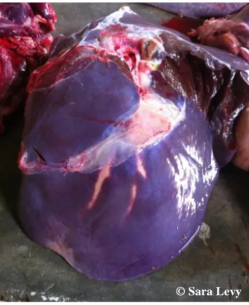
The most common form of Fasciolosis, both in animal and man, is chronic. In this form, chronic, pericanalicular, atrophying cirrhosis with formation of fibrous tissue in the walls of the bile ducts and surrounding liver tissue, including the portal canals, occurs due to mechanical irritation caused by immature flukes. This occurrence is followed by chronic hepatitis in which the fibrous deposit on the liver capsule becomes organized. Migration of the immature flukes usually occurs in the left lobe (Figure 9) (Gracey et al., 1999).
Figure 9 – Bovine liver with bile ducts’ thickening, on the left lobe.
The bile ducts become thicker due to fibrosis (Figure 10). In cattle, calcium deposits may be formed, inside the bile ducts (“pipe stem liver”) (Figure 11). These formations may cause occlusion of the later. This calcification doesn’t occur in sheep or goats (Gracey et al., 1999).
Figure 10- Bovine liver with hyperplasia of the bile ducts.
Figure 11- Calcium deposit (black arrow) and a Fasciola gigantica fluke (white arrow). Both
recovered from a bile duct of a bovine liver.
Atrophy of the liver is visible due to progressive fibrosis and scar tissue. This will cause gross distortion of the organ, which becomes thickened and shortened, this being most apparent in sheep (Gracey et al., 1999).
The lesions of F. gigantica are essentially the same as those of F. hepatica (Gracey et al., 1999).
3.5. Diagnosis
When a liver blockage coincides with a history of watercress consumption, Fasciolosis should be suspected (Roberts & Janovy, 2009).
The simplest way to diagnose Fasciolosis is to observe the adult parasite inside the bile ducts of the liver through necropsy or post mortem inspection (Almeida, 2010). Another direct
© Sara Levy
form of diagnosis is by finding the eggs in faeces (Roberts & Janovy, 2009). Hoffmann et al. (1934) developed a technique that is still used to this day, the faecal sedimentation. Although coproparasitological exams are a simple way of detecting a Fasciola spp. infection, weak or early stages of infection can pass undetected. For the eggs to be visualized in faeces, the flukes are required to be sexually mature already, in order to release eggs (Norbury, 2008).
Early diagnosis of Fasciolosis is needed in order to start treatment before liver damage is far too advanced (Roberts & Janovy, 2009). This can be achieved through immunodiagnosis.
These indirect techniques were developed to search antibodies produced against Fasciola and are characterized by high sensibility, specificity, speed and low cost (Almeida, 2010). Immunodiagnosis is also facilitated by the presence of a large variety of antigens, secreted and excreted by the parasites, in blood, faeces and milk, beside others, of infected hosts (Norbury, 2008).
Some immunologic search techniques comprehend complement fixation, immunofluorescence, immunoelectrophoresis, counterimmunoelectrophoresis, enzyme-linked immunosorbent assay (ELISA), and immunoelectrotransfer (Cordova et al., 1999; Carnevale et
al., 2001a; Carnevale et al., 2001b and Queiroz, 2005).
Serodiagnosis is crucial to diagnose and control Fasciolosis because it can be applied in every phase of the disease, contrary to coproparasitological exams, and it’s not invasive (Almeida, 2010). But, as yet, no test has 100% sensitivity and specificity (Rapsch et al., 2006).
The routine diagnosis for humans is based on egg detection and/or serological test (Norbury, 2008). Coproparasitological exams are important for follow-up after treatment (Almeida, 2010).
Ultrasound examination is an auxiliary method, vastly used in humans. It can show nodules or migratory tracks caused by the flukes, and biliary endoscopy can, sometimes, put the flukes in evidence (Fawzy et al., 1992).
3.5.1. Differential diagnosis between Fasciola hepatica and Fasciola gigantica
Since Fasciola species are polymorphic and can vary morphologically, depending on the definitive host, morphological analysis is not reliable to distinguish the two species (Stunkard, 1957; Lee & Zimmerman, 1993 and Lofty & Hillyer, 2003).
Also, differential diagnosis between F. hepatica and F. gigantica cannot be achieved through clinical, pathologic, coproparasitological or immunologic exams.
Nowadays, the only possible manner to differentiate the species is through genetic analysis. Through a simple and practical PCR-Restriction Fragment Length Polymorphism (RFLP) it’s possible to distinguish the species (Ai et al., 2011).
Marcilla et al. (2002) performed this technique using two restriction enzymes, Ava II and
Dra II, to differentiate Fasciola hepatica from Fasciola gigantica. The first was originated from
Bolivia, Spain and Egypt whereas the second was originated from Cape Verde, Burkina Faso and Egypt.
In China, there were also RFLP assays, using restriction enzyme Hsp92II and RcaI (Huang et al., 2004). In Iran, Fasciola of both species were also identified using TasI (Rokni et
al., 2010).
Because the species sometimes overlap, in several locations, identification is highly important (Marcilla et al., 2002).
3.6. Prevention and Control
The efficacy of the Fasciolosis control depends on preventive measures. These later have several aims: to reduce the number of parasites in the definitive host, to reduce the number of intermediary hosts and to make a proper management of the herds and flocks regarding the environment and the possibility of infection. In order to properly implant the measures, previous knowledge of the seasonal variations of animal infection, about the population dynamics of the intermediary host and dispersion forms of Fasciola must be acquired (Vignau et al., 2005).
The control of the snails includes the use of organic, inorganic and plant derived molluscicides and biological control using competitor snails, predators, snail parasites and commensals (Torgerson & Claxton, 1999). Drainage is the one permanent way to control or eliminate the molluscs (Mas-Coma et al., 2005), however in some locations, like Cape Verde, water is scarce so drainage is not even considered. Other method is to smooth out the banks of watercourses and remove marginal vegetation to prevent the formation of backwater pools where the snails may flourish (Mas-Coma et al., 2005).
pipes, to decrease the installation of mollusc population (Rosa et al., 2004).
Usually these procedures are almost impractical, insufficient and cost prohibited (Torgerson & Claxton, 1999).
Another possibility is the use of resistant livestock. A number of resistant animal breeds have been identified (Torgerson & Claxton, 1999).
The animals can be treated in anthelmintic, being Triclabendazole the drug of choice. There’s no vaccine against Fasciolosis yet (Mas-Coma et al., 2005).
Human Fasciolosis can be prevented by not eating raw watercress, lettuce and other metacercariae-carrying aquatic plants of wild or unknown source. These vegetables have to be produced in controlled conditions, away from animals and faecal contamination as well as snails (Mas-Coma et al., 2005). The vegetables, prior to eating, must be washed with citric acid, commercial vinegar, liquid soap or potassium permanganate to detach and kill all metacercariae (El-Sayad et al., 1997).
Some authors like Roberts and Janovy (2009) alert to the possibility of infection by drinking water containing metacercariae. Using washing units, that appropriately filter the water, can prevent this infection. Chemical treatment is also a possibility (Mas-Coma et al., 2005).
3.7. Economic losses related with Fasciolosis
Because a wide range of domestic animals is targeted by Fasciolosis, there’s a great impact on the economy of livestock industry. The clinical and lesion presentation of this disease leads to effects on the animal production and consequential economic losses (Norbury, 2008).
In sheep, the mortality rate, caused by acute Fasciolosis is 50 to 70% whereas in subacute is 5 to 20% and chronic is up to 50% (Vignau et al., 2005). But deaths account for only a part of this loss, other considerable factors are: reduced production and quality of wool, reduced lambing percentages, poor lambs’ growth rate, increased costs for replacement stock (Boray, 2007).
In bovine, economic losses are not related to deaths since only young animals die due to this disease (Vignau et al., 2005). Nevertheless, there are significant losses caused by reduced production and quality of milk and lower growth rates and lower feed conversion rates in fattening cattle (Boray, 2007). Condemnation of livers at slaughterhouses and expensive anthelmintic treatment also represent great economic contraction (Roberts & Janovy, 2009).
Boray (2007) refers that, in Australia, farmers spend, approximately, $10 million a year, on fluke drenches alone, and lost production costs a further $50 to 80 million, a year.
Centeio (2008) conducted a study in Cape Verde, where he calculated the average loss per liver being 2054,9 ECV. Based on that calculation, he demonstrated that Fasciolosis, occurred in 2004, registered in Praia and Assomada’s Slaughterhouses, signified an annual loss of 1.129.812 ECV.
In Ethiopia, Fasciolosis occurred in Soddo slaughterhouse, represented an annual loss of $4000 (Abunna et al., 2010).
Another study, also conducted in Ethiopia, showed a total economic loss of about 154.188 ETB, 215.000 ETB and 3.003.488,1408 ETB per annum, in cattle, due to fasciolosis in Ziway, Dire Dawa and Jimma slaughterhouses, respectively (Belay et al., 2012).
A study, made in Scotland, to determinate the losses regarding carcasses from infected animals, showed that carcass performance characteristics, assessed according to international standards agreed within the European Union, were deteriorated when liver fluke was present. Also, the analysis demonstrated that carcass conformation and fatness score deteriorate in cases of Fasciolosis, resulting lower carcass’ cold weight. All these aspects accentuate the economic loss because they decrease the value of the carcass. The differences in the average value of a normal carcass and a carcass from an animal with Fasciolosis, was £11. There was an overall reduction of 0.3% of the price paid per carcass when liver fluke was present (Sanchez-Vazquez & Lewis, 2013).
3.8. Fasciolosis in Cape Verde – Human and Animal
The two first reports of human Fasciolosis in Cape Verde were made in 1966, regarding a case of hepatic fasciolosis (Meira, 1966, cited by Almeida, 2010) and a case of cutaneous fasciolosis (Meira et al., 1966, cited by Almeida, 2010), both in Santiago.
Another case was of a Cape-Verdean citizen, 63 years old, that was admitted in a French hospital, with anorexia, weigh lost and complaining about severe epigastric pain. Although he presented a high number of eosinophils in blood, the parasitological exams came back negative. A gastric endoscopy revealed gastric wall thickening and the presence of several round formations, with a few millimeters in diameter. A CT (computerized tomography) scan showed,
apart from the thickening of the gastric wall, two nodules, with approximately 5 mm in diameter, in the hepatic surface. A surgery followed, in order to make a biopsy of the gastric and hepatic nodules and the results revealed presence of fluke’s eggs. The Fasciolosis diagnose was confirmed by immune-electrophoresis and the patient was treated with Triclabendazole. This case was interesting since the localization of Fasciola spp. was unusual (Nozais et al., 1998).
Nozais et al. (1998) also refer other 4 cases of patients that were infected in Cape Verde and one was an American that was returning to the United States after spending some time in Cape Verde.
In the 1990s, 58 patients were admitted to Praia’s hospital, in Santiago, with fever, anorexia, abdominal pain, hepatomegaly, eosinophilia, anemia and hepatic abscesses. To reach a diagnosis, immunologic exams were made in Instituto de Higiene e Medicina Tropical, in Lisbon, Portugal. These exams revealed a Fasciolosis prevalence of 44,83% (Grácio et al., 1998, cited by Almeida, 2010).
Graham et al. (2001) reported two cases of Fasciolosis, in the United States, of two men from Cape Verde. The first patient was a 67 old man that lived in the United States but went to visit his home country. He started developing abdominal pain, nausea, anorexia and had had a weight loss of 18 kg, three months before his return to the United States. After his arrival he was admitted to the surgical service at an urban hospital in Boston for evaluation of right upper quadrant tenderness. CT scan revealed multiple ill-defined hypodense lesions throughout both lobes of the liver and a small amount of ascites, this exam was followed by hepatic biopsy that demonstrated severe active hepatitis with eosinophilic abscesses, marked loss of parenchyma, and areas of fibrosis. After a month, the patient presented atrial fibrillation and congestive heart failure, apart from the still present abdominal pain, anorexia, and weakness. An abdominal ultrasound showed stones in the gallbladder, marked ascites, and a slightly heterogeneous liver. After that, stool samples were made and a single egg compatible with Fasciola spp. was found. To confirm the diagnosis, an ELISA test was performed, the results being positive for Fasciolosis. The patient was treated with Triclabendazole.
The second patient was a 33-year-old man. He went to the United States with a 10-month history of mild diffuse abdominal pain and intermittent diarrhea. The patient stated that he had consumed watercress while being on Cape Verde. He presented abdominal pain in the right upper
two poorly defined, irregular areas in the left and right lobes of the liver, with smaller cystic components that contained debris. The results of serum ELISA for F. hepatica were positive and the patient was treated with Triclabendazole (Graham et al., 2001).
In Portugal, a 15-year-old male patient from Cape Verde was admitted in hospital with fever, weight lost (20 kg), ascites and painful hepatomegaly. Blood tests revealed severe anemia and hypereosinophilia. The abdomen CT scan and ultrasound demonstrated heterogeneous hepatomegaly with focal nodular lesions with ramified appearance, on the right lobe of the liver, as well as ascites and bilateral pleural effusion. After biopsy a Fasciola spp. egg was found, and the faecal exam showed presence of this parasite’s eggs. The ELISA test came back positive for Fasciolosis (Oliveira et al., 2002).
Three patients with 41, 28 and 55 years, with Cape Verdean origin, were admitted in hospital with a history of fever (between 3 and 12 months), weigh lost and painful hepatomegaly. Blood tests revealed anemia, eosinophilia and high levels of alkaline phosphatase. Imagiology revealed nodular lesions in the liver and the faecal exam showed presence of Fasciola spp. eggs (Oliveira et al., 2002).
Semedo (2006) conducted a more broad study, from January to December of 2005, in S. Domingos, Santiago, Cape Verde. He investigated the prevalence of Fasciolosis in students from two primary schools as well as cattle and molluscs from that area. The results showed a prevalence of 0,42% between the students and 27,28% and 34,5% amongst molluscs and cattle, respectively.
Matos (2011) calculated the prevalence of Fasciolosis in bovines and molluscs in all Santiago’s counties, except Tarrafal. The counties with the highest prevalence of bovine Fasciolosis infection were Ribeira Grande (67%) and Santa Cruz (72%) whereas the ones with highest prevalence of molluscs’ infection were Santa Cruz (80%) and São Lourenço dos Órgãos (80%). This study was carried out in 2008, from May to June (Matos, 2008).
Pile et al. (2008) decided to investigate the monthly prevalence of Fasciolosis in patients that presented gastric and hepatic disorders in Santiago, Cape Verde. This study was made between March and December of 2005 and was also complemented with detection of Fasciola spp. in molluscs (Lymnaea natalensis) and cattle, near agricultural productions. The results indicated a Fasciolosis monthly prevalence of 33% amongst patients that presented the above-stated symptoms, and of 35,5% and 35,1% in molluscs and ruminants, respectively. What was