1
Integrated Masters in Bioengineering - Biomedical Engineering
branch
Dissertation
Manuela Jorge Estevinho Rodrigues
2
Manuela Jorge Estevinho Rodrigues
Development of liposomal formulations
for photodynamic therapy of cancer
i
ACKNOWLEDGMENTS
I would like to thank Doctor Cláudia Sousa Silva and Doctor Ana Catarina Pinto for the possibility of collaboration in this research project to develop liposomal formulations for photodynamic therapy of cancer.
I am very grateful to Doctor Ana Catarina Pinto, Dr. Luís Borges Rocha and Doctor Ana Paula Pêgo for the teaching, availability and support provided during the realization of work.
I am thankful to Bluepharma employees for all the help given.
I would like to show my gratitude to my family, particularly my mother and father, for all the support. Without them this work would not be possible.
ii This work was performed under the orientation of Doctor Ana Catarina Pinto and Dr. Luís Borges Rocha and under the co-orientation of Doctor Ana Paula Pêgo on the ambit of Dissertation, to obtain the degree of Bioengineering Master, of the Biomedical Engineering branch, during the 5th year of Integrated Masters in Bioengineering (2010/2011) of the Engineering Faculty of the Oporto University. It was developed at the Department of Business and Product Development of Bluepharma Indústria Farmacêutica S.A in Coimbra.
iii
GENERAL INDEX
Index of figures v Index of tables vi Abbreviations vii Abstract ixCHAPTER I – STATE OF THE ART 1
1- Photodynamic therapy 1
1.1- General considerations 1
1.2- Brief history of photodynamic therapy 3
1.3- Principle of photodynamic therapy 4
1.4- Mechanisms of tumour destruction by photodynamic therapy in
vivo
5
1.5- Photodynamic therapy photosensitizers 7
1.5.1- Topical and systemic photosensitizers for photodynamic therapy
12
1.5.2- Photosensitizers conjugates for photodynamic therapy 12 1.6- Clinical trials for cancer treatment by photodynamic therapy 13
2- Liposomes as nanoscale drug delivery systems 14
2.1- General considerations 14
2.2- Liposomes in photodynamic therapy 17
2.2.1- Conventional liposomes for photodynamic therapy 19 2.2.2- Passively targeted liposomes for photodynamic therapy 21 2.3- Methods for preparation of liposomal drug formulations 22
CHAPTER II – OBJECTIVE 25
CHAPTER III – MATERIALS AND METHODS 26
1.1- Materials 26
1.2- Methods 26
1.2.1- Encapsulation of a photosensitizer in liposomes 27
1.2.1.1- Lipid film hydration method 27
1.2.1.2- Reverse-phase evaporation method 30
1.2.1.3- Film loading method 30
iv 1.2.2- Non-encapsulated drug separation method 32 1.2.3- Physicochemical characterization of the prepared liposomes 32
1.2.3.1- Phospholipid quantification 33
1.2.3.2- Drug quantification 33
1.2.3.3- Validation of the method for non-encapsulated drug separation by size exclusion chromatography
34
CHAPTER IV – RESULTS AND DISCUSSION 35
1.1- Physicochemical characterization of the developed liposomal drug formulations
35
1.1.1- Mean size diameter and polydispersity index 35 1.1.2- Loading capacity and encapsulation efficiency 38
1.1.2.1- Phospholipid quantification 38
1.1.2.2- Drug quantification 38
1.1.2.3- Encapsulation parameters 38
CHAPTER V – GENERAL CONCLUSIONS AND FUTURE WORK 46
References 47
Annex I 51
v
INDEX OF FIGURES
Figure 1: The principle of photodynamic therapy 5
Figure 2: Pathways for PDT-mediated tumour destruction presenting vascular damage, direct tumour cell killing and host immune response as possible contributions
6
Figure 3: General porphyrin, chlorin, bacteriochlorin and phthalocyanine structures
8
Figure 4: Schematic illustration of the self-assembly process from individual phospholipids molecules (a) to bilayer membrane leaflets (b), followed by transformation into liposomes (c)
15
Figure 5: In vivo behaviour of the different types of liposomal delivery systems
20
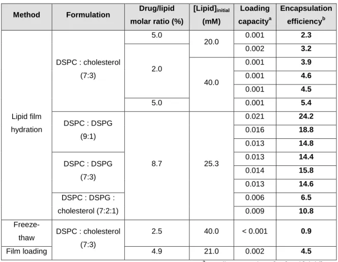
Figure 6: Comparison of the encapsulation efficiencies and loading capacities for DSPC : cholesterol (7:3) liposomes encapsulating PS molecule and prepared by different methods: lipid film hydration, freeze-thaw and film loading
40
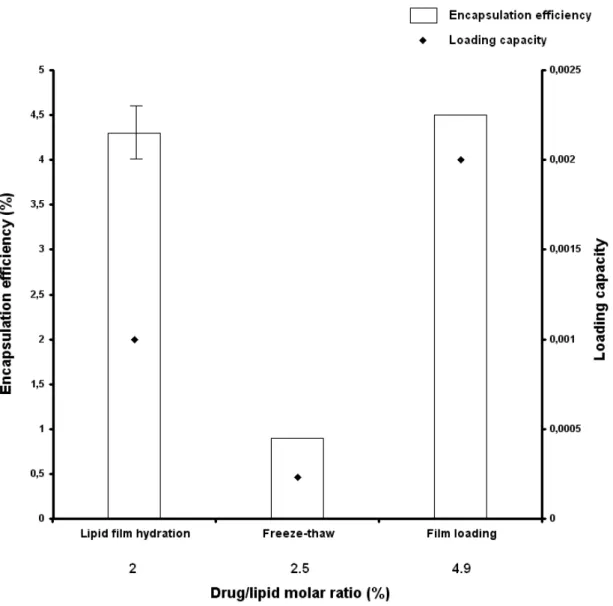
Figure 7: Comparison of the encapsulation efficiencies and loading capacities of DSPC : cholesterol (7:3) liposomes prepared by the lipid film hydration method
41
Figure 8: Encapsulation parameters (encapsulation efficiency and loading capacity) for the liposomal formulations with different lipid compositions prepared by the lipid film hydration method
44
Figure 9: Graphical representation of the validation of free drug separation by size exclusion chromatography for DSPC : DSPG (9:1) liposomes
45
Figure I: Calibration curve of standards S1-S5 prepared from KH2PO4 0.65
mM stock solution
51
Figure II: Calibration curve for Luz011c at 408 nm 53
vi
INDEX OF TABLES
Table 1: Distinction between necrosis and apoptosis – principal mechanisms of cell death in PDT
7
Table 2: Clinically available photosensitizers 11
Table 3: Examples of ongoing clinical trials for cancer treatment by PDT 13
Table 4: Example of a Visudine® liposomal formulation in PDT undergoing
clinical evaluation
18
Table 5: Description of the lipid film hydration method 22
Table 6: Description of the reverse-phase evaporation method 23
Table 7: Description of the film loading method 23
Table 8: Description of the freeze-thaw method 24
Table 9: Absorption wavelengths for drug quantification of the PSs used (Luz011c and Luz011)
34
Table 10: Experimental results of mean size diameter and PI obtained by DLS for all the liposomal drug formulations after the different preparation methods followed by the extrusion procedure
36
Table 11: Loading capacities and encapsulation efficiencies for all the liposomal formulations prepared by different procedures
39
Table 12: Encapsulation parameters (encapsulation efficiency and loading capacity) obtained for four liposomal drug formulations with different lipid compositions prepared by the lipid film hydration method
42
Table I: Absorbance values measured at 830 nm for standard solutions S1-S5 made from KH2PO4 0.65 mM stock solution
51
Table II: Exemplificative data for DSPC : DSPG (9:1) formulation – phospholipid concentration and total lipid concentration
52
vii
ABBREVIATIONS
Abs: absorbance AK: actinic keratosis
AMD: age-related macular degeneration ALA: 5-aminolevulinic acid
BCC: basal cell carcinoma BPD: benzoporphyrin derivatives
BPD-MA: benzoporphyrin derivative monoacid ring A CNV: choroidal neovascular degeneration
DLS: dynamic light scattering DNA:deoxyribonucleic acid
DPPC: dipalmitoylphosphatidylcholine DPPG: dipalmitoylphosphatidylglycerol DSPC: distearoylphosphatidylcholine DSPG: distearoylphosphatidylglycerol
EPR: enhanced permeability and retention effect FDA: Food and Drug Administration
HAL: hexaminolevulinate HBS: HEPES-buffered saline HDL: high density lipoproteins HPD: hematoporphyrin derivative
HPPH: 2-(1-hexyloxyethyl)-2-devinyl pheophorbide-alpha Inc.: incorporated
LDL: low density lipoproteins
logPOW: logarithm of the octanol/water partition coefficient
LT: total lipid Ltd.: limited
LUV: large unilamellar vesicle MACE: chlorin e6
M-ALA: methyl aminolevulinate MLV: large multilamellar vesicle MPS: monuclear phagocytic system
m-THPC: meso-tetra-hydroxylphenyl-chlorin MVV: multivesicular vesicle
viii PDD: photodynamic diagnosis
PDT: photodynamic therapy PEG: polyethylene glycol PI: polydispersity index PL: phospholipid PS: photosensitizer RNA: ribonucleic acid
ROS: reactive oxygen species SD: standard deviation
SUV: small unilamellar vesicle
TEM: transmission electron microscopy
Tm: transition temperature
U.S.: United States
USA: United States of America UV/Vis: ultraviolet-visible
ix
ABSTRACT
This work aimed the development of liposomal formulations with an encapsulated photosensitizer drug. Different passive encapsulation methods were tested, namely, the lipid film hydration method, the reverse-phase evaporation method, the freeze-thaw method and the film loading method. Additionally, different liposome membrane lipid compositions were used. Afterwards, the formulations encapsulating a hydrophobic photosensitizer (PS) were characterized in terms of average size, polydispersity index, loading capacity and encapsulation efficiency. Finally, the validation of the free drug separation method by size exclusion chromatography was made for the most promising liposomal formulation in order to confirm previous results.
Both the average size and size distribution of the vesicles encapsulating a PS were measured by dynamic light scattering (DLS).
For the determination of the encapsulation parameters (i.e. loading capacity and encapsulation efficiency), the PS quantification was made by UV/Vis spectroscopy, using an adequate calibration curve, and the total lipid quantification was performed by the colorimetric method of Bartlett for quantification of inorganic phosphate, being afterwards the concentration of lipid extrapolated from the obtained experimental phospholipid concentration.
Among the different preparation methods, the lipid film hydration method resulted in the best encapsulation parameters (encapsulation efficiency and loading capacity).
The presence of cholesterol seems to have a negative impact on the obtained encapsulation parameters and the presence of DSPG may have a favourable contribution. For that reason, the formulations DSPC : DSPG (9:1) and DSPC : DSPG (7:3) yielded the highest encapsulation efficiency and loading capacity values. Additionally, these formulations also exhibited appropriate average liposomal size for potential intravenous administration.
It is expected that the results obtained in the present work will prove useful in developing new and efficient methodologies for the preparation of liposomal formulations incorporating photosensitizing molecules for photodynamic therapy of cancer.
1
CHAPTER I – STATE OF THE ART
1- PHOTODYNAMIC THERAPY
1.1- General considerations
Photodynamic therapy (PDT) is a clinical technique that employs a light-sensitive drug (a photosensitizer, PS), in combination with light of a visible wavelength, to destroy target cells, especially cancerous or pre-cancerous cells [1]. An adequate concentration of molecular oxygen is also needed for anomalous tissue damage. If any one of these three components is missing, there will be no effect [2].
PDT requires single administration of a PS followed after a certain time interval by single irradiation with light of specific wavelength corresponding to an absorbance band of the sensitizer. This treatment, very frequently, does not require hospital admission. In comparison, characteristic curative radiotherapy regimes include daily irradiation for a total of 6-7 weeks (once more not requiring hospitalization). Chemotherapy schedules vary, but typically last for several months. Surgery, although a single procedure, requires general anaesthesia and hospitalization for one to several weeks. Cost-effectiveness comparisons have been made for palliative treatment of head and neck cancer with PDT versus extensive surgery or chemotherapy, and for PDT versus esophagectomy or endoscopic surveillance for patients with Barrett’s esophagus and high-grade dysplasia. PDT proved to be cost-effective and provided increased life expectancy, compared with other treatment options for these circumstances [3].
While PDT is a completely well-accepted treatment in clinical practice for some types of skin lesion (cancerous or not), it has yet to be explored for other forms of cancer. PDT is normally used either as a primary treatment (usually in skin conditions) or as an adjunctive treatment together with surgery, radiotherapy or chemotherapy [1].
PDT is a local, rather than systemic, treatment; it is appropriate only for localized disease [2]. Light of wavelengths used to excite current PSs can provoke photochemically induced tissue necrosis up to a maximum depth of 10 mm. This signifies that, for superficial illumination, the use of PDT as a primary treatment should be limited to small, accessible tumours. PDT can also be made in combination with surgery for palliative treatment of larger tumours [3].
2 The technique has the advantage of limited side effects, because phototoxicity is limited to sensitized cells in the area illuminated and the PS tends to accumulate in tumour cells [4]. In fact, some photosensitising drugs can reach higher concentrations in tumour tissue than in surrounding healthy tissue. The accurate mechanisms that drive this process are not totally understood, but the abnormal physiology of tumours, including poor lymphatic drainage, leaky vasculature, decreased pH, increased numbers of receptors for low-density lipoprotein and the abnormal stromal composition, might contribute to the selectivity of PSs [2]. Furthermore, the activation by light at a wavelength matching one of PSs absorbing wavelengths leads to the formation of reactive oxygen species (ROS), mainly the extremely reactive singlet oxygen (1O2), which travels very short distances and so photodamage mediated by PDT is mainly limited to the site of singlet oxygen generation [4].
Modern fiber-optic technology facilitates delivery of light, of the desired wavelength and fluence rate (light application rate), to tumours located almost anywhere in the body. Localized illumination enables specific tumour treatment without the destruction of critical normal tissues outside the treated area. By contrast, surgery and radiotherapy of tumours located close to critical structures can be very mutilating and lead to loss of function. PDT has the advantage that, although there is sometimes a significant ulceration of the illuminated area immediately after treatment, there is minimal long-term fibrosis, resulting in functional recovery without scarring [2]. PDT spares tissue architecture, providing a matrix for regeneration of normal tissue, since it does not damage subepithelial collagen and elastin and there is preservation of noncellular supporting elements [3].
Another advantage of PDT is that the treatment can be repeated in case of recurrence or appearance of a new primary tumour in the previously treated area, which is difficult with surgery or radiotherapy, without the risk of normal tissue damage [3]. PDT also offers the ability to treat large areas of diseased tissue, areas which are not accessible by surgery and preserves connective tissue within the treated area [1].
A limitation of PDT is that it cannot cure advanced disseminated cancer disease, because irradiation of the whole body with suitable doses is not possible (at least with current technologies). However, for advanced disease, PDT can improve quality of life and extend survival, because it is minimally invasive and it does not restrict the use of other subsequent treatments [2].
Currently, photodynamic diagnosis (PDD), which involves fluorescence to localize abnormal tissue, has been subjected to several clinical trials. In the fluorescence process, an outer electron excited by a photon of appropriate wavelength returns to its ground state emitting a lower energy photon. PDD reveals neoplastic
3 lesions that cannot be seen by means of conventional methods, representing an additional optical recognition criterion. The advantages features of fluorescence detection are the lack of background signal and specific targeting of a fluorochrome (molecule that makes use of the relaxation path mentioned above). Thus, PDD and PDT allow simultaneous diagnoses and therapy, improving cancer treatment efficiency [5], [6].
1.2- Brief history of photodynamic therapy
The first clinical application of PDT dates 1903, where von Tappeiner and Jesionek attempted unsuccessfully to treat basal cell carcinomas (BCCs) with topical eosin dye followed by exposure of the lesions to sunlight [3], [7]. Von Tappeiner and Jodlbauer later defined PDT as the dynamic interaction among light, a photosensitizing agent and oxygen resulting in tissue destruction [3].
After a hiatus of more than fifty years, in 1960 Lipson synthesised the first PDT drug which he named hematoporphyrin derivative (HPD) [3]. In 1975 Dougherty et al. reported that HPD in combination with red light could completely eradicate mouse mammary tumour growth [3]. Clinical trials were afterwards initiated with HPD to treat patients with bladder cancer and skin tumours. After these successful studies, several trials were initiated for a variety of cancers and PSs. This led to the approval of PDT using porfirmer sodium (Photofrin®; Axcan Pharma Inc., Mont-Saint-Hilaire, Canada) for the treatment of bladder cancer in Canada in 1993 [7]. This achievement was followed by approvals for PDT of tumours of lung and esophagus in the U.S. and other countries [7]. However, nearly all patients receiving Photofrin® acquire photosensitivity of their skin to direct sunlight, which may persist for one to three months and so this adverse reaction probably contributed to the low acceptance of PDT by the medical community, although severe reactions in patients have been rare [7].
Nowadays, others sensitizers are approved for clinical use, for example, 5-aminolevulinic acid (ALA, Levulan®; DUSA Pharmaceuticals Inc., Wilmington) is approved for actinic keratosis (AK), the methyl ester of ALA (M-ALA, Metvix®; Photocure ASA, Oslo, Norway) is approved for AK, Bowen’s disease, Basal cell carcinoma, and meso-tetra-hydroxyphenyl-chlorin (mTHPC, temoporfin, Foscan®; Biolitec Pharma Ltd., Dublin, Ireland) is approved for head and neck cancers. Thus, PDT is becoming an established treatment modality for localized cancers [3]. The term
4 “PDT” is being used also to describe non-cancer disorders photosensitized by a photosensitizing drug, including AK, psoriasis, acne, and age-related macular degeneration (AMD) [7].
Besides, PSs like ALA and porphyrin hexaminolevulinate (HAL) have obtained approval for the detection of malignant glioma and superficial bladder cancer, respectively, by fluorescence diagnosis in many European countries. Therefore, it is now possible to simultaneously diagnose a suspected cancerous lesion not visible with white light cystoscopy and conduct “curative” therapy, improving survival and quality of life [5], [6], [8].
1.3- Principle of photodynamic therapy
PDT is a developing modality for the treatment of superficial tumours, because the light used with a wavelength of 600-800 nm is not able to penetrate into the tissue more than 1 cm [9], [10].
The principle of PDT is based on the administration of a PS followed by local illumination of the tumour area at an adequate wavelength to activate the specific drug. Activation of the PS upon absorption of the light transforms the drug from its ground state (1PS) into an excited singlet state (1PS*) – Figure 1. From this state the drug may decay directly back to the ground state by emitting fluorescence, which can be used clinically for photodetection, or by internal conversion into heat. However, to obtain a therapeutic photodynamic effect, the excited singlet state must undergo electron spin conversion to its triplet state (3PS*) by a process called intersystem crossing (whereby the spin of the excited electron in 1PS* inverts to form an excited triplet state that has electrons with spin in a parallel conformation). In the presence of oxygen, the excited molecule can react directly with a substrate, such as the cell membrane or other cellular structures, by proton or electron transfer, to produce radicals and radical ions, which can further interact with oxygen to form oxygenated products (type I reaction). Alternatively, the energy of the excited PS can be directly transferred to oxygen to form singlet oxygen (type II reaction), which is the most damaging and cytotoxic agent produced during PDT, since it interacts efficiently with various biomolecules [3], [11], [12].
5
Figure 1: The principle of photodynamic therapy [9].
Both the type I and type II reactions occur simultaneously and the ratio between these processes depend on the type of PS used and also on the concentrations of substrate and oxygen. Due to the high reactivity and short-life of singlet oxygen and hydroxyl radicals, only molecules and structures that are close to the area of its production (i.e. areas of PS localization) are directly affected by PDT and destroyed [11].
1.4- Mechanisms of tumour destruction by photodynamic
therapy in vivo
The effectiveness of PDT in the treatment of cancer (e.g. tumour cell destruction) depends on the nature of PS, drug concentration, drug intracellular localization, total light dose (fluence), light application rate (fluence rate) and oxygen availability [10]. In general, the rates of singlet oxygen generation and therefore tissue oxygen consumption and depletion within the tumour are significant when tissue PS levels and fluence rate of light are high. An important parameter influencing the rate of tissue oxygen consumption is photobleaching of the PS (PS destruction / alteration by exposure to light or loss of the PS’s property of optical absorbance) because the reduction of PS levels also reduces the rate of photochemical oxygen consumption [13].
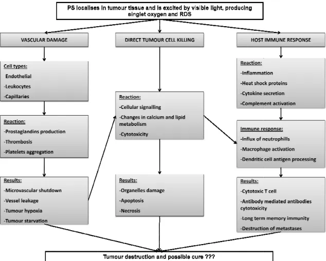
Three different mechanisms shown in Figure 2 have been assumed to reduce or frequently eliminate tumours when using PDT [11].
Figure 2: Pathways for PDT-tumour cell killing and host immun
Singlet oxygen produced by the photochemical reaction can directly kill tumour cells by the induction of apoptosis and/or necrosis (
extremely reactive and can diffuse only 0.01
[11]. Consequently, damage mediated by PDT is mainly limited to the site of singlet oxygen generation [3], [7], [
associated to the tumour, which can lead to thrombosis and haemorrhage in tumour blood vessels, resulting in indirect death via the induction of hypoxia and
the tumour. It has been shown that both PS accumulation and tumour cell kill decrease with the distance of tumour cells from the vascular supply
response may occur. After the acute inflammation and subsequent release of cytokines and stress response proteins induced in the
immune response by the attack of cytotoxic
at isolated locations. The inflammatory signalling after PDT initiates a regulated invasion of neutrophils, mast cells, and monocytes
outnumber resident cancer cells
-mediated tumour destruction presenting vascular damage, direct tumour cell killing and host immune response as possible contributions [11].
inglet oxygen produced by the photochemical reaction can directly kill tumour cells by the induction of apoptosis and/or necrosis (Table 1). Singlet oxygen is extremely reactive and can diffuse only 0.01-0.02 µm during its short lifetime [
]. Consequently, damage mediated by PDT is mainly limited to the site of singlet ], [11]. Alternatively, singlet oxygen damages
, which can lead to thrombosis and haemorrhage in tumour blood vessels, resulting in indirect death via the induction of hypoxia and
It has been shown that both PS accumulation and tumour cell kill decrease mour cells from the vascular supply [13]. Lastly, the host immune fter the acute inflammation and subsequent release of cytokines and stress response proteins induced in the tumour by PDT, they are able to initiate an the attack of cytotoxic T cells against the tumour and tumour cells
The inflammatory signalling after PDT initiates a
regulated invasion of neutrophils, mast cells, and monocytes / macrophages that may outnumber resident cancer cells too [13]. Therefore, the monocytes
6
presenting vascular damage, direct
inglet oxygen produced by the photochemical reaction can directly kill tumour Singlet oxygen is µm during its short lifetime [3], [7], ]. Consequently, damage mediated by PDT is mainly limited to the site of singlet damages vascular cells , which can lead to thrombosis and haemorrhage in tumour blood vessels, resulting in indirect death via the induction of hypoxia and starvation of It has been shown that both PS accumulation and tumour cell kill decrease the host immune fter the acute inflammation and subsequent release of cytokines able to initiate an against the tumour and tumour cells The inflammatory signalling after PDT initiates an immense rophages that may he monocytes can produce
7 antibodies that mediate cytotoxicity against tumour cells and then the organism acquires some long term memory immunity.
Table 1: Distinction between necrosis and apoptosis – principal mechanisms of cell death in PDT (data was compiled from [7], [11], [14]).
Necrosis or passive cell death Apoptosis or active cell death
Violent and quick process characterized by gross damage, spillage of intracellular
contents and presence of in vivo inflammation.
It is caused by physical or chemical damage and is considered to be an unprogrammed
process.
The intracellular targets are the plasma membrane and lysosomes.
Energy-requiring process highly regulated and controlled, characterized by nuclear condensation, cell shrinkage, bleb formation, and absence of inflammatory responses of the
affected tissue.
It is an indispensible process during normal development, tissue homeostasis, regulation
of the immune system...
Mitochondria and DNA are the likely targets for the apoptotic response.
Both implicated in the immunological responses to PDT
The three mechanisms can influence each other and the outcome of the PDT is dependent on all these mechanisms, being the relative contribution of each dependent on the treatment regimen given [3], [7], [11], [12], [15]. The combination of all these mechanisms in PDT is required for optimum long-term tumour regression, especially of tumours that may have metastasized [11].
1.5- Photodynamic therapy photosensitizers
Many PSs, for example, 21-thiaporphyrin and 21,23-dithiaporphyrin have been tested in vivo and in vitro in PDT experiences and despite several clinically approved PSs, none have shown ideal, safe and selective properties and, for this reason, recent studies have focused on the development and efficacy of new PSs like hexaminolevulinate derivative [11], [16], [12], [17]. The prerequisites for an ideal sensitizer include: i) simple, efficient and economical synthesis; ii) chemical and physical stability, chemical purity and long shelf-life; iii) solubility in biocompatible solvents or vehicles; iv) high absorption coefficient in the “phototherapeutic window”
8 (600-800 nm); v) short interval required between administration of the sensitizer and its maximal accumulation in tumour tissues; vi) singlet molecular oxygen sensitization (type II process) and/or superoxide generation (type I process) with a high quantum yield; vii) little or no dark toxicity; viii) low skin photosensitization; ix) controlled photobleaching; x) selective accumulation and prolonged retention in tumour tissues and xi) simplistic metabolism or rapid excretion after treatment [11], [12], [15], [18].
PSs can be classified in various ways, all with limitations. Three of those classifications are given below:
• Chemical structure
This is a widely accepted mode to characterize PSs for chemists but has limited utility in the clinical, because alterations in structures by the addition, subtraction or substitution of primary or side chains may sometimes enhance PS activity and may also create toxic substances, being useless for clinic. Fundamentally, most PSs are cyclic tetrapyrroles and are derivates of porphyrins, chlorins and bacteriochorins. Dyes, mainly those used in ink, are also a rich ground to develop PS [15], [18]. Figure 3 presents the chemical structures of these first families and an example of a dye (which corresponds to another type of PS family).
Figure 3: General porphyrin, chlorin, bacteriochlorin and phthalocyanine structures [7], [10].
NH N NH N NH N NH N NH N NH N Porphyrin Chlorin Bacteriochlorin NH N N N NH N N N Phthalocyanine
9
• Generation
Some attempts to classify PSs are based on when they were generated. First generation PSs are porphyrin based and include hematoporphyrin and its derivatives named hematoporphyrin derivatives. Second generation of PSs were developed based on the supposed deficiencies of the first generation drugs. Thus, second generation PSs demonstrate higher absorption in the 650-800 nm range where tissue penetration is optimal, have higher extinction coefficients of absorption in the red than first generation compounds and their tissue accumulation is much lower and therefore, the treatment can be carried out on the same day as the administration of the drug. Moreover, second generation PSs show lower toxicity [16]. These second generation of PSs have several structures including porphyrins, expanded porphyrins, chlorophyll derivatives and dyes, but most of the compounds are still very hydrophobic and show poor tumour selectivity. Third generation of PS contains fundamentally first and second generation PS conjugated to carrier molecules, which may specifically target the PS to the target cells, resulting in minimized accumulation in healthy tissues [16]. Many drugs of second and third generations are not commercially available [3], [7], [10], [12], [15].
• Targeting
Some PSs preferentially accumulate in tissue while others stay in the vascular supply. Some, such as Photofrin®, may initially circulate extensively than compartmentalize. Clinically, this indicates using vascular PS agents when targeting neovasculature. Though, all invasive tumours have neovasculature and using one PS for this indication may not be critical. An attempt may be made to classify PSs by what they specifically target. Thus, the hematoporphyrin derivatives are composed of monomers, dimmers and oligomers. The two smaller components are brought to mitochondria while the larger components are actively phagocytised by the cell membrane. Chlorin e6 (MACE) is brought to lysosomes by endocytosis. Phatalocyanines concentrate in mitochondria. Benzoporphyrin derivatives (BPD) accumulate in the Golgi apparatus. ALA goes into the cell membranes, lysosomes and mitochondria. By linking these PS to carriers, such as nanoparticles, the accumulation region can be altered significantly [15].
Note that, the important structural features for different intracellular localizations are the net ionic charge (which can range from -4 to +4), the degree of hydrophobicity expressed as the logarithm of the octanol / water partition
10 coefficient and the degree of asymmetry present in the PS molecule. Hence, hydrophobic PSs with two or less negative charges can diffuse across the plasma membrane and then relocate to other intracellular membranes. Less hydrophobic PSs with less than two negative charges tend to be more polar to diffuse across the plasma membrane and may be found preferably in lysosomes and in cytoplasm (in general, hydrophilic PSs are taken by endocytosis) [11], [14].
The clinically available PSs (approved and in trials) until the moment are given in Table 2.
11
Table 2: Clinically available photosensitizers (adapted from [19]).
Chemical family Product name Therapeutic substance Administration
route Manufacturer Disorder
Porphyrin Photofrin® Haematoporphyrin derivative (HPD) Intravenous Axcan Pharma Inc. Lung and esophageal cancers Photogem® Haematoporphyrin derivative (HPD) Intravenous Moscow Research Oncological Institute Bronchus, esophageal and colon cancers Levulan® 5-Aminolevulinic acid
(ALA) Topical DUSA Skin cancer, AK
Metvix®
Methyl
aminolevulinate (M-ALA)
Topical PhotoCure ASA
AK, Bowen’s disease, Basal cell carcinoma
Hexvix® Hexaminolevulinate
(H-ALA) Intravenous PhotoCure ASA Bladder cancer
Visudyne® Verteporfin (BPD-MA) Intravenous Novartis Pharmaceuticals Macular degeneration, Pathologic myopia, Ocular histoplasmosis Texaphyrin Antrin ® ,
Lu-Tex Lutexaphyrin Intravenous Pharmacyclis Breast cancer
Chlorin
Foscan® Temoporfin (mTHPC) Intravenous Biolitec Pharma
Ltd
Head and neck cancers LS11, Photolon® Talaporfin Intravenous Light Sciences Skin cancer, breast cancer, Uterus and rectum cancers LitxTM, ApoptosinTM, Laserphyrin Intravenous Photochlor 2-(1-Hexyloxyethyl) -2-devinyl pyropheophorbide-a (HPPH) Intravenous RPCI Lung and esophageal cancers Phthalocianines
Photosens® Phthalocyanine Intravenous General Physics
Institute Macular degeneration Pc4 Phthalocyanine Intravenous or intratumoral CWRU Mycosis Fungoides, Sezary syndrome
Bacteriochlorin Tookad
Palladium-Bacteriopheophorbide Intravenous The Weisman Institute of Science Prostate adenocarcinoma
12
1.5.1- Topical and systemic photosensitizers for photodymanic
therapy
Systemic PSs (administrated in a versatile way that enables binding between PS and serum proteins for effective PDT [3]) have the advantage to be accumulated in multiple lesions by a single administration whereas the topical PSs require their direct application in each lesion. However, topical PSs do not present the disadvantage of photosensitivity (toxic effect) generated by systemic PSs. The photosensitivity is generally a consequence of a slow rate clearance from the skin, persisting during various weeks after treatment. This fact was observed in clinical trials for hematoporphyrin and its more purified form Photofrin®-II [12].
In the clinic, when a topical PS like M-ALA is employed for cutaneous lesions, a series of intensely illuminated PDT sessions may be done, resulting in additionally cell death and increasing PDT efficacy, whereas for systemic PSs only a single powerfully illuminated session can be done [12], [15].
Many primary cutaneous lesions are likely to be treated with topical PSs because of easier application (by a cream / solution) and illumination (higher accessibility of skin to light exposure) and this is the treatment of choice for most patients [20]. In dermatological oncology, PDT is already a routine treatment, and its use will continue to increase. Excellent cosmetic outcomes make PDT suitable for patients with skin cancers. Skin pain during irradiation can be attenuated with local anaesthesia [2].
1.5.2- Photosensitizers conjugates for photodynamic therapy
The extended delocalised aromatic π electron system characteristic of PSs generally makes them extremely hydrophobic and consequently poorly water soluble and prone to aggregation in aqueous solution, which decreases their ability to generate singlet oxygen efficiently. In addition, currently clinically approved PSs have frequently poor bioavailability and unfavourable biodistribution, resulting in lower tumour specificity than the ideal and, thus, in undesirable side effects like prolonged skin photosensitivity and damage to surrounding healthy tissues. These drawbacks have lead to the development of conjugates, and supramolecular carriers like nanoparticles for the systemic delivery of PS (Vide 2.2.) [4], [21].
13 In fact, PSs often possess functional groups to which conjugation is possible by esterification or substitution. Many PSs conjugates have been designed to increase their bioavailability, solubility and target specificity. Distinct sets of proteins, such as receptors and transporters, are often overexpressed at the surface of cancer cells’ membrane, and conjugation of PS with solubilising and/or targeting moieties, including sugars, peptides, proteins and antibodies, is the underlying principle to increase target specificity of PDT by receptor-mediated endocytosis (for the cellular uptake of the conjugated PSs) [4].
1.6- Clinical trials for cancer treatment by photodynamic
therapy
In the future, PDT treatment regimens still have to be optimized and standardized for better therapeutic effectiveness and for increased safety [3]. Thus, various clinical trials are in development with the aim of determining the best treatment protocol in PDT for various cancer diseases (Table 3) [17].
Table 3: Examples of ongoing clinical trials for cancer treatment by PDT (data was compiled from [17]).
Product name
Therapeutic
substance Therapeutic Indication Sponsor Status
Metvix®
Methyl-5-aminolevulinate
hydrochloride
Basal cell carcinoma Roswell Park
Cancer Institute Phase I
-2-(1-Hexyloxyethyl) -2-devinyl
pyropheophorbide-a
Head and neck cancers Roswell Park
Cancer Institute Phase I
- Hexaminolevulinate Cervical intraepithelial
neoplasia PhotoCure Phase II
Pc4 Silicon phthalocyanine 4 Lymphoma; non-melanomatus skin cancer Case Comprehensive Cancer Center Phase I - 5-Aminolevulinic acid Non-melanomatus skin cancer Roswell Park
Cancer Institute Phase II
Hexvix® Hexaminolevulinate Bladder cancer PhotoCure Phase I
-
2-(1-Hexyloxyethyl) -2-devinyl
pyropheophorbide-a
Carcinoma of the oral cavity; oropharyngeal
cancer
Roswell Park
14
2- LIPOSOMES AS NANOSCALE DRUG DELIVERY
SYSTEMS
2.1- General considerations
One subject being investigated over this decade is the utilization of nanosystems in different scientific areas. These nanosystems include nanoparticles, which are particles synthesised in the nanoscale with promising applications in various fields of biomedicine, for example, in vectorization of anticancer drugs. They are biodegradable and biocompatible, enhance the drug’s biodisponibility and efficiency, reduce the drug’s toxicity and its side effects, allow a controlled drug delivery to the target site and enable an increase of the effective concentration of the drug in the target site [22]. Examples of these nanoparticles are quantum dots, liposomes and lipid nanoparticles, polymeric nanoparticles and dendrimers [22].
Only a limited number of the drug-loaded nanoparticles are successful for their clinical applications. An essential parameter of the delivery vehicle pertains to low or no toxicity of the carrier itself in vivo or in the environment as a by-product. Therefore, nanoparticles made-up using an assembly of natural biomolecules such as lipids, proteins, and carbohydrates are expected to be a suitable choice for clinical applications [23].
Among various lipid-based formulations, a classical example is liposomes. Liposomes are spherical self-closed structures, composed of phospholipid bilayers, which enclose part of the surrounding solvent into their interior (Figure 4) [23], [24]. The liposome bilayer can be composed of synthetic or natural phospholipids. Examples of phospholipids are: the charge-neutral phosphatidylcholine (neutral liposome), the negatively charged phosphatidic acid (anionic liposome) and the positively charged stearylamine (cationic liposome) [25]. The phospholipids are the major components of biological membranes, having a hydrophilic head and a hydrophobic tail. Thus, the lipid bilayer closes in on itself due to interactions between water molecules and the hydrophobic tails of the phospholipids. This process of liposome formation is spontaneous because the amphipathic phospholipids self-associate into bilayers [23], [26].
15
Figure 4: Schematic illustration of the self-assembly process from individual phospholipids molecules (a) to bilayer membrane leaflets (b), followed by transformation into liposomes (c). A single bilayer is typically ~5 nm thick and consists of neatly arranged individual lipid molecules with their hydrophobic tails facing each other and their hydrophilic headgroups facing toward internal and external aqueous medium (d) [25].
Liposomes are easily prepared by various methods (e.g. mechanical methods, methods based on replacement of organic solvents and methods based on size transformation or fusion of preformed vesicle) and can be mechanically stabilized in bloodstream by the inclusion of cholesterol, which also minimizes adsorption of plasmatic proteins by liposomes and controls liposome’s membrane fluidity [27].
Drug loading into liposomes can be achieved fundamentally through liposome formation in an aqueous solution saturated with soluble drug, the use of organic solvents and solvent exchange mechanisms and pH gradient methods, depending on the chemical nature of drug [26]. Hence, the encapsulated drug is inaccessible to metabolizing enzymes [27].
The mechanisms to load drugs into liposomes are the passive encapsulation and the active encapsulation. The passive encapsulation is applicable to both hydrophilic and hydrophobic drugs. For hydrophilic drugs, the encapsulation results from the hydration of the dry lipid film with an aqueous solution of drug. The spontaneous formation of liposomes passively captures the dissolved drug. For hydrophobic drugs, the compound is dissolved with the lipid constituents in a suitable organic solvent. Afterwards, the solvent is removed and the film hydrated with an aqueous solution, which result in the entrapment of the drugs within the lipid bilayer [28], [29]. These are the two most common passive encapsulation methods.
16 Active encapsulation takes advantage of the fact that certain weakly-basic drugs can exist either as neutral or as charged, dependent on the pH of their environment. The molecules are added to preformed liposomes, and in a neutral form, permeate the bilayer lipid through an increasing pH gradient (from outside to inside the liposome) or an ion capable of generating a pH gradient, as with ammonium sulfate or magnesium sulphate. The method of active encapsulation allows higher encapsulation efficiency than the passive encapsulation method [30], [31], [32].
Depending on the processing conditions and the chemical composition, liposomes are formed with one or several concentric bilayers. Liposomes are often distinguished according to their number of lamellae and size. They can be basically small unilamellar vesicles (SUVs, 20-100 nm), large unilamellar vesicles (LUVs, >100 nm), large multilamellar vesicles (MLVs, >0.5 µm), or multivesicular vesicles (MVVs, >1 µm) [27]. The preparation of SUVs starts usually with MLVs, which then are transformed into small vesicles using an appropriate manufacturing technique, e.g. extrusion methods or sonication [22].
Liposomes containing a drug (hydrophilic / hydrophobic) can be administrated by oral or intravenous route in cancer treatment. They generally reach the target site through bloodstream by passive or active (without or with the presence of a ligand on the surface of the lipid bilayer, respectively) targeting strategies. In the passive targeting, the liposome can accumulate in the tumour interstitium owing to a leaky microvasculature and an impaired lymphatic system supporting the tumour area. This effect is often called enhanced permeability and retention effect (EPR) [23], [26], [33]. However, due to the small size of liposomes, they can easily be eliminated by macrophages of the mononuclear phagocytic system (MPS), when liposomes are linked to serum opsonins (which facilitate phagocytosis). This opsonisation process can be reduced by polyethylene glycol (PEG) coating - PEGylation or PEG-coated, reducing clearance by MPS and increasing the circulation half-life [23], [26], [33].
The most widely used polymeric steric stabilizer is PEG, a water-soluble polymer that exhibits protein resistance, low toxicity, non-immunogenicity and antigenicity and can be prepared synthetically with high purity and in large quantities [16].
Given the advantages of liposomes, the major problems associated with them are their stability, poor batch-to-batch reproducibility, difficulty in sterilization and low drug loading capacity [22].
In conclusion, liposomes as drug carriers may avoid the side effects of the conventional cancer treatments and may be a non-invasive, more effective and safe way to treat the disease, leading to better quality of life of patients.
17 It should be noted that there are various medical applications of liposomes in different areas: drug delivery systems in the treatment of cancer, bacterial infections or ophthalmic disorders. Current clinical applications of gene delivery include liposomes. Also, other applications of liposomes include diagnostic imaging, vaccine adjuvant, photodynamic therapy, dermatology, hemoglobin or chelating agent transporter and enzyme replacement therapy [34].
2.2- Liposomes in photodynamic therapy
Having the concepts above in mind and considering that PDT aims to destroy tumour tissue without affecting healthy tissue (minimizing the risk of unwanted side-effects caused by damage to normal cells), it is still necessary to improve the efficacy and safety of PDT, because during clinical PDT, it is frequent practice to irradiate larger fields that correspond to healthy tissue with microscopic malignant foci and most PSs not seem to exhibit a high selective affinity for tumour tissue [9].
There are various ways to improve the effect of PDT and its safety. Hence, PSs can be loaded in liposomes, which are able to encapsulate hydrophobic as well as hydrophilic drugs without the loss or alteration of their therapeutic activity [16], [21]. Most PSs are usually hydrophobic because of the presence of aromatic rings, presenting low solubility in water. Therefore, liposomal formulations show the ability to decrease the tendency of PS to aggregate in aqueous media, prolonging the circulation of the drug in bloodstream and protecting it [35]. Besides, liposomes improve tumour-selective accumulation as a consequence of the above-mentioned EPR effect [16]. Conventional liposomes exhibit a plasma half-life which is too short for an efficient tumour uptake to occur, but liposomes PEGylated, i.e. long-circulating, and especially actively targeting liposomes, are a better choice in becoming truly tumoritropic carriers of PSs. They increase the tumour accumulation of PS, enhance the controlled release of PS and enhance the efficacy of PDT [9], [36].
Historically, one of the first nanoformulations of PSs was PS loaded into unilamellar liposomes. In the context of non-cancer PDT, Visudyne® (Verteporfin) was the first liposomal drug approved by the Food and Drug Administration (FDA) in 2000 for the treatment of age-related macular degeneration. Another application of this non-PEGylated formulation is in the subfoveal choroidal neovascular degeneration (CNV), which has shown promising results on Indian patients. The therapy was effective and
18 can cause stabilization or even improved vision [16]. A current Visudine® liposomal formulation in phase I clinical study is described in Table 4 [17].
Table 4: Example of a Visudine® liposomal formulation in PDT undergoing clinical evaluation (data was compiled from [17]).
Formulation name
Therapeutic
Substance Condition Intervention Sponsor Status
Visudyne® Liposomal benzoporphyrin derivative monoacid termed verteporfin Port Wine Stains Combine PDT and pulsed dye laser
treatment
University of California,
Irvine
Phase I
Foslip is a more recently developed third generation PS based on a dipalmitoylphosphatidylcholine (DPPC) / dipalmitoylphosphatidylglycerol (DPPG) liposomal formulation of 5,10,15,20-tetrakis(m-hydroxyphenyl)chlorin (mTHPC). Many studies have been published about Foslip, which present supposed absence of side effects, high efficacy and reduced damage of healthy tissue compared to the non-liposomal Foscan®. Newer studies are related to the potential use of an intratumoral injection of a liposomal formulation of Foslip in a mouse model of local recurrence of breast cancer and its photothrombic activity [16].
For ALA loaded in liposomes composed of phosphatidyl ethanoamine / cholesterol / sodium stearate at the molar ratio 2:1:2.5, improved skin penetration was reported. Likewise, inclusion of ALA esters, especially, of ALA hexyl esters, seemed to result in higher stability upon dilution with cell culture medium [16].
Another approach to tumour specific drug delivery is the use of folate modified liposomes (actively targeted liposomes). Folate receptors are often over-expressed on tumour cells. A comparison of the selectivity of free PS and mTHPC conjugated to the folate receptor for KB tumours with over-expression of folic acid receptors and HT-29 lacking folate receptors was performed. After intravenous injection, the folate specific uptake of conjugate PS was enhanced in KB tumours compared to the non-conjugated compound, and no significant difference between KB and HT-29 tumours was observed in case of free mTHPC. Furthermore, the ratio of tumour to normal tissue for conjugated PS showed a selectivity of 5:1 indicating that folate modified mTHPC is a possible approach for better selectivity in PDT of folate receptor positive tumours [16].
19
2.2.1- Conventional liposomes for photodynamic therapy
Most of the in vivo experiences with liposomal formulations have been done with conventional liposomes that were used as carriers for hydrophobic PSs. Conventional or unmodified liposomes are multilamellar or unilamellar vesicles composed of phospholipids and cholesterol. The latter improves the rigidity of the bilayer membrane and so, reduces the permeability for encapsulated molecules and enhances stability of the bilayer in the presence of biological fluids [9], [24].
Various reports comparing the PDT outcome of liposomal versus non-liposomal PSs under identical conditions, give strong evidence that a liposomal formulation can be advantageous because it enhances efficiency of PDT, i.e., it enhances the PS uptake in tumour tissue and the selectivity of tissue damage [9], [37].
Possibly, some interrelated aspects of PSs, liposomes, plasma proteins and tumour cells influence the final PDT outcome. It is verified that most hydrophobic PSs strongly aggregate in aqueous media. This aggregation significantly reduces the PS efficacy, because only monomeric species are considerably photoactive. Thus, it is known that a liposomal formulation can substantially decrease the extent of PS aggregation [9], [37].
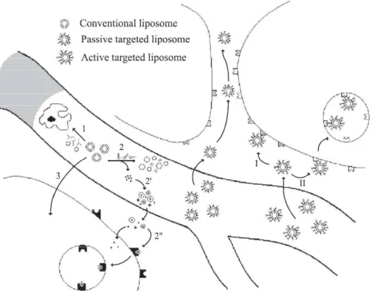
Also, the fate and pharmacokinetics of PSs encapsulated in liposomes are affected by the fact that liposomes show a short plasma half-life, in the range of minutes. Two different phenomena contribute to reduce the circulation time of conventional liposomes (Figure 5). Firstly, a lipid exchange between the liposomes and lipoproteins, particularly high density lipoproteins (HDL), leads to an irreversible and rapid disintegration of the liposome, which releases the PS in the bloodstream subsequently associated with lipoproteins and other plasma proteins. The associated molecules then enter the tumour cells mainly by endocytosis mediated by low density lipoproteins (LDL). Secondly and as mentioned before, conventional liposomes easily become opsonised by plasma proteins after which they are rapidly taken up by cells of the MPS. Consequently, they become concentrated in organs and tissues with a rich MPS like in the liver, spleen, bone marrow and blood circulation [9].
20
Figure 5: In vivo behaviour of the different types of liposomal delivery systems: conventional liposomes have a short plasma half-life either because they become absorbed by protein opsonins, followed by macrophage uptake (1) or because of lipid exchange with plasma proteins, followed by liposome disintegration and consequent PS release (2). The released PS might associate with plasma proteins (2’) and then enter the tumour cells mainly by LDL-receptor-mediated endocytosis (2’’). A small fraction of the conventional liposomes reaches the tumour tissue in its original formulation for intracellular uptake of the PS by direct binding to cell surface proteins (3). Passively targeted liposomes accumulate in the tumour interstitium without intracellular uptake. Actively targeted liposomes can either be directed to a non-internalising target (I) at the tumour cell or can enter the tumour cell by receptor-mediated endocytosis upon binding to an internalising receptor (II) [9].
In brief, due to a rapid disintegration and unspecific biodistribution, conventional liposomes are less able to establish elevated tumour-to-normal tissue ratios, hampering their generalised use as tumoritropic carriers of PSs [9].
21
2.2.2- Passively targeted liposomes for photodynamic therapy
Because of angiogenesis in malignant tissue, tumour vessel walls demonstrate an enhanced vascular permeability with fenestrae of a pore size of 100 to 1200 nm. Besides, as tumour tissue lacks a functional lymphatic system, extravasated macromolecules do not go back efficiently to the central circulation. This particular tumour architecture is at the origin of a spontaneous extravasation, accumulation and retention of some macromolecules. Provided that they circulate sufficiently long, the EPR effect allows liposomes to passively accumulate in tumour tissue at high concentrations. Thus, liposomes should be rendered “invisible” for lipoproteins and MPS. Many approaches based on surface modifications were explored to produce long-circulating liposomes with enhanced plasma stability [4], [9], [24], [33], [34].
While conventional liposomes are rapidly cleared from the bloodstream in ten minutes, the presence of glycolipids (e.g. monosialoganglioside) increases the circulation half-life to values up to 12 h. Inclusion of lipids with PEG-headgroups further prolongs the circulation half-life to the order of ten hours. Liposomes with prolonged circulation times due to alteration with glycolipids or PEGylated lipids are named sterically stabilized liposomes or stealth® liposomes [4], [9], [24], [34].
Only one in vivo study was performed to explore the PDT efficacy of long-circulating liposomes passively targeting a PS to tumour tissue. In this study, it was demonstrated a significant tumour regression and a high cure rate upon intravenous injection of benzoporphyrin derivative monoacid ring A (BPD-MA) encapsulated in glucuronide-modified liposomes and subsequent tumour illumination [25]. These long-circulating liposomes escaped from being trapped in MPS and accumulated extensively in the tumour (with an extent 3- to 4-fold higher than that of conventional liposomes with DPPG) [9].
It is shown that long-circulating liposomes, with their hydrophilic surface, do not interact efficiently with cells (Figure 5). Hence, one can speculate to what extent these extravasated liposomes accumulating in the tumour interstitium are able to transfer their PS content to tumour cells. This is significant as the cytotoxic singlet oxygen generated by the irradiated PS shows a very short migration radius. In the previous in
vivo study, it can be supposed that the liposomes were degraded under the huge
impact of singlet oxygen, launching the photocytotoxic principle into the restricted extracellular tumoral space to reach high local concentrations and that excited liposomal PSs can collapse the phospholipid barriers releasing themselves. However, in that study, the outcome could have been influenced by the use of a hydrophobic PS
22 (BPD-MA). It cannot be excluded that after a prolonged stay in the interstitial space, there occurred a limited but essential transfer of PS from the liposomes to the tumour cells [9].
Due to the inexistence of others in vivo studies, covering the efficacy of PDT using various PSs incorporated in passively targeted long-circulating liposomes, no general conclusions can be made concerning the general applicability of this type of liposomes as tumoritropic carriers for PSs [9].
2.3- Methods for preparation of liposomal drug formulations
Numerous procedures have been developed to prepare liposomes, but only a few of them are capable of encapsulating large quantities of a molecule with a specific physicochemical nature. Tables 5-8 present the description of the main passive encapsulation methods to prepare liposomal drug formulations. These procedures produce heterogeneous mixture of liposomes that after appropriate extrusion yield LUVs [38].
Table 5: Description of the lipid film hydration method (data was compiled from [28], [29], [39]).
Method of preparation Lipid film hydration
Chemical nature of the
molecule to be loaded Hydrophilic / Hydrophobic
Principle of the encapsulation
For hydrophilic drugs, the encapsulation results from the hydration of the dry lipid film with an aqueous solution of drug. The spontaneous formation of liposomes passively captures the dissolved drug. For hydrophobic drugs, the compound is dissolved with the lipid constituents in a suitable organic solvent. Afterwards, the solvent is removed and the film is hydrated with an aqueous solution at a temperature above the higher transition temperature of the lipids. For both cases, cycles of warming and mechanical agitation result in the entrapment of the drugs within the lipid bilayer.
Advantages Easy and fast procedure.
Disadvantages Low encapsulating efficiency, often less than 10%;
23
Table 6: Description of the reverse-phase evaporation method (data was compiled from [32], [38], [40]-[43]).
Method of preparation Reverse-phase evaporation
Chemical nature of the
molecule to be loaded Hydrophilic
Principle of the encapsulation
The lipid film previously prepared is redissolved in ether (organic phase). The drug solution buffer (aqueous phase) previously prepared is added directly to the organic phase. The system is then sonicated for emulsification of the two phases and the solvents removed under reduced pressure. Removal of the last traces of solvent transforms the gel into LUVs. The ratio of aqueous phase to organic phase is usually 1:3 for ether.
Advantages
High encapsulation efficiency up to 65 % can be obtained in a medium of low ionic strength. The method has been used to encapsulate small, large and biologically active macromolecules such as ribonucleic acid (RNA).
Disadvantages
Exposure of the compounds to be encapsulated to organic solvents and to brief periods of sonication, which can lead to the denaturation of some proteins or breakage of deoxyribonucleic acid (DNA) strands; limited by lipid solubility in organic phase.
Table 7: Description of the film loading method (data was compiled from [44]).
Method of preparation Film loading
Chemical nature of the
molecule to be loaded Hydrophobic
Principle of the encapsulation Empty liposomes are added to the drug film and then
sonicated and extruded.
Advantages Allows encapsulation of very hydrophobic drugs.
24
Table 8: Description of the freeze-thaw method (data was compiled from [41], [43], [45]).
Method of preparation Freeze-thaw
Chemical nature of the
molecule to be loaded Hydrophilic / Hydrophobic
Principle of the encapsulation
Sonication after film hydration and application of a series of freeze-thaw cycles, which break and re-fuse SUVs formed, replace the cycles of warming and mechanical agitation of the lipid film hydration method. Results in LUVs after extrusion.
Advantages
High encapsulation efficiencies approaching 90%; no detergents or solvents used; fast, simple and mild procedure.
Disadvantages Process inhibited by increasing the ionic strength of the
medium and by increasing the phospholipid concentration.
A method of encapsulation of drugs is acceptable from the pharmaceutical standpoint if it satisfies certain requirements, such as [40]:
• Yield well-defined and reproducible liposomes.
• Be quick, and lead to liposomes that retain the drug for long periods.
• Be applicable to liposomes prepared differently and not be influenced significantly by the liposomal lipid composition.
• Be suitable for various drugs that have similar physicochemical properties.
• Exhibit high encapsulation efficiency and loading capacity.
The three most important factors to be evaluated before selecting the method of preparation are the encapsulation efficiency, the final drug/lipid ratio (loading capacity) and appropriate drug retention properties. Thus, optimal liposomal formulations are those that will exhibit encapsulation efficiencies of 90% or more, employ inexpensive and relatively saturated lipids such as phosphatidylcholine and cholesterol (avoiding oxidation problems), and exhibit the highest possible loading capacity for economical reasons [40].
In this work, the liposomal preparation methods mentioned above were tested for encapsulation of two hydrophobic PSs and compared after physicochemical characterization of the developed formulations.
25
CHAPTER II - OBJECTIVE
The aim of this work is the development of a liposomal formulation exhibiting an efficient encapsulation of a hydrophobic PS in large unilamellar liposomes (LUVs) with an appropriate size for future intravenous administration. Liposomal formulations show the ability to decrease the tendency of hydrophobic PS to aggregate in aqueous media, prolonging the circulation of the drug in bloodstream and protecting it from premature degradation. Only the monomeric species are considerably photoactive and so liposomes enhance the PDT efficiency [9], [37]. Additionally, liposomes may enhance PS selective uptake and accumulation in tumour tissue, contributing to the selectivity of the treatment [9], [37].
In order to obtain liposomes encapsulating the PS, with the desired characteristics, different methods of preparing liposomes are tested, namely, the lipid film hydration method, the reverse-phase evaporation method, the freeze-thaw method and the film loading method. The liposome lipid composition is also varied by testing different types of phospholipids and different molar ratios of each component in the presence or absence of cholesterol. The more appropriate preparation method and liposome lipid composition will be the one that yields the highest loading capacity and encapsulation efficiency among the different conditions tested.
26
CHAPTER III – MATERIALS AND METHODS
1.1- Materials
The PS molecules Luz011c and Luz011 were kindly provided by Luzitin, SA (Coimbra, Portugal). Because these two molecules are extremely similar in terms of structure and physicochemical properties (molecular weights of 1136.15 and 1134.15 g/mol, respectively, and logarithm of the octanol/water partition coefficient (logPOW) of 2.7), differences in their behaviour when incorporated in a liposome formulation are not expected. Their use in the experiments was determined by their availability at the moment. Phospholipids distearoylphosphatidylcholine (DSPC) and distearoylphosphatidylglycerol (DSPG) were purchased from Lipoid (Ludwigshafen, Germany). Cholesterol, Sephadex G50 and Cremophor EL were supplied by Sigma-Aldrich (St. Louis, USA). Potassium di-hydrogen phosphate and perchloric acid 70% were purchased from Panreac (Barcelona, Spain). All others reagents and solvents used were supplied by Merck (Darmstadt, Germany). Polycarbonate membranes used in the extrusion were obtained from Avestin (Mannheim, Germany). The water used was internal ultra-pure water from a Purelab Ultra ultrapure water system (ELGA Process Water, Marlow, United Kingdom).
1.2- Methods
All the experimental steps involving the PS were performed in dim light conditions, because the PS is activated in the presence of sunlight, producing singlet oxygen that will destroy the bilayer of the liposome by peroxidation and will favor the release of the PS from the liposome.
The compound is also sensible to the temperature, which was controlled. The solid PS and its stock solution in chloroform were stored at -18°C under nitrogen. The preparation of liposomes was performed at room temperature, except in the stages where it was necessary to raise the temperature slightly above the transition temperature (Tm) of the lipids. The Tm corresponds to the temperature required to induce a change in the lipid physical state of a more ordered gel phase (solid phase), when the carbon chains are fully extended and with tilted structure, to a disordered liquid crystal phase, in which the carbon chains of the lipid are less oriented and more
![Figure 3: General porphyrin, chlorin, bacteriochlorin and phthalocyanine structures [7], [10]](https://thumb-eu.123doks.com/thumbv2/123dok_br/15719944.1070350/19.892.217.677.664.1100/figure-general-porphyrin-chlorin-bacteriochlorin-phthalocyanine-structures.webp)
![Table 3: Examples of ongoing clinical trials for cancer treatment by PDT (data was compiled from [17])](https://thumb-eu.123doks.com/thumbv2/123dok_br/15719944.1070350/24.892.120.788.714.1137/table-examples-ongoing-clinical-trials-cancer-treatment-compiled.webp)
![Table 4: Example of a Visudine ® liposomal formulation in PDT undergoing clinical evaluation (data was compiled from [17])](https://thumb-eu.123doks.com/thumbv2/123dok_br/15719944.1070350/29.892.104.785.271.456/example-visudine-liposomal-formulation-undergoing-clinical-evaluation-compiled.webp)
![Table 5: Description of the lipid film hydration method (data was compiled from [28], [29], [39])](https://thumb-eu.123doks.com/thumbv2/123dok_br/15719944.1070350/33.892.129.770.711.1151/table-description-lipid-film-hydration-method-data-compiled.webp)
![Table 7: Description of the film loading method (data was compiled from [44]).](https://thumb-eu.123doks.com/thumbv2/123dok_br/15719944.1070350/34.892.126.773.788.978/table-description-film-loading-method-data-compiled.webp)