Vol. 51, Special Number: pp. 97-102, December 2008
ISSN 1516-8913 Printed in Brazil BRAZILIAN ARCHIVES OF
BIOLOGY AND TECHNOLOGY
A N I N T E R N A T I O N A L J O U R N A L
Biodosimetry for Dose Assessment of Partial-Body
Exposure: A Methodological Improvement
Thiago Salazar Fernandes1*, David C. Loyd2and Ademir Amaral1 1
Grupo de Estudos em Radioproteção e Radioecologia; Departamentode Energia Nuclear; Universidade Federal de Pernambuco; Av. Professor Luiz Freire, 1000; 50740-540; thiagosalazar@hotmail.com; Recife - PE - Brasil.
2
Health Protection Agency/Radiation Protection Division; Chilton, Didcot, OX11 0RQ, United Kingdom
ABSTRACT
This study has explored the possibility of combining culture times with extending the duration for which Colcemid is present in cell culture in order to obtain better dose estimations following partial-body exposures. Irradiated and unirradiated blood was mixed to simulate a partial-exposure. Dicentric frequencies and resultant dose estimations were compared from 48 and 72 h cultures with Colcemid added at the beginning, after 24 h or for the final 3 h. The frequencies of dicentrics in first division cells increased with the cell culture time, providing better dose estimations. Unwanted excessive contraction of chromosomes caused by prolonged contact with Colcemid was measured and ways to avoid this are discussed. It is suggested that the combination of a lower than usual concentration of this drug combined with its earlier addition and longer culture time may provide metaphases better suited for interpreting partial-body exposures.
Key words: Biodosimetry, partial-body exposure, dicentrics, chromosome contraction
*
Author for correspondence INTRODUCTION
Most human exposures to ionizing radiation occur to part of the body and as a result, peripheral blood samples contain a mixture of exposed and unexposed lymphocytes (IAEA, 2001; Fernandes et al., 2006; Heimers et al., 2006).
For biodosimetry based on the scoring of chromosome aberrations (dicentrics, rings and fragments), it is important to consider only first-division (M1) lymphocytes. However, following partial-body irradiations, the irradiated fraction of cells may not have enough time to reach the first metaphase in traditional 48 h cell cultures because they may be selectively delayed or held for longer at check points during the cell cycle (Amaral, 2002; Hoffmann et al., 2002; Hone et al., 2005;
Heimers et al., 2006). Irradiated cells may also be selectively removed by apoptosis.
Considering this, two mathematical methods were proposed to interpret the aberration frequencies in terms of partial-body dose, known as the Qdr and contaminated Poisson (CP) (Sasaki and Miyata, 1968; Dolphin, 1969; Lloyd et al., 1973; IAEA, 2001). The usual time of cell culture is 48 h, because with longer times many cells enter into their second or later cell cycles so that selective elimination of chromosomal damage starts to occur by mitotic non-disjunction.
Nevertheless, if culture time is prolonged to 72 h to compensate for the delay, there is the possibility of slow growing irradiated T cells or other
radiosensitivities, coming to M1 (Han and Daday, 1978; Wuttke et al., 1993).
The technique of harlequin staining (Fluorescence plus Giemsa – FPG) allows unambiguous identification of cells in the M1 stage (Scott and Lyons, 1979). An alternative to FPG is to add Colcemid earlier to prevent fast growing cells from escaping the mitotic block (Hayata et al., 1992; Kanda et al., 1994; Senthamizhchelvan et al., 2006). This could be combined with longer culture times in order to permit the slower cells to reach M1.
Prolonged contact with Colcemid has been reported as causing excessive chromosome contraction. However, this may be overcome by
reducing the concentration of this drug
(Senthamizhchelvan et al., 2006).
This paper examines two different cell culture durations combined with the addition of Colcemid at three different times to evaluate how varying these parameters might improve the accuracy of dose assessment following partial-body exposures.
MATERIAL AND METHODS
Samples and Irradiation
Venous blood from a 28-year-old healthy non-smoking male donor was collected with informed consent and according to the local ethics protocol. Blood was irradiated at 37 ºC with 4.0 Gy 250 kVp X-rays (HVL 1.2 mmCu) acutely (0.7 Gy/min). The remaining tubes were treated identically but received zero doses. All tubes were then held for 2 h at 37 °C and then a mixture of 70% irradiated 30% unirradiated blood was made.
Cell Culture
From this mixture, whole blood lymphocyte cultures were set up using a standard protocol
(IAEA, 2001), in Eagle’s MEM with
Bromodeoxyuridine, 20% Foetal Bovine Serum, Phytohaemagglutinin (PHA) and antibiotics. Replicate cultures were incubated for 48 and 72 h and for each Colcemid (0.5 µg/mL) was added at 0, 24 or 3 h before termination.
After hypotonic treatment with 0.075 M KCl, cultures were fixed by the standard method with methanol:acetic acid (3:1) and cells were dropped onto microscope slides (IAEA, 2001).
Chromosome analysis
Replicate slides from each culture were stained with fluorescence plus Giemsa (FPG) and Fluorescence in Situ Hybridization (FISH) highlighting chromosomes 2 in green with FITC, chromosomes 3 in red with Texas Red, and using DAPI blue as counterstaining (Finnon et al., 1995; IAEA, 2001).
Additionally, other slides were stained with Giemsa for scoring of chromosome aberrations (Fernandes et al., 2008).
All the microscope analyses were carried out by one person on coded slides and strict scoring criteria were adopted whereby metaphases had to be complete with 46 centromeres and 46 or more objects present in the spread (IAEA, 2001). Aberrations recorded from the Giemsa and FPG stained cells were unstable chromosome types; dicentrics, centric rings and excess acentrics. For the FPG material the scoring was confined to M1 metaphases (IAEA, 2001). For each data point, scoring was terminated when 30 complete dicentrics, i.e. with their accompanying acentric fragments and in M1 metaphases, had been found. In the FISH stained material, Karyotyping and
FISH Imaging MetaSystems Isis software
(Germany) was used to capture the images and to
measure the lengths of the highlighted
chromosomes 2 and 3.
Statistical analysis
The chi-squared test for homogeneity of proportions was used to test for significance of difference between the Colcemid addition time points, and also the differences between incubation times. The Student’s t-test was used for significance of the difference in chromosome lengths.
Dicentric frequencies were used to estimate doses by reference to a dose-effect curve previously calibrated in the same laboratory with the same X-ray source, filtration and geometry. This curve fitted to the linear quadratic model: Y= 0.0005 (±0.0005) + 0.046 (± 0.005) D + 0.065 (± 0.003)
D2; where Y = dicentrics per cells and D = dose in
gray (Gy). The standard method considered the dose estimation without correction to the irradiated fraction of the body. In addition, the 70% was used to calculate dose to the irradiated fraction. For
estimation of the partial-body dose, two
RESULTS
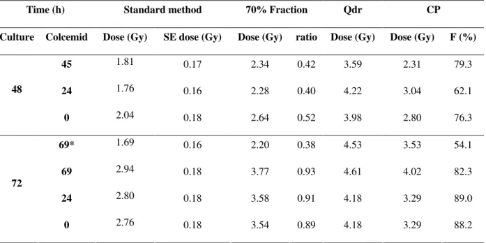
Table 1 (column 3) shows whole-body dose estimates obtained for each time point by referring the dicentric frequencies to the X-ray calibration curve. As expected, these values underestimate the dose for 48 h culture, but it also shows that they increase for longer cultures (72 h).
Column 5 shows the estimated dose to the irradiated fraction, where better estimations were obtained in 72 h. Columns 7 and 8 show the partial-body doses estimated by the Qdr and CP methods and from the latter the estimated
irradiated fraction (F) is also derived (column 9).
The Qdr method estimates approximately the real dose in 48 h cultures, and overestimates were obtained in 72 h. Better estimates using the CP method were obtained at 72 h, however, this
method provided better estimation of F in 48 h cell
culture.
Earlier addition of Colcemid (0 and 24 h) in order to reduce the confounding presence of non-M1 metaphases without the need for FPG technique carries the risk of excessive chromosome condensation, as shown in Fig. 1-B with painted 2 (light gray) and 3 (dark gray) chromosomes. Analyzing 50 metaphases, the length of pairs of chromosomes 2 and 3 from 72 h cell culture with 69 h Colcemid were 9.2 and 7.9 µm, respectively. In 72 h culture with 24 h Colcemid, the lengths reduced to values of 6.4 and 5.8 µm, statistically significant (p < 0.05).
DISCUSSION
It is axiomatic in biodosimetry that aberration scoring should be confined to M1 metaphases, because in prolonged cultures beyond the customary 48 h the dicentric frequency in M1 cells can increase. Hone et al. (2005), for example, have shown that the dicentric yield remains constant up to 51 h, but rises by about 50% to a constant value beyond 60 h.
The effect of culture time on aberration frequencies may be exacerbated in the situation of a partial-body exposure (Amaral, 2002). The irradiated fraction of cells may be selectively delayed in response to mitotic stimulation with PHA, slower progression around the cell cycle, or apoptotic elimination (Hoffmann et al., 2002; Hone et al., 2005).
The present study has shown (Table 1) improvement in dose estimates in the case of simulated partial-exposure, when the time for cell culture is prolonged from 48 to 72 h. Thus, this allows more time for the irradiated fraction of cells to reach metaphase.
In practice, following most partial-body
overexposures, knowledge about the irradiated fraction of the body (column 5 in Table 1) is generally not straightforward, unless there was reliable independent information on the irradiated volume, e.g. radiotherapy and very occasionally industrial radiation accidents. In these cases, precise exposure geometry may be well defined or obtained by questionnaire.
Figure 1 - Pairs of chromosomes 2 (light gray) and 3 (dark gray) painted after FISH. Normal length chromosomes (A) and highly contracted chromosomes induced by Colcemid (B).
Table 1 - Estimation of absorbed dose by standard method, considering the irradiated fraction (70%) and using the Qdr and CP methods.
* Without FPG staining / SE = Standard error / F (%) = Irradiated fraction.
Qdr is the only method which provides better dose estimates for 48 h cultures. On the other hand, for this same culture time, CP method provides better
estimations of F, which may be of vital importance
to the medical team in planning the therapy of highly irradiated persons.
Colcemid at 69 h produced post-M1 lymphocytes. Those dicentrics that do pass through to daughter cells should be distinguishable by the absence of a fragment, although it is possible for some to retain fragments in daughter cells. Therefore, searching for M1 cells becomes laborious and time consuming due to the contamination with the later division cells.
This problem can be solved by adding Colcemid earlier to arrest most of the cells in M1 (Hayata et al., 1992; Kanda et al., 1994). However, this approach and using the standard Colcemid concentration (0.5 µg/mL) leads to an excessive contraction of the chromosomes (Fig. 1-B) that could make the cytogenetic analysis more difficult, especially for less experienced technicians.
The practice of earlier addition of Colcemid was introduced some years ago particularly in some Japanese laboratories. Sasaki et al. (1989), used a 10 times lower concentration (0.05 µg/mL) than that used for this experiment and do not refer to the phenomenon of chromosome contraction.
Hayata et al. (1992) used a slightly lower concentration of Colcemid of 0.043 µg/mL also added at the start of 48 h lymphocyte culture and they do not mention problems of chromosome contraction nor is it apparent in their published photomicrographs.
An even lower concentration (0.02 µg/ml) of
Colcemid was added at 24 h by
Senthamizhchelvan et al. (2006), who reported that the metaphase spreads were adequate for the identification of dicentrics.
These papers, therefore, suggest that both the concentration of Colcemid and its addition time are important factors and the correct combination
makes it possible to avoid chromosome
contraction.
However, it has to be cautioned that reducing the concentration of Colcemid could lead to an insufficient amount for effective mitotic arrest. Then, cells will progress into second and further cycles, diluting the dicentric frequency and particularly for partial-body exposures, leading to an underestimation of absorbed dose.
Therefore, it is important to find a window of Colcemid concentration that is low enough to avoid chromosome contraction but high enough to accumulate exclusively M1 metaphases. This possibility requires more investigation.
Time (h) Standard method 70% Fraction Qdr CP
Culture Colcemid Dose (Gy) SE dose (Gy) Dose (Gy) ratio Dose (Gy) Dose (Gy) F (%)
45 1.81 0.17 2.34 0.42 3.59 2.31 79.3
24 1.76 0.16 2.28 0.40 4.22 3.04 62.1
48
0 2.04 0.18 2.64 0.52 3.98 2.80 76.3
69* 1.69 0.16 2.20 0.38 4.53 3.53 54.1
69 2.94 0.18 3.77 0.93 4.61 4.02 82.3
24 2.80 0.18 3.58 0.91 4.18 3.29 89.0
72
Blood samples from just one donor were intentionally used for this experiment in an attempt to confirm whether the interpretation of partial exposure being dependant on culture time is a phenomenon that exists per se.
Clearly, inter-individual variability in cell cycling speeds would vary among different donors. However, the experience from biodosimetry would suggest that this is not relevant because of the reproducibility of dose effect curves calibrated with blood from a small panel of donors and used to estimate the dose from other persons who have been exposed to ionizing radiation.
CONCLUSIONS
Prolonged cell culture time and earlier addition of Colcemid was shown to be better suited for estimating dose in cases of partial-exposures. Using this protocol, a good agreement was obtained with the actual dose used circumventing the need for mathematical calculations (Qdr and CP methods). Adding Colcemid earlier can cause excessive chromosome condensation but this can be eliminated by reducing its concentration.
ACKNOWLEDGMENTS
The authors would like to thank: Alan Edwards, Jayne Moquet and Pat Hone (Health Protection Agency – HPA), for their contribution during this research; Liz Ainsbury (HPA), for the English language revision; CNPq and CAPES for supporting this work.
RESUMO
Este trabalho avaliou a estimativa da dose de radiação simulando uma exposição parcial do corpo através da irradiação in vitro de amostras de sangue misturadas com amostras não irradiadas. Foi observado que o prolongamento do tempo de cultura permite que a real fração de linfócitos em M1 contendo aberrações cromossômicas seja detectada, propiciando melhores estimativas de dose, sem a necessidade de correções matemáticas.
REFERENCES
Amaral, A. (2002), Trends in biological dosimetry: an overview. Braz Arch Biol Technol,, 45, 119-124. Dolphin, G. W. (1969), Biological dosimetry with
particular reference to chromosome aberration analysis. A review of methods. In: Handling of Radiation Accidents, IAEA, Vienna, pp. 215–224. Fernandes, T. S.; Amaral, A.; Cavalcanti, M. B.; Braga,
L. R.; Melo, R. A. (2006), Unstable chromosome aberrations and micronuclei analyses in the biomonitoring of workers occupationally exposed to ionizing radiation. Int J Low Radiat., 3, 299-309. Fernandes, T. S.; Lloyd, D. C.; Amaral, A. (2008), A
comparison of different cytological stains for biological dosimetry. Int J Radiat Biol., 84, 703-711. Finnon, P.; Lloyd, D. C.; Edwards, A. A. (1995),
Fluorescence in situ hybridization detection of chromosomal aberrations in human lymphocytes: applicability to biological dosimetry. Int J Radiat Biol., 68, 429-435.
Han, T.; Daday, B. (1978), T lymphocyte dependency of B lymphocyte blastogenic response to phytomitogens. Immunology, 34, 625-629.
Hayata, I.; Kajima, J.; Okabe, N. (1992), Distinction of metaphases in the first cell cycle for automated system in radiation dosimetry. Radiat Phys Chem.,
39, 517-520.
Heimers, A.; Brede, H. J.; Giesen, U.; Hoffmann, W. (2006), Chromosome aberration analysis and the influence of mitotic delay after simulated partial-body exposure with high doses of sparsely and densely ionizing radiation. Radiat Environ Biophys., 45, 45-54.
Hoffmann, G. R.; Sayer, A. M.; Littlefield, L. G. (2002), Higher frequency of chromosome aberrations in late-arising first-division metaphases than in early-arising metaphases after exposure of human lymphocytes to x-rays in G0. Int J Radiat Biol., 789,
765-772.
Hone, P. A.; Edwards, A. A.; Lloyd, D. C.; Moquet, J. E. (2005), The yield of radiation-induced chromosomal aberrations in first division human lymphocytes depends on the culture time. Int J Radiat Biol., 81, 523-529.
Kanda, R.; Jiang, T.; Hayata, I.; Kobayashi, S. (1994), Effects of Colcemid concentration on chromosome aberration analysis in human lymphocytes. J Radiat Res., 35, 41-47.
Lloyd, D. C.; Purrot, R. J.; Dolphin, G. W. (1973), Chromosome aberration dosimetry using human lymphocytes in simulated partial body irradiation.
Phys Med Biol., 18, 421-431.
International Atomic Energy Agency (IAEA). (2001), Cytogenetic analysis for radiation dose assessment. in
Sasaki, M. S.; Miyata, H. (1968), Biological dosimetry in atom bomb survivors. Nature (London)., 220,
1189-1193.
Sasaki, M. S.; Kobayashi, K.; Hiedas, K.; Yamada, T.; Ejima, Y.; Maezawa, H.; Furusawa, Y.; Ito, T.; Okada, S. (1989), Induction of chromosome aberrations in human lymphocytes by monochromatic X-rays of quantum energy between 4.8 and 14.6 keV.
Int J Radiat Biol., 56, 975-988.
Senthamizhchelvan, S.; Pant, G. S.; Rath, G. K.; Julka, P. K.; Nair, O.; Joshi, R. C.; Malhotra, A.; Pandey, R. M. (2006), Biodosimetry using chromosome aberrations in human lymphocytes. Radiat Prot Dosim., 5, 1-5.
Scott, D.; Lyons, C. Y. (1979), Homogeneous sensitivity of human peripheral blood lymphocytes to radiation induced chromosome damage. Nature
(London), 278, 756-758.
Wuttke, K.; Streffer, C.; Müller, W. U. (1993), Radiation induced micronuclei in subpopulations of human lymphocytes. Mut Res., 286, 181-188.