VELOCITY FIELDS OF BLOOD FLOW IN MICROCHANNELS
USING A CONFOCAL MICRO-PIV SYSTEM
RUI LIMA1, 2, TAKUJI ISHIKAWA1, SHUJI TANAKA3, MOTOHIRO TAKEDA1, 4, KEN-ICHI TSUBOTA1, SHIGEO WADA5, TAKAMI YAMAGUCHI1
1) Dept. Bioeng. & Robotics, Grad. Sch. Eng., Tohoku Univ., 6-6-01 Aoba, 980-8579 Sendai, Japan.
2) Dept. Mechanical Tech., ESTiG, Braganca Polyt., C. Sta. Apolonia, 5301-857 Braganca, Portugal.
3) Dept. Nanomechanics, Grad. Sch. Eng., Tohoku Univ., 6-6-01 Aoba, 980-8579 Sendai, Japan.
4) Div. Surgical Oncology, Grad. Sch. Medicine, Tohoku Univ., 2-1 Seiryo-machi, Aoba-ku, 980-8575 Sendai, Japan.
5) Dept. Mechanical Science and Bioeng., Grad. Sch. Eng., Osaka Univ., Toyonaka, 560-8531 Osaka, Japan.
The in vitro experimental investigations provide an excellent approach to understand complex blood flow phenomena involved at a microscopic level. This paper emphasizes an emerging experimental technique capable to quantify the flow patterns inside microchannels with high spatial and temporal resolution. This technique, known as confocal micro-PIV, consists of a spinning disk confocal microscope, high speed camera and a diode-pumped solid state (DPSS) laser. Velocity profiles of pure water (PW), physiological saline (PS) and in vitro blood were measured in a 100µm glass square and rectangular polydimethysiloxane (PDMS) microchannel. The good agreement obtained between measured and estimated results suggests that this system is a very promising technique to obtain detail information about micro-scale effects in microchannels by using both homogeneous and non-homogeneous fluids such as blood flow.
1.
Introduction
Detailed knowledge on flow velocity profiles of blood flow in
microchannels is essential to provide a better understanding on the blood
rheological properties and disorders in microvessels. Despite the high amount of
research in microcirculation, there is not yet any detailed experimental
information about flow velocity profiles, RBCs deformability and aggregation in
microvessels. This lack of knowledge is mainly due to the absence of adequate
techniques to measure and quantitatively evaluate fluid mechanical effects at a
microscopic level [1, 2].
or conventional particle image velocimetry (PIV). However, due to limitations
of those techniques to study effects at a micro-scale level, Meinhart and his
colleagues [3] have proposed a measurement technique that combines the PIV
system with an inverted epi-fluorescent microscope, which increases the
resolution of the conventional PIV systems [3, 4]. More recently, considerable
progress in the development of confocal microscopy and consequent advantages
of this microscope over the conventional microscopes have led to a new
technique known as confocal micro-PIV [5-9]. This technique combines the
conventional PIV system with a spinning disk confocal microscope (SDCM).
Due to its outstanding spatial filtering technique together with the multiple point
light illumination system, this kind of microscope has the ability to obtain
in-focus images with optical thickness less than 1
µ
m, task extremely difficult to be
achieved by using a conventional microscope. As a result, by combining SDCM
with the conventional PIV system it is possible to achieve a PIV system with not
only extremely high spatial resolution but also with capability to generate 3D
velocity profiles.
The main purpose of this paper is to investigate the ability of the confocal
micro-PIV system to measure velocity fields of both homogeneous and
non-homogeneous fluids in two different kinds of microchannels, i. e., 100
µ
m glass
square and rectangular (300
µ
m wide, 45
µ
m deep) PDMS microchannel.
2.
Materials and methods
2.1.
Working fluids and microchannels
Four working fluids were used in this study: pure water (PW), physiological
saline (PS) and PS containing about 17%(17Hct) and 20% (20Hct) of human red
blood cells (RBCs). All fluids were seeded with 0.1 to 0.15% (by volume)
1-µ
m-diameter red fluorescent solid polymer microspheres (R0100; Duke
Scientific, USA). The blood was collected from a healthy adult volunteer, where
ethylenediaminetetraacetic acid (EDTA) was added to prevent coagulation. The
RBCs where separated from the bulk blood by centrifugation and aspiration of
the plasma and buffy coat and then washed twice with PS. The washed RBCs
where diluted with PS to make up the required RBCs concentration by volume.
All blood samples were stored hermetical at 4ºC until the experiment was
performed at room temperature (25 to 27ºC).
Two microchannels were used in this study. The first was 100
µ
m
×
100
µ
m
borosilicate glass square microchannel fabricated by
Vitrocom
. The square
microchannel was mounted on a slide glass with thickness of approximately 120
microchannel (300
µ
m wide, 45
µ
m deep) fabricated by a soft lithographic
technique at the Esashi, Ono and Tanaka Laboratory, Department of
Nanomechanics, Tohoku University [10].
2.2.
Confocal micro-PIV experimental set-up
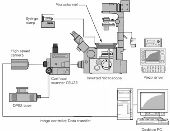
The confocal micro-PIV system used in our experiment consists of an
inverted microscope (IX71, Olympus, Japan) combined with a confocal
scanning unit (CSU22, Yokogawa, Japan) and a diode-pumped solid state
(DPSS) laser (Laser Quantum Ltd, England) with an excitation wavelength of
532 nm. Moreover, a high-speed camera (Phantom v7.1, U.S.A.) was connected
into the outlet port of the CSU22. The microchannels were placed on the stage
of the inverted microscope where the flow rate of the working fluids was kept
constant by means of a syringe pump (KD Scientific Inc., U.S.A.). For the
square microchannel the flow rate was 0.15
µ
l/min (Re = 0.014), whereas for the
PDMS rectangular microchannel the flow rate was 0.22
µ
l/min (Re = 0.021).
Fig. 1. Confocal micro-PIV experimental set-up.
information about the experimental set-up, used in the present study, has already
been described previously [9].
3. Results and discussion
3.1. Comparison between Experimental and Analytical Results
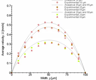
By using the optical sectioning ability of the confocal system it was
possible to obtain series of optical sectioned images along z axis. Figures 2 and
3 shows a comparison between analytical solutions [12] and average fluid
velocities of 100 PIV image pairs at several optical sectioned images for both
microchannels. Generally, the experimental results from Figures 2 and 3 show
good agreement with the analytical solution, especially the results from the
middle plane where we found errors less than 5%. As one moves out of the
middle plane the deviations start to increase and at locations close to the wall the
errors can be slightly larger than 10%. We believe that these latter errors are
mainly attributable to "second-order effects" such as the surface roughness of
the wall and background noise generated from particles adhered into the wall.
The results from Figure 3 also show that the velocity patterns were
approximately parabolic which is characteristic of laminar flow in a square
microchannel. In contrast, the velocity profiles from the PDMS microchannel
(see Figure 4) were markedly flat in the central region, which is characteristic of
laminar flow in a rectangular microchannel with low aspect ratio (h/w = 0.15).
Fig. 3. Comparison between experimental data and analytical solutions at several optical sectioned images in a rectangular PDMS microchannel. The working fluid is PS.
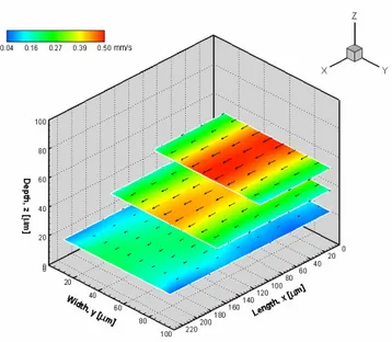
3.2. Velocity fields of in vitro blood flow
Besides the employment of PW and PS, in this study PS containing around
17% and 20% of suspended blood cells was also used in order to evaluate the
potentialities of the confocal micro-PIV system to investigate the flow behaviour
of
in vitro
blood in both microchannels.
Fig. 4. Velocity vector fields from several optical sectioned images along z axis, in a 100 µm glass square microchannel. The working fluid is in vitro blood with 17% Hct.