Leonardo de Souza VasconcellosI, Andy PetroianuII, Juliana Ribeiro RomeiroIII, Wilson Campos Tavares
JuniorIV, Vivian ResendeV
Correlation between the values of circulating blood
elements with the size of spleen in the presence of
schistosomal splenomegaly
1Acta Cir Bras. 2018;33(12):1103-1109
Abstract
Purpose: To evaluate a possible relationship between the size of the spleen and values of
circulating blood elements in patients with schistosomatic splenomegaly.
Methods: Sixty one patients with hepatosplenic schistosomiasis mansoni underwent a
clinical exam and peripheral venous blood was collected for a hemogram. The erythrocyte, hemoglobin, hematocrit, leukocyte, and platelet values were determined. All patients underwent abdominal ultrasound to measure the spleen. The hematological test results were compared to the size of the spleen.
Results: The size of the spleen varied from 14.0 to 28.4 (19.9 ± 3.7) cm according to the
ultrasound image. Thrombocytopenia was observed 58 (95%) patients, leukopenia in 55 (90%) patients, and anemia in 32 (52.4%) patients. Leukopenia was proportional to splenomegaly.
Conclusion: Schistosomal splenomegaly leads to leukopenia in direct proportion to the size
of the spleen.
Key words: Schistosomiasis mansoni. Splenomegaly. Leukopenia. Thrombocytopenia. Anemia.
DOI: http://dx.doi.org/10.1590/s0102-865020180120000008
IPhD, Associate Professor, Complementary Propaedeutics Department, School of Medicine, Universidade Federal de
Minas Gerais (UFMG), Belo Horizonte-MG, Brazil. Scientific, intellectual, conception and design of the study; acquisition, analysis and interpretation of data; technical procedures; statistical analysis; manuscript preparation and writing.
IIPhD, Full Professor, Department of Surgery, Faculty of Medicine, UFMG, Researcher 1B CNPq, Belo Horizonte-MG, Brazil.
Scientific, intellectual, conception and design of the study; acquisition, analysis and interpretation of data; technical procedures; manuscript preparation and writing; critical revision; final approval.
IIIPhD, School of Medicine, UFMG, Belo Horizonte-MG, Brazil.Acquisition of data, technical procedures. IVMD, MSc, Clinical Hospital, UFMG, Belo Horizonte-MG, Brazil.Acquisition of data, technical procedures.
VPhD, Associate Professor, Department of Surgery, School of Medicine, UFMG,Belo Horizonte-MG, Brazil.Scientific,
■
Methods
This study was approved by the Research Ethics Committee from Universidade Federal de Minas Gerais (UFMG), logged under protocol number ETIC 006/08, as well as by the Teaching, Research, and Extension Board from the UFMG Clinical Hospital, logged under protocol number 065/09.
This study was performed on 61 patients with hepatosplenic schistosomiasis, 36 men and 25 women. The age of the patients varied from 18 to 65 (42 ± 11) years. Regarding the skin color, 17 were caucasian, 30 light-skinned black, and 14 dark-light-skinned black. All of the patients, upon undergoing a physical exam, presented hepatosplenomegaly. Patients with any other clinically manifested disease or who were undergoing complementary exams were excluded. All patients were advised as regards the purpose of this research and only those who agreed to the terms of the study and who signed the Free and Informed Consent Form were included.
For the hemogram dose, 4 ml of peripheral venous blood was collected, aimed at quantifying the erythrocyte, leukocyte, and thrombocyte series. The collection was conducted by a vacuum blood collection system in EDTA tubes. The samples were processed in a specific automation device, by electric impedance and flow cytometry (Cell Dyn 3000® System, Abbott Diagnostics
Division, Mountain View, California, USA), and the results were manually confirmed by a hematoscopy exam, performed by a Clinical Pathologist.
Anemia was considered when hemoglobin was less than 13.0 g/dl in men and 12.0 g/dl in women. Thrombocytopenia was classified as serum platelets of less than 150.000/mm3. Leukopenia was considered when total serum leukocyte values were below 4.000/mm3 14.
All of the patients were submitted
■
Introduction
Symmers-Bogliolo is a typical hepatosplenic lesion of Schistosomiasis mansoni (SM), which culminates in presinusoidal portal hypertension1.After five to 15 years of schistosomatic hepatopathy, scheral congestive splenomegaly, hyperdynamic porto-systemic collateral circulation, and the reduction in the number of peripheral blood elements begin to appear2,3.
The association between scheral congestive splenomegaly and hematological alterations of leukopenia, plateletopenia, and anemia is still not fully understood, considering that the blood elements remain within the spleen without being destroyed, as occurs in leukosis and hypersplenism4,5. By contrast, in emergency, such as severe infections and intense bleeding, leukocytes and platelets are released in the quantity that the organism needs6,7. In this sense, patients with intense leukopenia begin to present leukocytosis during septic complications at the same level as people with a normal spleen8. As regards thrombocytopenia, no schistosomal patient, even those with platelet levels of below 10.000/mm3, presents hemorrhagic disorders, purple spots, or bruises, and their bleeding time is normal9.Thus, surgical procedures are performed on these patients with no difficulty in vascular coagulation and without the need for a transfusion of platelet concentrates10. Therefore, leukopenia and thrombocytopenia are only laboratorial, with no clinical manifestation and without hypersplenism. The spleen plays the role of storing the blood elements, without destroying them, and releases them into systemic circulation when necessary10.
to a bidimensional ultrasound study15. The exams were conducted by a single ultrasound technician, following the same evaluation standard for the spleen and other abdominal organs, including the portal system. The size of the spleen was defined by measuring its craniocaudal length16. The size of the spleen was correlated with the values of the hematological exams.
The statistical tests were carried out using the Statistical Package for Social Sciences (SPSS) version 12.0 for Windows® (SPSS
Incorporation, Chicago, Illinois, USA, 2005). Spearman’s correlation coefficient was used to compare the size of the longitudinal axis of the spleen with the hematological parameters17. The significance level was set at p < 0.05 corresponding 95% CI.
■
Results
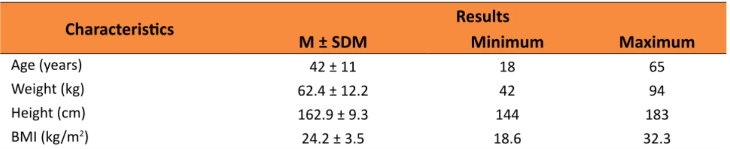
Data referent to age, weight, height, and body mass index (BMI) of the 61 patients are presented in Table 1.
Table 1 –Charcteristics of 61 patients with splenomegaly caused by Schistosomiasis mansoni.
Characteristics Results
M ± SDM Minimum Maximum
Age (years) 42 ± 11 18 65
Weight (kg) 62.4 ± 12.2 42 94
Height (cm) 162.9 ± 9.3 144 183
BMI (kg/m2) 24.2 ± 3.5 18.6 32.3
Mean ± standard deviation of mean (M ± SDM); BMI = Body Mass Index.
Table 2 presents the sizes of the spleens
measured by ultrasound and the evaluated hematological results.
Table 2 –Hematological exams and craniocaudal length of the spleen of 61 patients with splenomegaly
caused by Schistosomiasis mansoni.
Parameters Results
M ± SDM Minimum Maximum Reference Intervals
Red blood cells (106/mm3) 4.24 ± 0.60 2.76 5.40 4.5 – 5.5 (M)
3.8 – 5.4 (F)
Hemoglobin (g/dL) 11.2 ± 2.10 7.2 16.0 13.0 – 17.5 (M)
12.0 – 16.0 (F)
Hematocrit (%) 34.4 ± 5.10 25.0 47.3 40 – 50 (M)
36 – 46 (F) Total leukocytes (cels/mm3) 2.710 ± 1.348 900 7.600 4.000 – 11.000
Platelets (cels/mm3) 55.918 ± 33.253 15.000 167.000 150.000 – 450.000
Craniocaudal length of spleen (cm) 19.9 ± 3.7 14.0 28.4 8 – 12 Mean ± standard deviation of mean (M ± SDM); M = Male, F = Female.
At the ultrasound exam, no patient presented parenchymal alterations of cirrhosis, tumor, or thrombosis of the main veins of
whose measurements of the craniocaudal length varied between 14 and 28.4 (19.9 ± 3.7) cm.
Regarding the hematological results, the thrombocytopenia was the most common alteration, which was observed in 58 (95%) patients. Leukopenia and anemia were observed in 55 (90%) and in 32 (52.4%) of the cases, respectively.
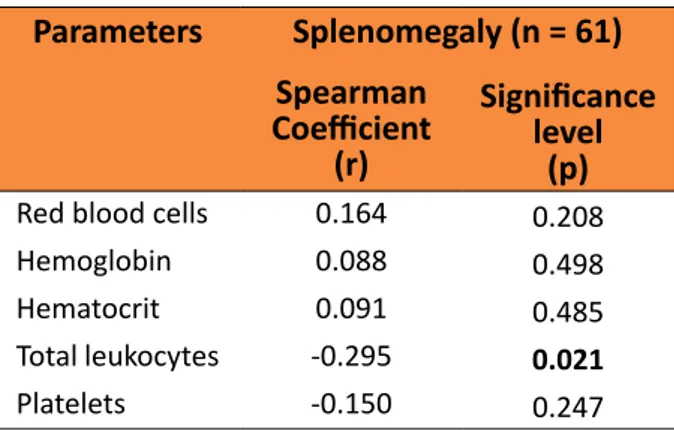
The correlations between the size of the spleen and the hematological parameters are presented in Table 3.
Table 3 – Correlation between splenomegaly
and results from hematological exams from 61 patients with splenomegaly caused by Schistosomiasis mansoni.
Parameters Splenomegaly (n = 61)
Spearman
Coefficient
(r)
Significance
level (p)
Red blood cells 0.164 0.208
Hemoglobin 0.088 0.498
Hematocrit 0.091 0.485
Total leukocytes -0.295 0.021
Platelets -0.150 0.247
Proportionality was observed between leukopenia and the splenic dimensions, larger spleens were accompanied by proportionally more intense leukopenia (p = 0.021). In the other evaluated hematological parameters, no difference or proportionality was found. No difference between the sexes nor between the patients with different skin colors. No alterations stemming from patient age and BMI were found.
■
Discussion
Among the exams available to determine the size of the spleen, the ultrasound
is the most common18-20. Splenic dimensions can vary according to age, nutritional state, anthropometric dimensions, and the presence of diseases. In healthy adults, the normal spleen measures approximately 8 cm to 12 cm along its craniocaudal length, of 4 cm to 7 cm in width (anteroposterior), and 3 cm to 4 cm in thickness (laterolateral)15,16.
Since the measures were taken by a single professional in all patients, according to health protocols of the World Health Organization (WHO)16, potential errors were maintained constant, and the dimensions followed a similar pattern21-23 .
According to the literature, the cytopenias associated with splenomegaly and portal hypertension of any etiology occur in a percentage that varies between 30% and 70% of the cases4,5,23 and, specifically in schistosomiasis, between 50% and 95% of the cases20-,22. According to the literature, the majority of the patients presented pancytopenia4-6. Among the cytopenias observed, plateletopenia was the most common, followed by leukopenia and anemia. These findings were also reported in other studies24-30 .
Although pancytopenia is common among patients with spenomegaly, the reduction of erythrocyte, leukocyte, and thrombocyte series is not always associated with the size of the spleen10. Cytopenia does not necessarily occur in all peripheral blood elements, as the patient may present only anemia or leukopenia or thrombocytopenia, as well as the reduction of only two of the three series27. This situation also remains unexplained in current knowledge, which ignores the etiopathogenesis of the reduction of each one of the series4,10.
In this study, the spelomegaly was correlated only to leukopenia. Such findings were similar to those from Khishen et al.24,
a correlation with thrombocytopenia. By contrast, Wadenvik et al.5 verified that the
dimensions of the spleen, measured by ultrasound, had a correlation with the low levels of hemoglobin and platelets but not with the count of circulating leukocytes. Neither thrombocytopania, present in 95% of the patients evaluated, nor the intensity of anemia proved to be correlated to the size of the spleen. Such results contradict studies conducted by Martins et al.25, who found a correlation
between splenomegaly and plateletopenia. However, these authors evaluated neither the leukocyte nor the erythrocyte series. Leite et al.26 also observed a correlation
between spenomegaly and thrombocytopenia in 55 patients in the hepatosplenic stage of Schistosomiasis mansoni.
According to Eichner7, augmented spleens are not necessarily abnormal, much like hyperfunctioning spleens are not always augmented. Grover et al.23 observed no
hematological alterations in the presence of splenomegalies stemming from different causes, while Gielchinsky et al.6 found
pancytopenia in the presence of small spleens, with no perceivable disorders.
The divergence in findings in the literature reinforces how difficult it is to understand the physiopathology of pancytopenia in patients with splenomegaly4. Most authors erroneously describe cytopenia as necessarily pertaining to a medical condition of hypersplenism4,5. This idea began with studies that considered hypersplenism as an increase in hemocatheretic activity resulting from structural hyperplasia of the spleen, always accompanied by a destructive autoimmune mechanism of the blood elements29,30. However, this situation rarely occurs10.The more common is splenomegaly, associated only with an increase in the storage of blood elements without provoking any clinical disorder8,9,12. This concept is reinforced by the fact that partial
and subtotal splenectomies have normalized the number of peripheral blood elements, thereby maintaining the splenic tissue. In this sense, the immune response hypothesis no longer contains a substrate10.
A weakness aspect of this study was the use of ultrasound to determine the longitudinal measurement of the spleen, rather than the volumetric dimensions of the organ, since the process of leukocyte accumulation and other blood components in the spleen occurs in the three dimensions. In fact, ideally, splenic index calculated for the splenic volume, using the craniocaudal dimension, width, and thickness, would be the most reliable measurement for diagnosing splenomegaly. However, according to literature18-20,31, a single measurement, for example the craniocaudal length, is not different to emphasise the splenic index from the one calculated for the splenic volume.
■
Conclusion
The size of the spleen was proportionally correlated with leukopenia when in the presence of schistosomal splenomegaly.
■
References
1. McManus DP, Dunne DW, Sacko M, Utzinger J, Vennervald BJ, Zhou XN. Schistosomiasis. Nat Rev Dis Primers. 2018;4:13. PMID: 30093684.
2. Gryseels B, Polman K, Clerinx J, Kestens L. Human schistosomiasis. Lancet. 2006;368:1106-18. PMID: 16997665.
3. Coura JR, Amaral RS. Epidemiological and control aspects of schistosomiasis in Brazilian endemic areas. Mem Inst Oswaldo Cruz. 2004;99:13-9. PMID: 15486629. 4. Petroianu A, De Oliveira AE, Alberti LR.
Hypersplenism in schistosomatic portal hypertension. Arch Med Res. 2005;36:496-501. PMID: 16099328.
6. Gielchinsky Y, Elstein D, Hadas-Halpern I, Lahad A, Abrahamov A, Zimran A. Is there a correlation between degree of splenomegaly, symptoms and hypersplenism? Br J Haematol. 1999;106:812-6. PMID: 10468878.
7. Eichner ER. Splenic function: normal, too much and too little. Am J Med. 1979;66:311-20. PMID: 371397.
8. Petroianu A, da Silva RG, Simal CJ, de Carvalho DG, da Silva RA. Late postoperative follow-up of patients submitted to subtotal splenectomy. Am Surg. 1997;63:735-40. PMID: 9247444.
9. Petroianu A, Resende V, da Silva RG. Late follow-up of patients submitted to subtotal splenectomy. Int J Surg. 2006;4:172-8. PMID: 17462342.
10. Petroianu A, Oliveira AE, Alberti LR. “Hiperesplenismo” em hipertensão porta por esquistossomose mansônica. Rev Bras Hematol Hemoter. 2004;26:195-201. doi: 10.1590/S1516-84842004000300009. 11. Petroianu A. Tratamento cirúrgico da
hipertensão porta na esquistossomose mansoni. Rev Soc Bras Med Tropical. 2003;36:253-65. doi: S0037-86822003000200010.
12. Petroianu A, Resende V, da Silva RG. Late postoperative follow-up of patients undergoing subtotal splenectomy. Clinics (Sao Paulo). 2005;60:473-8. PMID: 16358137.
13. Petroianu A, Rezende Neto JB. Tratamento de hemorragia intestinal grave decorrente de hipertensão porta, por meio de esplenectomia subtotal e anastomose esplenorrenal proximal. Rev Col Bras Cir. 2008;35:264-8. doi: 10.1590/S0100-69912008000400010.
14. Van den Bossche J, Devreese K, Malfait R, Van de Vyvere M, Wauters A, Neeis H, De Schouwer P. Reference intervals for a complete blood count determined on different automated haematology analysers: Abx Pentra 120 Retic, Coulter Gen-S, Sysmex SE 9500, Abbott Cell Dyn 4000 and Bayer Advia 120. Clin Chem Lab Med. 2002;40:69-73. PMID: 11916274.
15. Cairo Working Group 1992. The use of diagnostic ultra-sound in schistosomiasis-attempts at standardization of methodology. Acta Tropica. 1992;51:45-63. PMID:
1351355.
16. Akpata R, Neumayr A, Holtfreter MC, Krantz I, Singh DD, Mota R, Walter S, Hatz C, Richter J. The WHO ultrasonography protocol for assessing morbidity due to Schistosoma haematobium. Acceptance and evolution over 14 years. Systematic review. Parasitol Res. 2015;114:1279-89. doi: 10.1007/ s00436-015-4389-z.
17. Jacobson BC, Rowland DY. A simpler approach to biostatistics. Gastrointest Endosc. 2011;73:570-4. PMID: 21353855. 18. Lambertucci JR, Cota GF, Pinto-Silva RA,
Serufo JC, Gerspacher-Lara R, Drummond SC, Antunes CM, Nobre V, Rayes AA. Hepatosplenic schistosomiasis mansoni in field-based studies: a combined clinical and sonographic definition. Mem Inst Oswaldo Cruz. 2001;96:147-50. PMID: 11586441. 19. Cota GF, Pinto-Silva RA, Antunes
CM, Lambertucci JR. Ultrasound and clinical investigation of hepatosplenic schistosomiasis: evaluation of splenomegaly and liver fibrosis four years after mass chemotherapy with oxamniquine. Am J Trop Med Hyg. 2006;74:103-7. PMID: 16407352. 20. Voieta I, de Queiroz LC, Andrade LM, Silva
LC, Fontes VF, Barbosa A Jr, Resende V, Petroianu A, Andrade Z, Antunes CM, Lambertucci JR. Imaging techniques and histology in the evaluation of liver fibrosis in hepatosplenic schistosomiasis mansoni in Brazil: a comparative study. Mem Inst Oswaldo Cruz. 2010;105:414-21. doi: 10.1590/S0074-02762010000400011. 21. Barkun AN, Camus M, Green L, Meagher
T, Coupal L, De Stempel J, Grover SA. The bedside assessment of splenic enlargement. Am J Med. 1991;91:512-8. PMID: 1951414. 22. Santos GT, Sales DM, Leão ARS, Santos
JEM, Aguiar LAK, Brant PE, Shigueoka DC, Colleoni RN, D’Ippolito G. Reprodutibilidade da classificação ultra-sonográfica de Niamey na avaliação da fibrose periportal na esquistossomose mansônica. Radiol Bras. 2007;40:377-81. doi: 10.1590/S0100-39842007000600005.
23. Grover SA, Barkun NA, Sackett DL. The rational clinical examination. Does this pacient have splenomegaly? JAMA. 1993;270:2218-21. PMID: 8411607.
is contraindicated for thrombocytopenia secondary to portal hypertension. Surg Gynecol Obstet. 1985;160:233-8. PMID: 3975794.
25. Martins RN, Cleva R, Gouveia EM, Ghosn NB, Herman P. Correlação entre esplenomegalia e plaquetopenia na forma hepatoesplênica da esquistossomose mansônica. ABCD Arq Bras Cir Dig. 2010;23:254-8. doi: 10.1590/ S0102-67202010000400010.
26. Leite LAC, Domingues AL, Lopes EP, Ferreira R de C, Pimenta A de A Filho, da Fonseca CS, Dos Santos BS, Lima VL. Relationship between splenomegaly and hematologic findings in patients with hepatosplenic schistosomiasis. Rev Bras Hematol Hemoter. 2013;35:332-6. PMID: 24255616.
27. Maia MD, Lopes EPA, Ferraz AAB, Barros FMR, Domingues ALC, Ferraz EM. Evaluation of Splenomegaly in the Hepatosplenic form of Mansonic Schistosomiasis. Acta Tropica. 2008;101:183-6. PMID: 17336262.
28. Drummond SC, Pereira PN, Otoni A, Chaves BA, Antunes CM, Lambertucci JR. Thrombocytopenia as a surrogate marker of hepatosplenic schistosomiasis in endemic areas for Schistosomiasis mansoni. Rev Soc Bras Med Trop. 2014;47(2):218-22. PMID: 24861297.
29. Nelson EW, Mone MC. Splenectomy in high risk patients with splenomegaly. Am J Surg. 1999; 178:581-96. PMID: 10670877.
30. Thijs L, Messiaen P, van der Hilst J, Madoe V, Melis C, Van Eyken P, Vanmoerkerke I, Janssens F. Hepatic schistosomiasis with massive splenomegaly: a case report and literature review. Acta Gastroenterol Belg. 2018;81:93-6. PMID: 29562382.
31. Indiran V, Singh NV, Prasad TR, Maduraimuthu P. Does coronal oblique length of spleen on CT reflect splenic index? Abdom Radiol. 2017;42:1444–8. PMID: 28130582.
Correspondence:
Andy Petroianu
Avenida Afonso Pena, 1626/1901 30.130-005 Belo Horizonte - MG Brasil Tels.: (55 31)98884-9192 / 3409-9948 [email protected]
Received: Aug 23, 2018 Review: Oct 24, 2018 Accepted: Nov 25, 2018
Conflict of interest: none
Financial sources: FAPEMIG, CNPq, and PRPq-UFMG