UNIVERSIDADE DE LISBOA
FACULDADE DE CIÊNCIAS
DEPARTAMENTO DE QUÍMICA E BIOQUÍMICA
Cloning, Expression and Purification of a
Sea Urchin adhesive protein towards the
development of a new biomedical adhesive
Mestrado em Bioquímica
Especialização em Bioquímica Médica
Ana Catarina Alcarva Pontes
Dissertação orientada por
Doutor Carlos Cordeiro e Doutora Romana Santos
Acknowledgments
A
GRADECIMENTOS
Gostaria de agradecer aqui a todos aqueles que me apoiaram e me acompanharam, quer a nível profissional como pessoal, nesta jornada mais longa do que o esperado. Quero salientar a importância que a minha escolha de fazer a tese no laboratório de Enzimologia teve no meu futuro. Foi aí que me foram proporcionadas todas as condições para começar a crescer como cientista. Só desta forma me foi possível perseguir aqueles que eram os meus objetivos desde que decidi começar a minha formação académica na Faculdade de Ciências, entrar num programa doutoral.
Não foi no entanto um caminho fácil de percorrer, e por isso estou ainda mais grata por todo o apoio que me foi dado por colegas de trabalho, amigos e familiares, àqueles que sempre me disseram que eu ia conseguir apesar das minhas dúvidas e àqueles que não me deixaram desistir: Ao professor Carlos Cordeiro pelo apoio científico que me proporcionou durante o recorrer do meu ano no laboratório de Enzimologia, pelo seu espírito crítico e pela constante disponibilidade. Para além do supracitado, estou especialmente grata pelo apoio pessoal que me prestou não só durante esse ano mas como no que se seguiu. Mais importante do que aqueles que nos apoiam nas nossas decisões são os que não o fazem para nosso próprio benefício. Sem a sua ajuda, sem a motivação que me transmitiu, e sem a sua persistência este projeto teria ficado inacabado. Há diferentes tipos de professores, líderes de grupo, orientadores, e diferentes formas de os avaliar, mas, os melhores são aqueles que adaptam a sua estratégia de ensino perante o aluno que têm, os que vêm o aluno como uma pessoa e não como um meio necessário para produzir resultados.
Aos restantes membros do grupo de Enzimologia, com especial atenção à professora Ana Ponces, Marta Silva e Gonçalo Costa. Ao Martin por me ter permitido tirar o tempo necessário para acabar esta há muito adiada etapa. À minha colega de laboratório e amiga Raquel que teve todos os dias ao meu lado no laboratório (e fora dele) sempre disponível a ajudar em tudo o que estivesse ao seu dispor. À Quica, pelas longas conversas (científicas e não só) e pelo duro espírito crítico e racional que bem a caracteriza. À Joana e ao Nuno por serem as minhas bolhas de ar fresco. À minha mãe e irmã pela imensa paciência que tiveram todos os dias em ouvir-me e “aturar-me” nos dias bons e nos dias maus ficando sempre, sempre do meu lado.
Table of Contents v
T
ABLE OF CONTENTS
Agradecimentos ... 4 Table of contents ... v Resumo ... viii Summary ... xTable List ... xii
Figure List ... xiv
Introduction ... 1
I -Introduction ... 3
1 -The Sea Urchin ... 4
2 -1.1 The stem ... 5
1.2 The disc ... 6
1.3 Biochemistry of the adhesive ... 6
Methods ... 15
II -Collection of P. lividus and footprint material ... 17
1 -Total RNA Extraction and RT-PCR ... 17
2 -Nectin gene amplification using PCR ... 17
3 -Cloning using TOP10 E. coli cells ... 19
4 -Plasmid DNA extraction from recombinant E. coli ... 19
5 -Expression vector and nectin ligation ... 19
6 -Sequencing results ... 19
7 -Protein separation by 1D SDS-PAGE ... 20
8 -In-Gel Digestion ... 20
9 -Maldi-MS/MS ... 20
10 -Results and Discussion ... 24
III -Sequencing and cloning of the nectin gene ... 26
1 -1.1 P. lividus adhesive tube feet discs mRNA extraction ... 26
1.4 Nectin cDNA ... 26
1.5 Nectin cloning ... 28
Table of Contents
vi 1.7 Nectin Purification ... 30
References ... 34 IV
-Resumo
viii
R
ESUMO
Os ouriços do mar vivem em zonas de elevado hidrodinamismo, e para sobreviverem dispõe de órgãos adesivos, os pés ambulacrários, especializados na locomoção e adesão ao substrato. Embora se fixem fortemente ao substrato, estes órgãos fazem-no de forma temporária, colando-se e descolando-se repetidamente. Os pés
ambulacrários orais estão extraordinariamente bem adaptados à adesão temporária, sendo constituídos por um disco apical achatado, com o qual se fixam ao substrato, e um caule extensível que liga o disco à carapaça do ouriço. O disco é o local de
produção da secreção adesiva, que fixa o ouriço ao substrato, bem como da secreção “desadesiva” com o qual se descola do mesmo. Até aos anos 90, foi demonstrado que a adesão temporária dos ouriços resulta da presença de um sistema duo-glandular na epiderme do disco, composto por dois ou mais tipos de células que produzem
separadamente secreções adesivas e “desadesivas”. A secreção adesiva permanece fixa ao substrato sob a forma de pequenos círculos (material adesivo secretado) enquanto a secreção “desadesiva” não fica incorporada nestes círculos, atuando provavelmente como um enzima.
Foi demonstrado recentemente que o disco dos pés ambulacrários dos ouriços do mar adere com tenacidades (força adesiva/ unidade de área) semelhantes às de outros adesivos marinhos e sintéticos, sendo eficaz em substratos com diferentes rugosidades e propriedades químicas. Estes resultados reforçam o potencial
biotecnológico do adesivo secretado pelos ouriços do mar, uma vez que, dada a sua resistência, eficácia e versatilidade em meios aquosos e em vários tipos de substratos, poderá ter aplicações industriais e biomédicas. Apesar destas promissoras aplicações, pouco se conhece sobre os mecanismos moleculares responsáveis pela adesão temporária dos ouriços do mar. A caracterização bioquímica do adesivo secretário pelo ouriço do mar Paracentrotus lividus (Pl) e do proteoma dos discos adesivos dos pés ambulacrários da mesma espécie foi recentemente realizada e várias proteínas que parecem estar envolvidas na sua adesão temporária foram identificadas.
Neste trabalho, descrevo a purificação da Pl Nectin, uma das possíveis proteínas candidatas à existência de adesão. Para tal, o gene da Pl Nectin foi clonado e sequenciado usando primers desenhados com base nas sequências peptídicas obtidas previamente. O cDNA correspondente foi clonado em plasmídeos e expresso em bactéria. Por fim, a purificação da proteína por cromatografia de afinidade foi tentada, sem sucesso no entanto, apesar de todos os nossos esforços.
Summary
x
S
UMMARY
Sea urchins inhabit wave-swept shores being subjected to substantial hydrodynamic forces. They thus, rely on adhesive organs, the adoral tube feet, specialized in
locomotion and anchoring. These adhesive appendages attach strongly but reversibly to the substratum, detaching and re-attaching voluntarily. Adoral tube feet are well designed for temporary adhesion, possessing an enlarged and flattened apical disc, with which they attach and detach from the substratum, connected to the animal body by an extensible tether, the stem. The tube foot disc produces the adhesive secretion that fastens the animal to the substratum and the de-adhesive secretion that allows it to move. Before the 90s it was only known that the temporary nature of sea urchin
adhesion is due to the presence of a duogland adhesive system in the disc epidermis which comprises 2 or more cell types capable of producing separately adhesive and de-adhesive secretions. After detachment, the adhesive secretion remains on the substratum as a footprint (circle of secreted adhesive). As for the de-adhesive
secretion it is not incorporated in the footprint, indicating that it might act as an enzyme. It was recently shown that the tube feet discs attach with similar tenacity (adhesive force per unit area) to other marine and commercial adhesives, being effective on several substrata with variable chemistries and roughness. These results reinforce the biotechnological potential of the adhesive secreted by sea urchins, because given its resistance, effectiveness and versatility on aqueous media and several types of substrata, it may find both industrial and biomedical applications. However, despite these promising applications, little is known on the molecular mechanisms behind tube foot temporary adhesion.
The biochemical characterization of the adhesive secreted by the sea urchin
Paracentrotus lividus (Pl) and the proteome of the tube foot adhesive discs of the same
species was recently done and several proteins that seem to be involved in the temporary adhesion were pin-pointed.
Here I describe the purification of the Pl Nectin, one of the possible candidates for this adhesion. To achieve this goal the Pl Nectin gene was cloned and sequenced using primers designed based on the previously obtained peptide sequences. The
correspondent cDNA was cloned in plasmids and expressed in bacteria. Finally, the purification of the protein by affinity-chromatography was attempted but failed despite all of out tries.
Table List
xii
T
ABLE
L
IST
Table II-1 - Reagents and volumes used for the PCR ... 18
Table II-2 – Primers pairs used in the reactions ... 18
Table II-3 – Amplification Conditions used for Nec1 and Nec1-2 fragments ... 18
Table II-4 – Tested amplification conditions to amplify Necfull ... 18
Table II-5 – Reagents and volumes used for the PCR ... 19
Table III-1 – Set of primers used and the corresponding amplified fragments ... 26
Figure List
xiv
F
IGURE
L
IST
Figure 1 – The sea urchin Paracentrotus lividus ... 4 Figure 2 – Scanning electron microscopy photograph of a disc-ending tube foot of the
echinoid Paracentrotus lividus attached to a smooth glass substratum ... 5 Figure 3 - One dimension protein profile of the adhesive secretion separated in 12.5%
(lane 2), 8% (lane 3), and 15% (lane 4) polyacrylamide gels ... 7 Figure 4 – Electron microscopy images ... 8 Figure 5 - One and two dimension protein profile from the tube feet discs of
Paracentrotus lividus separated in 12.5% polyacrylamide gels ... 9
Figure 6 - Phosphorylated and glycosylated proteome of Paracentrotus lividus adhesive discs ... 10 Figure 7 – Nectin and its domains and the fragments amplified with each pair of
primers ... 26 Figure 8 - Agarose gel with different Nectin fragments’ PCR amplification. FL – Full
length nectin ... 27 Figure 9 - Agarose gel confirming Nec1-2 amplification ... 27 Figure 10 – Cloning of the Nec1-2 insert. ... 28 Figure 11 – Representation of the six domains present in Nectin emphasising the first
four domains that the used shortened form contain. ... 28 Figure 12 – Digested expression vector and Nec1-2 with the restriction enzymes SalI
and NotI ... 28 Figure 13 – pGEX4T.1_Nec1-2 extracted from DH5α, RII and C41 cells ... 29 Figure 14 – SDS-PAGE profiles of the optimized expression conditions using RII and
C41 ... 29 Figure 15 – Nec1-2 purification attempt using Glutathione Sepharose beads ... 30 Figure 16 – Purification of glutathione S-transferase using Glutathione Sepharose
1
I
NTRODUCTION
-I - -Introduction
3
Introduction
1
-Sea urchins are common inhabitants of the wave-swept shores, being subjected to substantial hydrodynamic forces and therefore rely on adhesive organs, the adoral tube feet, specialized in locomotion and anchoring. These adhesive appendages attach strongly but temporarily to the substratum, being able of voluntary detach and reattach1. Adoral tube feet are well designed for temporary adhesion, possessing an enlarged and flattened apical disc, connected to the animal’s body by an extensible tether, the stem. The tube foot disc is important because it produces the adhesive secretion that fastens the animal to the rocks, where it usually inhabits, as well as the de-adhesive secretion that allows it to move1.
The literature published until the 90’s focused on the histology and ultrastructure of adoral tube feet, revealing that the reversible nature of sea urchin adhesion is due to the presence of a duogland adhesive system in the disc epidermis which comprises 2 or more cell types capable of producing separately adhesive and de-adhesive secretions. After detachment, the adhesive secretion remains on the substratum as a footprint (circle of secreted adhesive). As for the de-adhesive secretion it is not incorporated in the footprint, indicating that it might act as an enzyme2,3. It was shown that the tube feet discs attach with a tenacity (adhesive force per unit area) similar to other marine and commercial adhesives, being effective on several substrata with variable chemistries and roughness3–5. Therefore, these results reinforced the biotechnological potential of the adhesive secreted by sea urchins, because given its resistance, effectiveness and versatility on aqueous media and several types of substrata, it may find both industrial and biomedical applications1. However, despite these promising applications, little is known on the molecular mechanisms behind tube foot temporary adhesion.
The first biochemical characterization of the adhesive secreted by the sea urchin Paracentrotus lividus6 was done in 2009 following the characterisation on the proteome of the tube foot adhesive discs of the same species, and on the phospho- and glycoproteome of the disc (unpublished results), post-translational modifications usually present in adhesive proteins from other marine organisms. These results pin-pointed 5 proteins, that seem to be involved in sea urchin temporary adhesion: 2 proteins homologous to Echinonectin and Nahoda protein from Strongylocentrotus
purpuratus, both identified with 3 peptides. Their sequence analysis shows that they
are probably secretory proteins and contain domains usually associated to proteins involved in cell-substrate adhesion.
I - Introduction
4 The involvement of these 5 proteins in sea urchin temporary adhesion was further confirmed by immunolabelling using antibodies raised against the adhesive material of other sea urchin species.
The Sea Urchin
2
-There are mainly two different strategies that marine organisms use to adhere to a substrate: permanent and temporary adhesion. Some permanent adhesion mechanisms are quite well understood, like the cement that mussels and barnacles use or the viscous film limpets use to move and adhere. The same is not true for sea urchins that attach strongly but temporarily to the substratum, which adhesion mechanisms are far from being understood. This temporary adhesion is based on their ability to attach strongly to the substrate and then detach easily and voluntarily from it, before reinitiating another attachment-detachment cycle7. Echinoderms possess a unique water-vascular system, also called ambulacral system, formed by canals and appendages, such as the tube feet. Although sea urchins possess many hundreds of tube feet spread all over their body, the only ones specialized in locomotion and temporary attachment are those located at the adoral surface (Figure 1).
Figure 1 – The sea urchin Paracentrotus lividus (A) using its adoral tube feet to adhere to the substrate; (B) General view of non-attached adoral tube feet using scanning electron microscopy. CA - disc central area; CD - disc central depression; CG - circular groove; D - disc; S - stem (Adapted from 4,8)
Adoral tube feet are extremely well designed for temporary adhesion: they possess an enlarged and flattened apical disc, which is connected to the animal body by an extensible tether, the stem. Together they form the stem, allowing the tube foot
I - Introduction
5 to extend, flex and retract, whereas the disc makes contact and adheres to the substrate4.
To attach, the disc makes contact with the substrate and releases the content of adhesive granules contained inside specialized secretory cells, thus initiating the attachment process. To detach the tube foot, a second type of secretory cells releases de-adhesive granules that are believed to have enzymes, which break the bonds established between the adhesive and the disc, leaving a circle of adhesive material (footprint) attached to the substrate4. As the tube foot disc produces the adhesive secretion, that is responsible for the attachment, and the de-adhesive secretion, that allows the animal to move, the stem must bear the tension the animal is through due to the present hydrodynamism where it usually inhabits7,9.
1.1 The stem
The sea urchin tube feet stems (Figure 2) were shown to possess an ideal balance of extensibility (117–166%), strength (23–60 MPa) and stiffness (89–328 MPa), which together produce a material with adequate toughness (2.5–15.7 MJ m-3) (Table 3) to absorb the impact of waves and currents, and thus to resist the environmental challenges in which sea urchins live5,10. Of these mechanical properties, only the stiffness of extended stems varied significantly between the studied temperate species, being two times higher in Paracentrotus lividus than in Arbacia lixula and
Sphaerechinus granularis10. Some of the measured stem mechanical properties were found to increase with the strain rate, suggesting that in the natural environment, stem mechanical properties are increased when tube feet are subjected to bigger tension (such as waves) compared to smaller tension (such as self-imposed protraction), providing an adaptive advantage to sea urchins5,10.
Figure 2 – Scanning electron microscopy photograph of a disc-ending tube foot of the echinoid
Paracentrotus lividus attached to a smooth glass substratum (adapted from 1). The picture also shows an adhesive footprint (F) left by another tube foot after detachment. D, disc; S, stem
I - Introduction
6
1.2 The disc
The morphology of sea urchin tube feet discs are very similar between species, presenting two well-defined ciliated areas with different functions. The peripheral area of the disc is narrow and has a sensory function, having longer and clustered cilia that belong to sensory cells, which contact with the substratum and sense whether it is suitable for attachment. The large central area is directly involved in temporary adhesion through the stimulation of shorter and uniformly distributed cilia also from sensory cells, which interact with the nearby adhesive secretory cells via the nerve plexus of the disc. De-adhesive secretory cells are also present in this central area and detachment occurs following stimulation of their sub-cuticular cilia4.
1.3 Biochemistry of the adhesive
The first preliminary biochemical characterization of the adhesive material was done using P. lividus tube feet footprints6. The footprints are composed by inorganic residues (45,5%), proteins (6,4%), neutral sugars (1,2%), and lipids (2,5%). Similarly to other marine adhesives, sea urchin footprints are insoluble, requiring significant amounts of reducing agents and denaturants to be solubilized.
1.3.1 Proteomics of the adhesive
Although the carbohydrates can also play an important role on the sea urchin’s temporary adhesion, more importance was given to the proteins present on the footprints. The protein fraction of P. lividus adhesive secretions was further characterized in terms of amino acid composition, highlighting a bias toward 6 amino acids (glycine, alanine, valine, serine, threonine, and asparagine/aspartic acid), together with higher levels of proline (6.8%) and half-cystine (2.6%) than the average eukaryotic proteins6. These traits are common to marine adhesives and can be the reason for the high adhesive strength, cohesion and insolubility that is characteristic of these adhesives. The cysteine residues may be involved in intermolecular disulfide bonds reinforcing the cohesive strength of the adhesive and contributing to the insolubility of marine adhesives2,11. Small side chain amino acids (Ser, Gly, Ala, Pro) are also characteristic of both permanent and nonpermanent marine adhesives11, so maybe these amino acids may also play a role on the high cohesive strength of marine adhesives.
The sea urchin footprints are composed by at least 13 proteins (Figure 3), of which 6 could be identified by MALDITOF/ TOF MS conjugated with homology driven
I - Introduction
7 database search: alpha and beta tubulin, actin, and histones H2A, H2B, H3 and H4. Their presence is most likely due to remains of epidermal cellular material in the adhesive, although the possibility that these proteins actually belong to sea urchin adhesive bulk cannot be discarded. For the remaining unidentified proteins, the obtained MS/MS spectra were further processed by automated de-novo peptide sequencing, but again no homologies were found, suggesting that these proteins might be either novel or highly modified6.
Figure 3 - One dimension protein profile of the adhesive secretion separated in 12.5% (lane 2), 8% (lane
3), and 15% (lane 4) polyacrylamide gels; lane 1 corresponds molecular mass markers and numbers on the right side of lanes 3 and 4 indicate gel bands excised for in-gel tryptic digestion and protein identification by mass spectrometry. (Adapted from 6,12)
1.3.2. Proteomics of the disc
After the aforementioned, the first sea urchin adoral tube feet proteome characterization was achieved in P. lividus. As it was mentioned before it’s in the adoral tube feet that the adhesive and de-adhesive secretory cells are located, enclosing the adhesive and de-adhesive proteins precursors12. The sea urchin tube feet discs present a complex histological structure that facilitates the adhesiveness (Fig. 1B,C), being composed by an inner myomesothelium surrounding the water-vascular lumen (cavity that contains a fluid maintained at a sufficient hydrostatic pressure to allow tube foot movement), a connective tissue layer, a nerve plexus and an outer epidermis covered externally by a cuticle4. The myomesothelium is arranged to form the retractor and levator tube foot muscle systems, needed for tube foot mobility (protraction, flexion and retraction). The connective tissue layer encloses collagen fibers, mesenchymal cells and skeletal elements (calcified structures composed of two superposed structures, a distal rosette and a proximal frame), which support the whole disc. As for the nerve tissue, it is present at the base of the disc in
I - Introduction
8 the form of a nerve ring from which depart several radial branches, made up of neuritis, that run mainly in a plane parallel to the apical surface of the tube foot disc.
The epidermis is located just above the nerve plexus, coated by a well-developed, multilayered glycocalyx, the cuticle. The peripheral epidermis role has a sensory role whereas the central epidermis is involved in the temporary attachment. The central epidermis has four types of cells that form clusters, separated by connective tissue protrusions: support cells, sensory cells and adhesive and de-adhesive secretory cells4. Adhesive cells have basal enlarged cell bodies that send out long apical processes that reach the distal surface of the disc.
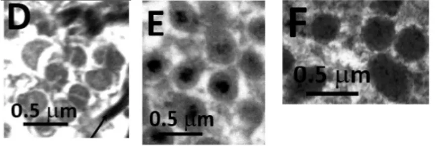
There are two types of adhesive secretory cells in P. lividus, the first type have large homogeneous granules and are located in the central area of the disc (Figure 4D) whereas the second type enclose smaller and more complex granules and are located in all the rest of the central area (Figure 4E). There is only one type of de-adhesive cells in the disc, they contain small, membrane-bound elliptic secretory granules with a small apical subcuticular cilium (Figure 4F)4. In the cell body of these cells, developing secretory granules are closely associated with Golgi membranes and rough endoplasmic reticulum cisternae, suggesting that these organelles are involved in the synthesis of the granule contents, which will be released, upon nerve stimulation, at the tip of microvillar-like cell projections, which form a tuft at the cell apex7. Adhesive cells are non-ciliated and therefore are thought to interact with the sensory cells via the nerve plexus, whereas de-adhesive cells are believed to be controlled by direct stimulation of their apical cilia7,9.
Figure 4 – Electron microscopy images (D) of the large homogeneous granules that are located in
the central area of the disc, (E) the smaller and more complex granules located in all the rest of the central area, (F) and the small, membrane-bound elliptic secretory granules with a small apical subcuticular cilium present on the only type of de-adhesive cells in the disc (adapted from 13)
Therefore, given the expected protein complexity, tube feet disc protein extracts were separated using 1D and 2-D electrophoresis (Figure 5). The resolution of each band present in a 1-D is relatively low which means that each band has more than one protein. For this reason an extra separation step was performed by injecting each
I - Introduction
9 band’s tryptic digest in a nano-flow HPLC coupled to a MALDI plate spotter. 2-D spots’ tryptic digests were also further separated using homemade microcolumns packed with two different reversed phase-like materials (POROS R2 and graphite). As the genome of the sea urchin, P. lividus has not yet been sequenced, two search algorithms (Mowse and Paragon) and four protein databases (UniProt/SwissProt database joined together with the sequenced purple sea urchin Strongylocentrotus purpuratus predicted proteins database, the nonredundant Uniref100 database and the translated
Paracentrotus lividus ESTs database from different developmental stages such as
unfertilized egg, gastrula, blastula and pluteus).
Figure 5 - One and two dimension protein profile from the tube feet discs of Paracentrotus lividus
separated in 12.5% polyacrylamide gels; horizontal lines in the 1-DE and black circles in 2-DE indicate, respectively, the bands and spots excised for in-gel tryptic digestion and protein identification by mass
spectrometry. (Adapted from 6,12)
This strategy resulted in the identification of 328 non-redundant proteins, belonging to 157 protein clusters. These results were complemented with an analysis of the tube feet disc proteome uncovered by 2D-PAGEwith specific fluorescent stains in order to label phospho- and glycoproteins (Figure 6), which are recurrent post-translational modification (PTMs) in marine permanent and nonpermanent adhesive proteins, resulting in the identification of 69 non-redundant proteins from a total of 92 excised spots, from which 44 were exclusively phosphorylated, 18 were exclusively glycosylated and 31 stained for both PTMs12.
A considerable fraction of the identified proteins in the total tube feet disc proteome are involved either in major cellular processes (35%) or cellular metabolism (23%). However, many proteins implicated in other processes such as development
I - Introduction
10 and regeneration (16%), nerve-related events (12%), immunological response (9%), muscle-related events (4%) and sensory perception (3%) were also found. Adhesive proteins were only found to represent 2% of the total proteome12.
Among these putative adhesive proteins are two P. lividus egg/embryonic secreted cell adhesion proteins, Nectin and Toposome that had never been reported in the adult adhesive organs. Nectin was detected both in the tube feet proteome and the phospho- and glyco-subproteomes. In the 2-D total proteome, Nectin formed a cluster of seven spots in the central upper part of the gels, which is in accordance with P.
lividus Nectin predicted molecular mass and isoelectric point (pI). However, there was
one isolated spot that presented the same apparent molecular mass but a more acidic pI. In the subproteome analysis of P. lividus tube feet discs with fluorescent stains, specific for phospho- and glycoproteins, Nectin, was also present in a cluster in the same relative position and all the spots were simultaneously phosphorylated and glycosylated, although with variable degrees of both PTMs.
Figure 6 - Phosphorylated and glycosylated proteome of Paracentrotus lividus adhesive discs
This is in agreement with the identification of proteins involved in amino acid PTMs. Within these there is a Serine/threonine-protein phosphatase, which indicates the existence of proteins with phosphorylated serine and/or threonine residues. This is further confirmed by the identification of a molecular chaperone Endoplasmin that is believed to regulate phosphoprotein phosphatase activity during the processing and transport of secreted proteins. In addition, several Protein disulfide-isomerases were also identified, which are resident proteins of the ER known to be important for protein
I - Introduction
11 folding, disulphide bond formation of secreted proteins and regulation of the extent of proteins glycosylation. The presence of Protein disulfide-isomerases corroborates previous data, since reducing conditions were shown to be determinant to solubilize sea urchin adhesive secretions and high levels of cysteine were also quantified in the footprints6. Therefore, these results seem to indicate that the Nectin present in adult P. lividus tube feet discs is highly homologous to the Nectin present in P. lividus eggs and
embryos but contains some sequence differences. Up to date, the few published papers on P. lividus Nectin show that this protein is secreted during embryogenesis, from cytoplasmic granules into the hyaline layer that surrounds the embryo, where it becomes part of the embryonic extra cellular matrix. Moreover, when coated on polystyrene substrates, purified egg P. lividus Nectin [Q70JA0] has been shown to significantly increase the binding of dissociated cells, thus confirming its involvement in the adhesion of sea urchin embryonic cells to the substrate14. Therefore, the presence of P. lividus Nectin in tube feet discs seems to indicate that this protein adhesive abilities are also being used by adult sea urchins for substrate attachment. This hypothesis is further corroborated by the fact that temporary adhesives, as those secreted by sea urchins, are typically composed by a mixture of proteins and carbohydrates, and therefore P. lividus Nectin (which contains six galactose-binding discoidin-like domains that can bind molecules bearing galactose and N-acetylglucosamine carbohydrate moieties) could have also a cohesive role, ensuring the cohesion of the adhesive components12. As for P. lividus Toposome, a modified calcium-binding iron-less transferrin, it was identified in five 1-D PAGE bands and on one 2-D PAGE spot in a relative position that is in accordance with its reported molecular mass and pI. However, in terms of PTMs there seems to be a discrepancy between egg/embryonic and adult Toposome since in eggs/embryos this protein is highly glycosylated15 which was not the case in adult tube feet. Again, there seem to be some sequence differences along the development of sea urchins, which is in accordance with previous data showing that during development Toposome is proteolytically processed into species of smaller molecular mass at the expense of the mature glycoprotein. Similarly to Nectin, Toposome is also a secreted protein that in eggs is present on the surface, as well as stored in granules, and has been reported as being responsible for the adhesive integrity of the sea urchin blastula under active regulation of calcium15.
Additionally, two other proteins, Transglutaminase A chain and Galectin-8 were also identified in tube feet discs, and although usually associated with coagulation, can have a role in sea urchin temporary adhesion12. In fact, there is recent evidence that
I - Introduction
12 barnacle cement polymerization and blood clotting occur by similar enzymatic mechanisms. Barnacle adhesive polymerization was shown to begin with the activation of cement structural and proteolytic precursors by trypsin-like serine proteases, a process that maximizes bonding interactions, facilitating their assembly and rearrangement with the surface, followed by covalent cross-linking, brought about by hemocyte-released transglutaminase, which reinforces the cement, resulting in an insoluble mesh of interwoven fibrous proteins16.
In fact, a P. lividus EST with significant homology with Transglutaminase A chain was identified in the tube feet discs and therefore it can be hypothesized that as in barnacles this protein might be released into the adhesive to ensure its insolubility. As for Galectin-8, a secreted β-galactoside-binding protein, there is recent evidences that it is present not only in P. lividus adult tube feet discs but also in embryos, where it acts as a physiological modulator of cell adhesion, increasing adhesion when immobilized and decreasing it when present in excess as a soluble ligand17. Given its mode of action, it can be hypothesized that the identified Galectin-8 can have a role in the regulation of tube feet disc attachment and detachment. In fact, the current knowledge on de-adhesive secretions is scarce.
However, based on the recurrent presence of an adhesive footprint on the substrate after detachment, de-adhesive secretions are believed to function as enzymes, causing the discard of the outermost-layer of the cuticle, the so-called fuzzy coat2. Since sea urchin temporary adhesives are made of a mixture of proteins and carbohydrates (free or conjugated) it is likely that de-adhesive enzymes such as proteases and glycosylases could trigger de-adhesion by degradation of the secreted adhesive. However, none of these types of enzymes were found in the tube feet discs proteome, indicating that other approaches are needed to elucidate on the composition of echinoderm de-adhesive secretions. However, a preliminary quantification of the proteolytic activity of protein extracts from tube feet discs versus stems, has shown that discs contain a significantly higher amount of proteases (unpublished data), indicating that the later are probably unknown or highly modified thus preventing a successful identification by an homology-driven database searching strategy.
I - Introduction
13
M
ETHODS
-II - Methods
17
Collection of P. lividus and footprint material
1
-Sea urchins from the species Paracentrotus lividus (Lamark 1816) were collected at low tide on the west coast of Portugal (Estoril, Cascais) After collection, the animals were transported to the “Vasco da Gama Aquarium” (Oeiras) and kept in open-circuit tanks at 15°C and 33‰ salinity.
The sea urchins were placed in small plastic aquariums (3L) containing artificial seawater (Crystal Sea, Marine Enterprises International, Baltimore, MD, USA) in order to collect the adhesive material. These aquaria were covered internally with removable glass plates to which animals were allowed to attach. The animals were manually detached causing a certain number of tube feet to break by their disc. The broken discs were then removed with tweezers and stored at -80ºC.
Total RNA Extraction and RT-PCR
2
-Total RNA was extracted from sea urchin adhesive discs using two different methods of RNA extraction, TRIzol Reagent with PureLink RNA Micro Kit (Invitrogen) and RNeasy Mini kit (Quiagen) (the procedure consists on the lysate preparation with TRIzol Reagent, phase separation and on the binding, washing and sample elution) following the manufacturers’ instructions (the extracted RNA purity and concentration values were similar for both methods). Special attention was given to the tissue homogenization step. A mechanical homogenization process (with a rotor) was preferred over a chemical homogenization process (e.g. protease K). The RNA was kept in ice in aseptic conditions during the procedure and RNase-free tubes and water were used to prevent RNA degradation. After RNA extraction, the RNA was precipitated with ethanol and sodium acetate and later quantified. cDNAs were prepared from the purified RNA using the SuperScript III First-Strand Synthesis System for RT-PCR (Invitrogen) according to the manufacturer’s instructions.
Nectin gene amplification using PCR
3
-The PCR reactions were carried in a total volume of 25uL in each tube in a iCyclerTM Thermal Cycler (Bio-Rad) using the reagents mix shown in Table II-1.
II - Methods
18
Table II-1 - Reagents and volumes used for the PCR
Mix µL H2O 16,15 Buffer GC 5 dNTPs 10mM 0,5 Primer Forward (10mM) 1 Primer Reverse (10mM) 1 DMSO 0.1 Phusion 0,25 DNA 1
Table II-2 – Primers pairs used in the reactions
Fragment length Primer pairs
Nec1 771bp Nec_F1d_ATG GCG ATA TCA CAT AAT GCG TTA ATG G (57ºC) Nec_R2 CTG GCG ACC CTG CGT AAT AAC TC (58ºC)
Nec1-2 2019bp Nec_F1d_(5´ SalI) 5´GTC GAC ATG GCG ATA TCA CAT AAT GC Ne_R3d – (3´NotI) 3` GCG GCC GCG GTG GAA GCC GTT ATA
Nec_full 2956 bp Nec_F1d_ATG GCG ATA TCA CAT AAT GCG TTA ATG G (57ºC) Nec_R1d - CTC ACT CTG TGA GAG GAC CTT CC (58ºC)
Table II-3 – Amplification Conditions used for Nec1 and Nec1-2 fragments
Table II-4 – Tested amplification conditions to amplify Necfull
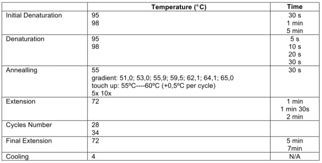
Temperature (°C) Time
Initial Denaturation 98 1 min Denaturation 98 20 sec Annealling 55 30 sec Extension 72 1min30 sec Cycles Number 34
Final Extension 72 5 min
Cooling 4 N/A Temperature (°C) Time Initial Denaturation 95 98 30 s 1 min 5 min Denaturation 95 98 5 s 10 s 20 s 30 s Annealling 55 gradient: 51,0; 53,0; 55,9; 59,5; 62,1; 64,1; 65,0 touch up: 55ºC----60ºC (+0,5ºC per cycle) 5x 10x 30 s Extension 72 1 min 1 min 30s 2 min Cycles Number 28 34
Final Extension 72 5 min
7min
II - Methods
19
Table II-5 – Reagents and volumes used for the PCR
Cloning using TOP10 E. coli cells
4
-Invitrogen TOPO® TA Cloning® kit was used for the cloning according to the manufacturer’s instructions.
Plasmid DNA extraction from recombinant E. coli
5
-The kit -ThermoScientific minpreps was used according to the manufacturer’s instructions.
Expression vector and nectin ligation
6
-The obtained pCR4 TOPO plasmids and the template pGEX 4T.1 were digested using SalI and NotI Fast digest (Thermo scientific) followed by purification in agarose gel. The digested plasmid and the desired insert were ligated using T4 DNA ligase (Thermo scientific) according to the manufacturer’s instructions.
Sequencing results
7
-The sequencing was done by the company Stabvida. Mix µL Buffer GC 5 Buffer HF 5 dNTPs 10mM 0,5 Primer Forward (10mM) 0,5 1 Primer Reverse (10mM) 0,5 1 DMSO 0 0,1 0,2 MgCl2 50mM 0 0,1 0,25 Phusion 0,25 pfu 1 DNA -0,5 -1 -diluições 1:10 e 1:100
II - Methods
20
Protein separation by 1D SDS-PAGE
8
-
For 1-D PAGE, 25 µg of total protein was separated using 7 cm 10% polyacrylamide gels in a Mini-PROTEAN II gel system (Bio-Rad) at a constant voltage of 100 V. Gels were then stained with Coomassie Blue Colloidal18. Briefly, after fixing the proteins in the gels for 18 h with a solution of 50% (v/v) ethanol, 3% (v/v) phosphoric acid, gels were preincubated for 1 h with 34% (v/v) methanol containing 3% (v/v) phosphoric acid and 17% (w/v) ammonium sulphate. Coomassie BlueG-250 (Sigma) was then added [0.35% (w/v)] to the previous solution and staining of the gels continued for 100 h more. Prior to image acquisition with a densitometer (LabScan, GEHealthcare), gels were washed with deionized water to remove background stain.
In-Gel Digestion
9
-Protein bands were manually excised from the gels using a disposable scalpel, washed in Milli-Q H2O, and distained in 50% acetonitrile (ACN) and subsequently 100% ACN. Disulfide bonds were reduced with 10 mM DTT and alkylated with 50 mM iodoacetamide. The dried gel pieces were swollen in a 50 mM NH4HCO3 digestion buffer containing 6.7 ng/µL of trypsin (modified porcine trypsin, sequencing grade; Promega, Madison, WI, USA) on an ice bath. After 30 min, the supernatant was removed and discarded, 20 µL of 50 mM NH4HCO3 was added to the gel pieces and digestion was allowed to proceed at 37°C overnight. After digestion, the remaining supernatant was removed and stored at −20°C until use19.
Maldi-MS/MS
10
-Protein digests were desalted and concentrated as previously described20,21. Home-made microcolumns were made by packing POROS R2 chromatographic resin (PerSeptive Biosystems, Foster City, CA, USA) or graphite powder (activated charcoal; Sigma-Aldrich, St. Louis, MO, USA) in a constricted GELoader tip (Eppendorf, AG, Hamburg). A syringe was used to force liquid through the columns by applying gentle air pressure. The columns were equilibrated with 20 µL of 2% trifluoroacetic acid (TFA) and the peptide digests were added first to R2 microcolumns and the flow through transferred directly to graphite microcolumns. Then, the columns were washed with 20 µL of 2% TFA and the peptides were eluted with 0.8 µL of α-cyano-4-hydroxycinnamic acid solution (CHCA, Sigma-Aldrich, St. Louis, MO, USA; 10 mg/µL in 70% ACN, 0.1% TFA) directly onto the MALDI target.
II - Methods
21 Mass spectrometry (MS) and tandem mass spectrometry (MS/MS) spectra were acquired on an Applied Biosystems 4700 Proteomics Analyzer MALDI-TOF/TOF (Applied Biosystems, Foster City, CA, USA) in both MS and MS/ MS mode. Positively charged ions were analyzed in positive reflectron mode and the collision gas used for fragmentation was atmospheric air. Each MS spectrum was obtained with a total of 1,000 laser shots accumulations (eight subspectra consisting of 125 laser shots each) and was externally calibrated using six spots of the standard mixture (Calibration Mixture 2, Applied Biosystem, Foster City, CA, USA). Five s/n best precursors from each MS spectrum were selected for MS/MS analysis. For MS/MS spectra, a maximum of 5,200 laser shots were accumulated, each subspectrum consisting of 65 shots (maximum of 80 subspectra). Raw data were generated by the 4000 Series Explorer Software v3.0 RC1 (Applied Biosystems, Foster City, CA, USA) and all contaminant m/z peaks originating from human keratin, trypsin autodigestion, or matrix were included in the exclusion list used to generate the peptide mass list used in database search.
The interpretation of the combined MS+MS/MS data was carried out using the GPS Explorer software (Version 3.5, Applied Biosystems, Foster City, CA, USA). Peptide mass maps and sequences obtained were searched against the National Center for Biotechnology Information (NCBI) database with no taxonomic restriction (6,572,387 entries, June 6, 2008) and against the purple sea urchin Strongylocentrotus
purpuratus database (42,420 entries; ftp://ftp.ncbi.nih.gov/genomes/Strongylocentrotus_purpuratus/protein/protein.fa.gz) using an in-house MASCOT server (Version 2.0). The search was performed using monoisotopic peptide masses and the following criteria: one missed cleavage, p<0.05 significance threshold, 50 ppm peptide mass tolerance, 0.25 Da fragment mass tolerance, carbamidomethylation of cysteine as fixed modification, and methionine oxidation as variable modification. Significant hits were visually inspected to eliminate false positives.
MS/MS spectra of the unidentified proteins were further analyzed by the DeNovo Explorer™ software (Version 3.5, Applied Biosystems, Foster City, CA, USA) using the following settings: trypsin as enzyme, carbamidomethylation of cysteine as fixed modification, methionine oxidation as variable modification, 0.2 Da fragment tolerance. This software automatically generates candidate sequences, assigning them a score between 0 and 100, which is an indication of the degree of matching between the theoretical fragmentation pattern and the fragmentation spectra of the SAM
II - Methods
22 peptides. In order to minimize randomness, we only considered peptides with scores higher than 70 and with at least two spectra with identical candidate sequence for the same gel band in two replicate gels. These de novo-derived sequences were submitted to Basic Local Alignment Search Tool (BLAST) searches at
http://blast.ncbi.nlm.nih.gov/Blast.cgi, using the following settings: no redundant
protein sequence database, taxonomy restricted to S. purpuratus (taxid: 7668), and blastp as algorithm.
R
ESULTS AND
D
ISCUSSION
-III – Results and Discussion
26
Sequencing and cloning of the nectin gene
1
-1.1 P. lividus adhesive tube feet discs mRNA extraction
The mRNA from P. lividus adhesive tube feet discs was extracted in order to synthesize the double-strand cDNA.
1.4 Nectin cDNA
To amplify the mRNA for nectin precursor of Paracentrotus lividus different strategies were followed. We used different sets of primers (Error! Reference source
not found.) that allowed the amplification of smaller parts of the protein, as shown in
Figure 7, since the amplification of the full-length nectin was not being successful due to its big size ( Figure 8).
Figure 7 – Nectin and its domains and the fragments amplified with each pair of primers Table III-1 – Set of primers used and the corresponding amplified fragments
Primers Fragments Nec_F1 - GGCCCGGTAATGCATAAGGGCAACACG (27bp Tm78,1 Gc%59,3) Nec_F2 - ACAAATTCCGGACAACGCCA 3‘ (20bp Tm64,6 GC%50) Nec_F3 - CCGACGACATTGGCGGGTTC 3‘ (20bp Tm69 GC65%) Nec_R1- GGCCACGCGTCGACTAGTACTT 3' (22bp Tm64,6 GC%59,1) Nec_R2 - CTGGCGACCCTGCGTAATAACTC 3' (23bp Tm66,3 Gc%56,5) Nec_R3 - 'TTTGAGAGCACCACTCAGGG 3‘ (20bp Tm59,4 GC%55)
Full lenght - Nec_F1+Nec_R1
(Frag. 3593bp) Nec1 - Nec_F1+Nec_R2 (Frag. 930bp) Nec2 - Nec_F2+Nec_R3 (Frag. 1543bp) Nec3 - Nec_F3+Nec_R1 (Frag. 1466bp) Nec1-2 - Nec_F1+Nec_R3 (Frag. 2048bp)
III – Results and Discussion
27
Figure 8 - Agarose gel with different Nectin fragments’ PCR amplification. FL – Full length nectin
III – Results and Discussion
28
1.5 Nectin cloning
The successfully amplified Nec1-2 fragment was cloned into a pGEX4T.1 plasmid (Figure 10).
Table III-2 - Pair of primers used to amplify Nec1-2
Primers Amplified
product
Insert Name Nec F1d (5´Sal I) 5´GTC GAC ATG GCG ATA TCA CAT AAT GC
Nec R3d (3´Not I) 3` GCG GCC GCGGTG GAA GCC GTT ATA
2019bp Nec_1-2
Figure 10 – Cloning of the Nec1-2 insert.
Figure 11 – Representation of the six domains present in Nectin emphasising the first four domains that the used shortened form contain.
III – Results and Discussion
29
Figure 13 – pGEX4T.1_Nec1-2 extracted from DH5α, RII and C41 cells
The amplified fragment was first inserted into a PCR4 Topo vector as shown in Figure 10. This plasmid was then restricted with SalI and NotI so it could be inserted in a pGEX4T.1 expression plasmid previously restricted with the same enzymes. Both DNA fragments were then purified from an agarose gel (Figure 12) and ligated successfully. The plasmid was sent to sequencing and after sequence confirmation, used to transform DH5α, RII and C41 cells (Figure 13).
1.6 Nectin Expression
The protein expression of Nec1-2 was optimized using different Escherichia coli strains, namely, BL21, BL21 plus, C41 and RII. The expression results were evaluated by SDS-PAGE and mass spectrometry.
III – Results and Discussion
30 The expression was possible to optimize for C41 and RII although it was unsuccessful for BL21 and BL21 plus. Nec1-2 has a molecular weight of 75kDa, adding the glutathione S-transferase (GST) tag’s weight (26kDa) that is expressed with it, it gives 101kDa, which corresponds to the band we see being overexpressed in Figure 14. When looking at it more carefully one concludes that the best conditions for Nec1-2 expression is 3 hours induction with IPTG using both C41 and RII cells.
1.7 Nectin Purification
I tried to purify Nec1-2 using Glutathione SepharoseTM 4 Fast Flow, an affinity chromatography medium for purification of GST-tagged proteins produced using the pGEX series of expression vectors. Despite a lot of tries the purification was not possible to optimize Figure 15 although the procedure was working properly as we can see in Figure 16.
Figure 15 – Nec1-2 purification attempt using Glutathione Sepharose beads
III – Results and Discussion
31 There is more than one possible explanation behind the purification problem that can justify it. One of them is that due to their proposed adhesive character of the domains that compose the protein, the GST tag gets trapped inside the protein not being able to adhere to the glutathione sepharose beads.
The Galactose-binding domain-like, the domain found repeated four times in Nec1-2 can be found in several different protein families, being one of its most common function to bind to specific ligands, such as cell-surface-attached carbohydrate substrates and phospholipids. The structure of the galactose-binding domain-like members usually consists of a beta- sandwich in which the strands that make the sheets exhibit a jellyroll fold. Even in cases when the sequence similarity between different family members is low, there is still a high degree of similarity in their beta-sandwich and their loops.
One option that could be tried next would be purification with Gelatin Sepharose as optimized in 22, since this purification technique for Paracentrotus lividus embryonic Nectin shows very good results, it’s possible that it can also work with its recombinant. Our purpose is to obtain a functional and active form of Nectin, and the use of this purification method would assure that we would get an active form – only the active form will bind to the Gelatin Sepharose and be consequentially purified.
One of our assumptions in this project was that the shortened recombinant of Nectin we used, Nec1-2 would keep, at least by some extent some of the full-length protein activity/ function, which contains six galactose-binding domain-like domains in contrast with the Nec1-2 that contains only the first four. This assumption is valid because as it was previously discussed, one of the main characteristics of these domains is being very similar in structure between themselves.
What also should be taken in account is that maybe Nectin’s function in sea urchin discs’ composition is not to be adhesive by itself, but to connect sugars and inorganic parts of the glue together by making use of its ability to bind galactose and similar extracellular matrix proteins. This would explain the high percentage of these compounds in the footprint.
Another potential application for the domains that compose nectin would be their use as tags in recombinant proteins in order to purify them, since their purification can be potentially achieved with something as cheap and accessible as Gelatin Sepharose.
III – Results and Discussion
32 Even though these problems can arise, it is still of fundamental interest to purify and characterize unknown proteins, once that’s the only way to know if it has any future potential.
34
R
EFERENCES
1. Santos, R., Hennebert, E., Coelho, A. V. & Flammang, P. in Functional Surfaces
in Biology, Vol. 2 (ed. Gorb, S. N.) 2, 205–238 (Springer Netherlands, 2009).
2. Flammang, P., Michel, a, Cauwenberge, A., Alexandre, H. & Jangoux, M. A study of the temporary adhesion of the podia in the sea star asterias rubens (Echinodermata, asteroidea) through their footprints. J. Exp. Biol. 201 (Pt 16, 2383–95 (1998).
3. Santos, R., Gorb, S., Jamar, V. & Flammang, P. Adhesion of echinoderm tube feet to rough surfaces. J. Exp. Biol. 208, 2555–2567 (2005).
4. Santos, R. & Flammang, P. Morphology and tenacity of the tube foot disc of three common European sea urchin species: a comparative study. Biofouling 22, 173–185 (2006).
5. Santos, R. & Flammang, P. Estimation of the attachment strength of the shingle sea urchin, Colobocentrotus atratus, and comparison with three sympatric echinoids. Mar. Biol. 154, 37–49 (2008).
6. Santos, R. et al. First insights into the biochemistry of tube foot adhesive from the sea urchin Paracentrotus lividus (Echinoidea, Echinodermata). Mar.
Biotechnol. 11, 686–698 (2009).
7. Flammang, P. Adhesion in echinoderms. Echinoderm studies (1996).
8. Santos, R. & Flammang, P. Is the adhesive material secreted by sea urchin tube feet species-specific? J. Morphol. 273, 40–48 (2012).
9. Flammang, P. & Jangoux, M. Functional morphology of coronal and peristomeal podia in Sphaerechinus granularis (Echinodermata, Echinoida). Zoomorphology
113, 47–60 (1993).
10. Santos, R. & Flammang, P. Morphometry and mechanical design of tube foot stems in sea urchins: A comparative study. J. Exp. Mar. Bio. Ecol. 315, 211–223 (2005).
11. Flammang, P. in Biological Adhesives 183–206 (2006).
12. Santos, R., Barreto, A., Franco, C. & Coelho, A. V. Mapping sea urchins tube feet proteome - A unique hydraulic mechano-sensory adhesive organ. J.
Proteomics 79, 100–113 (2013).
13. Santos, R. A multidisciplinary analysis of sea-urchin temporary adhesion: morphology, biomechanics and proteomics – a review. Cah. Biol. Mar. 54, 479– 489 (2013).
14. Matranga, V., Ferro, D. Di, Zito, F., Cervello, M. & Nakano, E. A new extracellular matrix protein of the sea urchin embryo with properties of a substrate adhesion molecule. Dev. Biol. 173–178 (1992).
15. Noll, H. et al. The toposome, essential for sea urchin cell adhesion and development, is a modified iron-less calcium-binding transferrin. Dev. Biol. 310, 54–70 (2007).
16. Dickinson, G. H. et al. Barnacle cement: a polymerization model based on evolutionary concepts. J. Exp. Biol. 212, 3499–3510 (2009).
17. Karakostis, K. et al. Molecular characterization and biological activities of a newly identified galectin-8 from P.lividus embryo.
18. Neuhoff, V., Arold, N., Taube, D. & Ehrhardt, W. Improved staining of proteins in polyacrylamide gels including isoelectric focusing gels with clear background at nanogram sensitivity using Coomassie Brilliant Blue G-250 and R-250.
Electrophoresis 9, 255–62 (1988).
19. Bennett, K. L. et al. Chemical cross-linking with thiol-cleavable reagents combined with differential mass spectrometric peptide mapping--a novel approach to assess intermolecular protein contacts. Protein Sci. 9, 1503–18 (2000).
20. Gobom, J., Nordhoff, E., Mirgorodskaya, E., Ekman, R. & Roepstorff, P. Sample purification and preparation technique based on nano-scale reversed-phase columns for the sensitive analysis of complex peptide mixtures by matrix-assisted laser desorption/ionization mass spectrometry. J. Mass Spectrom. 34, 105–16 (1999).
21. Larsen, M. R., Cordwell, S. J. & Roepstorff, P. Graphite powder as an alternative or supplement to reversed-phase material for desalting and concentration of peptide mixtures prior to matrix-assisted laser desorption/ionization-mass spectrometry. Proteomics 2, 1277–87 (2002).
22. Zito, F., Burke, R. D. & Matranga, V. Pl-nectin, a discoidin family member, is a ligand for betaC integrins in the sea urchin embryo. Matrix Biol. 29, 341–5 (2010).