Clinical Case Study
GE Port J Gastroenterol 2019;26:430–437
Fatty Liver Caused by Glycogen Storage
Disease Type IX: A Small Series of Cases
in Children
Catarina Leuzinger Dias
aInês Maio
bJosé Ricardo Brandão
cEdite Tomás
dEsmeralda Martins
a, eErmelinda Santos Silva
a, baInstituto de Ciências Biomédicas Abel Salazar, Porto, Portugal; bGastroenterology Unit, Paediatrics Division, Child and Adolescent Department, Centro Materno-Infantil do Norte, Centro Hospitalar Universitário do Porto, Porto, Portugal; cPathologic Anatomy Division, Hospital Geral de Santo António, Centro Hospitalar Universitário do Porto, Porto, Portugal; dPaediatrics Division, Centro Hospital de Tâmega e Sousa, Porto, Portugal; eMetabolic Diseases Unit, Pediatrics Division, Child and Adolescent Department, Centro Materno-Infantil do Norte, Centro Hospitalar Universitário do Porto, Porto, Portugal
Received: September 19, 2018
Accepted after revision: December 18, 2018 Published online: March 14, 2019
Ermelinda Santos Silva
Departamento da Criança e do Adolescente, Centro Materno-Infantil do Norte © 2019 Sociedade Portuguesa de Gastrenterologia
Published by S. Karger AG, Basel
DOI: 10.1159/000496571
Keywords
Glycogen storage disease type IX · Non-alcoholic fatty liver disease · Steatohepatitis · Children
Abstract
Background: The prevalence of non-alcoholic fatty liver
dis-ease (NAFLD) affecting children and adolescents has in-creased dramatically in recent years. This increase is most probably related to the obesity pandemic and the high con-sumption of fructose. However, hepatic steatosis has some rare causes (e.g., some metabolic diseases) of which clini-cians should be aware, particularly (but not only) when pa-tients are non-obese or non-overweight. Differential diagno-sis is notably important when pathologies have a specific treatment, such as for glycogenosis type IX (GSD-IX). Aims: To contribute to the knowledge on the differential diagnosis of NAFLD in paediatric age and to the clinical, biochemical, molecular, and histological characterisations of GSD-IX, a rare metabolic disorder. Methods: We performed a retro-spective study of a small series of cases (n = 3) of GSD-IX di-agnosed in the past 6 years, who were currently being fol-lowed up in the Units of Gastroenterology or Metabolic
Dis-eases of the Paediatric Division of our hospital and whose clinical presentation was NAFLD in paediatric age. Results: Three male patients were diagnosed with NAFLD before 2 years of age, 2 with confirmed diagnosis before the age of 3 years (alanine aminotransferase [ALT], liver ultrasound, and molecular analysis) and 1 whose diagnosis was confirmed at 11 years (ALT, liver ultrasound, liver histology, and molecular analysis). None of the patients were obese or overweight, and the daily fructose consumption was unknown. The out-come was favourable in all 3 patients, with follow-up periods ranging from 2 to 6 years. Conclusion: The decision on how far the search for secondary causes of NAFLD should go can be difficult, and GSD-IX must be on the list of possible causes.
© 2019 Sociedade Portuguesa de Gastrenterologia Published by S. Karger AG, Basel
Fígado gordo causado por glicogenose tipo IX: Uma pequena série de casos em crianças
Palavras Chave
Glucogenose tipo IX · Fígado gordo não-alcoólico · Esteatohepatite · Crianças
Resumo
Introdução: A prevalência de fígado gordo não-alcoólico
(NAFLD) afetando crianças e adolescentes, aumentou enormemente nos últimos anos. Este aumento está muito provavelmente relacionado com a pandemia de obesi-dade e o elevado consumo de fructose. No entanto, a es-teatose hepática pode ser causada por algumas entidades raras (por exemplo, doenças metabólicas) para as quais os clínicos devem estar alertados, particularmente (mas não só) quando os doentes não são obesos nem têm excesso de peso. O diagnóstico diferencial é especialmente im-portante quando as patologias têm um tratamento espe-cífico, como é o caso da Glicogenose tipo IX (GSD-IX).
Ob-jetivos: Contribuir para o conhecimento dos diagnósticos
diferenciais de NAFLD em idade pediátrica, e contribuir para a caracterização clínica, bioquímica, molecular e his-tológica da GSD-IX, uma doença metabólica rara.
Metodo-logia: Estudo retrospectivo de uma pequena série de
ca-sos (n = 3) de GSD-IX diagnosticados nos últimos 6 anos, atualmente em seguimento nas Unidades de Gastren-terologia ou de Doenças Metabólicas do Serviço de Pe-diatria do nosso hospital, e cuja forma de apresentação clínica foi NALFLD em idade pediátrica. Resultados: Três doentes de sexo masculino foram diagnosticados com NAFLD antes dos 2 anos de idade, dois com diagnóstico confirmado antes dos 3 anos (alanina aminotransferase [ALT], ecografia hepática, e estudo molecular), e um cujo diagnóstico foi confirmado aos 11 anos (ALT, ecografia e histologia hepáticas, e estudo molecular). Nenhum dos doentes era obeso ou tinha excesso de peso, e o consumo diário de frutose era desconhecido. A evolução foi fa-vorável nos três doentes, com um tempo de seguimento que variou entre 2 e 6 anos. Conclusões: A decisão sobre até onde a pesquisa de causas secundárias de NAFLD deve ir pode ser difícil, e a GSD-IX deve estar na lista de causas possíveis. © 2018 Sociedade Portuguesa de Gastrenterologia
Published by S. Karger AG, Basel
Introduction
Non-alcoholic fatty liver disease (NAFLD) is currently the most common cause of fatty liver both in children and adults [1, 2] and is already the most common cause of chronic liver disease in children and adolescents [3]. Un-favourable lifestyles leading to obesity and a high-fruc-tose diet may be the causes [4]. NAFLD is characterised by fatty infiltration of the liver (more than 5% of hepato-cytes) in the absence of secondary causes, such as alcohol or drug consumption, infections, malnutrition, and
ge-netic/metabolic diseases. Therefore, the diagnosis of NAFLD is one of exclusion, and it is particularly impor-tant to exclude diseases with specific treatment [5]. Among the metabolic diseases to be considered are some glycogen storage diseases (GSDs) [6].
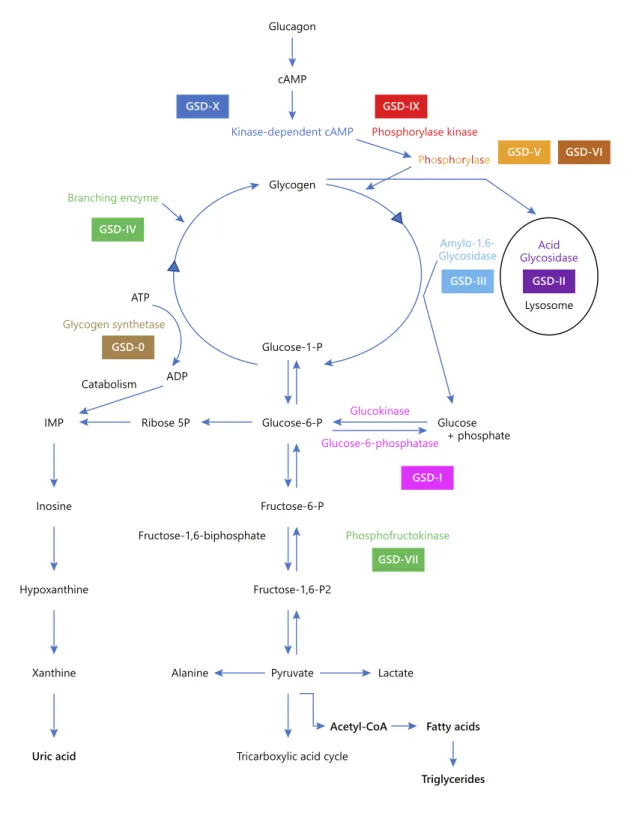
The GSDs are a group of inherited metabolic disorders that result from a defect in any of the various enzymes required for the synthesis or degradation of glycogen (Fig. 1). They are divided into two groups: GSDs with liver involvement (GSD-0, I, III, IV, VI, IX) or those as-sociated with neuromuscular disease or both. The glyco-gen storage disease type IX (GSD-IX) is one of the most common forms of GSD (25% of cases; estimated frequen-cy of 1/100,000) and result from a defect in an enzyme required for the degradation of glycogen. The enzymatic blockage leads to elevated pyruvate levels, which will be converted to lactate and acetylcoenzyme A. The latter is the starting point of the liver lipogenesis pathway with fatty acids and triglyceride synthesis. Although the main substrate accumulated in the target organs is glycogen, there may be a predominance of lipids, particularly in the liver [7, 8].
GSD-IX is both genetically and clinically heteroge-neous. It is caused by deficiency in hepatic phosphorylase kinase (PhK), which is composed of four subunits (α, β, γ, and δ) with each one encoded by a different gene. GSD-IX subtypes are identified according to the affected sub-unit (GSD-IXa, GSD-IXb, GSD-IXc, and GSD-IXd) and exhibit different modes of transmission: X-linked reces-sive for the PHKA2 gene (subtype α, 75% of all GSD-IX) and autosomal recessive for the PHKB (subtype β) and PHKG2 (subtype γ) genes. The correlation genotype-phenotype is not clear but patients with PHKG2 muta-tions (GSD-IXc) have been reported to have the most se-vere phenotypes, and the most common subtype (GSD-IXa) has been characterised with a wide variability. Even if the majority of the patients have a benign course in the long term, with mild symptoms in childhood improving with age, some may progress to liver cirrhosis [9, 10] or develop liver adenomas and hepatocarcinoma in adult-hood [11].
Case Report
We describe a small series of cases (n = 3) of GSD-IX, subtype α (GSD-IXa), diagnosed and managed in our outpatient clinic in the past 6 years and whose clinical presentation was fatty liver dur-ing paediatric age. Data were collected from clinical records. We analysed the clinical, analytical, histological, and molecular genet-ics parameters.
Kinase-dependent cAMP Phosphorylase kinase Phosphorylase cAMP Glycogen Amylo-1,6-Glycosidase Glycogen synthetase ATP ADP Catabolism Ribose 5P Glucose-1-P Acid Glycosidase Lysosome Glucose-6-P Glucose-6-phosphatase Glucokinase Glucose + phosphate GSD-X GSD-IX GSD-V GSD-VI GSD-II GSD-I GSD-VII GSD-III GSD-IV GSD-0 Branching enzyme Alanine Lactate
Acetyl-CoA Fatty acids
Triglycerides Fructose-6-P Phosphofructokinase Fructose-1,6-biphosphate Fructose-1,6-P2 Pyruvate
Tricarboxylic acid cycle IMP Inosine Hypoxanthine Xanthine Uric acid Glucagon
Fig. 1. Simplified scheme for the synthesis and degradation of glycogen and its enzymatic defects in several types of glycogenesis.
Results Case 1
A male infant, the first and only child of a healthy non-related couple, was born after a 40-week pregnancy, with
a weight of 3,080 g (p10–50) and a length of 48.5 cm (p10). At 18 months of age, he was hospitalised for bacterial pneumonia, and had hepatosplenomegaly and elevated transaminases (aspartate aminotransferase [AST]: 67 IU/L; alanine aminotransferase [ALT]: 53 IU/L). AST
Table 1. Baseline clinical and analytical features of patients at admission to the referral centre
Case 1 Case 2 Case 3 Reference
values
Clinical features
Sex M M M
Age (year of birth) 11 years (2000) 15 months (2010) 18 months (2013)
Parental consanguinity no no no
Family history negative negative negative
Weight, kg/percentile 43.6/p50–85 11.0/p50–85 11.0/p50 Length or height, cm/percentile 150.5/85 80/p50–85 82/p50 BMI, kg/m2/percentile 19.25/p50–85 17.2/p50–85 16.4/p50–85
Hepatomegaly no yes (5 cm) yes (3.5 cm)
Splenomegaly no no no Biochemical parameters Total bilirubin, mg/dL 0.28 0.16 0.26 0.20–1.00 Conjugated bilirubin, mg/dL 0.10 0.06 0.10 0.00–0.20 AST, IU/L 26 150 158 10–34 ALT, IU/L 16 219 60 10–44 γGT, IU/L 13 35 24 10–66 CPK, IU/L 136 95 NA 24–204 Glucose, mg/dL 77 66 74 70–105 Bicarbonates, mmol/L 17.5 17.9 15.4 22.0–29.0 Lactate, mmol/L 1.2 1.28 1.63 0.5–2.20 Uric acid, mg/dL 4.7 3.4 4.0 2.0–5.5 Total cholesterol, mg/dL 174 174 196 0–200 LDL cholesterol, mg/dL 94 116 136 0–130 HDL cholesterol, mg/dL 70 13 25 35–55 VLDL cholesterol, mg/dL 10 45 35 3–56 Triglycerides, mg/dL 50 227 174 40–160
Other exams performed after
Hepatitis A, B, and C CMV EBV negative IgG + IgM – IgG + IgM – negative IgG – IgM – IgG – IgM – negative IgG+ IgM – IgG + IgM – Serum alpha-1-antitrypsin, mg/dL normal normal normal
Serum ferritin, ng/mL 22 NA NA 12.8–454
Transferrin saturation rate, % 10 NA NA 15–45
Serum caeruloplasmin, mg/dL 26 NA NA 16–36
24-h urinary copper, μmol/d 1.086 ND ND 0.040–0.050
Copper in liver tissue, µg/g 24.82 ND ND <40
Sweat test ND normal ND
LAL activity (in leucocytes) normal ND normal
Molecular studies
PhKA2 gene mutation c.1054C>T
(p.352X) exon 11 in hemizygosity mutation c.892C>T (p. R298X) exon 9 in hemizygosity mutation c.706G>T (p.E236*) exon 7 in hemizygosity
BMI, body mass index; CMV, cytomegalovirus; EBV, Epstein-Barr virus; LAL, lysosomal acid lipase; NA, non-available; ND, not done.
and ALT eventually normalised, but hepatosplenomegaly persisted and led to admission to our outpatient clinic when he was 25 months old. Upon admission, comple-mentary investigation was normal and included creatine phosphokinase, alpha-1-antitrypsin, hepatitis C virus an-tibodies, ceruloplasmin, serum ferritin, transferrin satu-ration, and serum lipids. Throughout the following years, spleen dimensions returned to normal, but the liver re-mained slightly enlarged with hyper-echogenicity. When the patient was 5 years old, the enzymatic activity in the leucocytes for glycogen storage disease type III (GSD-III) was normal. He was discharged by his attending physi-cian, and performing annual abdominal ultrasound ex-aminations was recommended.
By the age of 11 years, the abdominal ultrasound main-tained a diffuse liver hyper-echogenicity, a pattern sug-gesting steatosis. The laboratory tests performed are pre-sented in Table 1. Liver histology revealed macro- and micro-steatosis without inflammatory infiltrates or fibro-sis (Fig. 2). The copper level in the liver tissue was normal. The molecular study confirmed the previously described mutation c.1054C>T (p. 352X) exon 11 in hemizygosity on the PHKA2 gene. Dietary measures were prescribed. Currently, at 18 years of age, the patient is asymptomatic; transaminases and other liver function tests remain nor-mal; the liver maintains a pattern of steatosis in ultra-sound with no adenomas; and the spleen is in the upper limit of normal size. His mother has normal ALT and has not undergone liver ultrasound or molecular study.
Case 2
A male infant, the second child of a healthy non-relat-ed couple, was born after a 37-week pregnancy with a weight of 2,750 g (p10–50) and a length of 46.5 cm (p10– 50). He developed physiological jaundice that resolved within a week without phototherapy. During his first months, he had several episodes of wheezing, which were treated with montelukast and fluticasone in aerosol. At 11 months of age, during a wheezing episode, his transami-nases (AST 158 IU/L; ALT 155 IU/L) and triglycerides (180 mg/dL) were elevated. At 15 months, he was admit-ted to our hospital for hepatomegaly (6 cm below the cos-tal grid, smooth surface, and thin edge) and persistently elevated transaminases; abdominal ultrasound showed an enlarged and hyperechogenic liver with a steatosis pat-tern; the spleen was normal in size. The laboratory tests highlighted the slightly low fasting glycemia and bicar-bonate, discreetly increased triglycerides and normal se-rum uric acid (Table 1). Cardiac evaluation was normal. Molecular study showed a previously described causal mutation on the PHKA2 gene [c.892C>T (p. R298X – exon 9)] with a hemizygosity pattern, confirming GSD-IXa. Dietary measures were prescribed. Two years later, hepatomegaly remained the same, although the transam-inases normalised. Bicarbonates were slightly low and uric acid elevated. Currently, at 8 years old, he has no signs of progression to chronic liver disease or adenomas. His older sister (16 years old) is asymptomatic, with nor-mal ALT and liver ultrasound. His mother has nornor-mal
200 μm 100 μm
a b
Fig. 2.a, b Liver histology in case 1 (at 11 years of age) showing macro- and micro-steatosis; hepatocytes with preserved morphology, and dimensions within normal ranges, without inflammatory infiltrate or fibrosis (H&E 100×).
ALT and has not undergone liver ultrasound or molecu-lar study.
Case 3
A male infant, the first and only child of a healthy non-related couple, was born after a 39-week high-risk preg-nancy due to placental displacement, with a weight of 3,300 g (p10–50) and a length of 51 cm (p50). In the first week, he developed jaundice (total bilirubin 17.3 mg/dl) that was treated with phototherapy. During the first 8 months, he had multiple infections: urinary tract infec-tion caused by Escherichia coli, acute gastroenteritis, fever of undetermined origin, septicaemia caused by Neisseria
meningitidis, upper respiratory infection caused by Coro-navirus, cystitis caused by Proteus mirabilis, acute
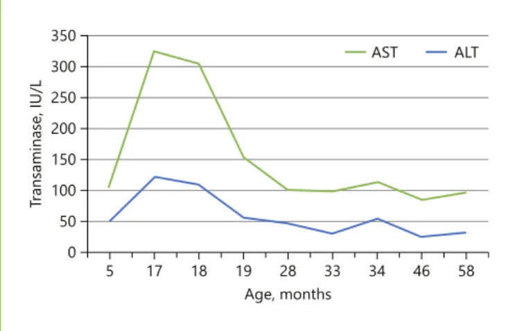
bron-chiolitis followed by acute otitis media and an episode of bacteraemia of unknown origin. This overwhelming number of infections led to an investigation on humoral and the cellular immunity defects. A study performed out of context of infection excluded a complement deficiency and revealed normal serum immunoglobulins and im-munophenotyping of lymphocytes. Throughout all these infectious episodes, the patient had palpable hepatomeg-aly and consistently elevated transaminases. At 18 months, abdominal ultrasound showed an enlarged liver with a pattern suggestive of steatosis and a normal-sized spleen. Table 1 presents the laboratory tests performed. One year later, molecular study confirmed the new and unrecog-nized mutation c.706G>T (p. E236*) exon 7 in hemizy-gosity in the PhKA2 gene, which leads to the production of a truncated protein. Diet measures were prescribed. Recurrent infections ceased after the first year of life. Cur-rently, at 5 years of age, the patient is doing well, with
slight hepatomegaly (2 cm below the costal margin) and normal ALT (Fig. 3) and other liver function tests, and maintains a liver steatosis pattern on ultrasound. His mother is a carrier of heterozygosity, with normal ALT and liver pattern on ultrasound.
Discussion/Conclusion
NAFLD is one of the comorbidities of obesity, and its incidence is increasing dramatically. In addition, NAFLD has been associated with a certain dietary pattern, even without obesity or overweightness. Nonetheless, fatty liv-er can also be secondary to a large numbliv-er of othliv-er enti-ties [5]. Therefore, a child with fatty liver, regardless of the child’s body mass index, is a challenging issue. The challenges are in the following order: when to suspect NAFLD, how to confirm the diagnosis, how to accom-plish its staging, and how to rule out secondary causes.
None of our 3 patients was obese or overweight. How-ever, we were not able to establish with reasonable accu-racy their daily consumption of fructose. All 3 had hepa-tomegaly and elevated transaminases. For this reason, they underwent an abdominal ultrasound that confirmed an enlarged liver with hyper-echogenic pattern. These findings were suggestive of fatty liver.
ALT is currently the best screening method for fatty liver in children aged ≥10 years, with 88% sensitivity and 26% specificity for values >2 times the normal value [12]. Liver ultrasound may show a suggestive pattern, although sensitivity is low. Liver histology is the gold standard for diagnosis, but it is too invasive as a screening method and has some limitations [13]. Magnetic resonance imaging (MRI) and spectroscopy (MRS) were validated and have showed to be accurate to detect and quantify hepatic ste-atosis in both adults [14] and children [15]. In fact, MRI-estimated proton density fat fraction is currently the most accurate test to quantify liver steatosis and can already be considered the gold standard [16]. However, it is usually unavailable and cannot be considered a ‘non-invasive’ procedure in young children because it requires sedation. For staging NAFLD, liver histology is still the best meth-od for distinguishing NAFL from non-alcoholic steato-hepatitis and for revealing the presence or absence of ad-vanced fibrosis/cirrhosis, whereas some non-invasive methods are still in development [16].
Only 1 of our 3 patients (case 1) had fatty liver con-firmed through histology at 11 years old, when his age and the increased level of 24-h urinary copper prompted us to investigate for Wilson’s disease [17]. In the other 2
58 46 34 33 28 Age, months 19 18 17 5 Tra nsaminase, IU/L 350 AST 300 250 200 150 100 50 0 ALT
(cases 2 and 3), the diagnosis was presumptive and based on increased ALT and suggestive liver pattern in the ul-trasound. The execution of MRI or MRS and liver biopsy was not considered because the benign clinical status of the patients did not justify the performance of invasive examinations. Moreover, the confirmation of a secondary cause that can be associated with lipids accumulation in the liver, such as GSD-IXa [7, 8], reinforces this presump-tion.
The guidelines are scarce concerning the ruling out of secondary causes of fatty liver. The North American So-ciety of Pediatric Gastroenterology, Hepatology and Nu-trition (NASPGHAN) guidelines for diagnosis and treat-ment of NAFLD (2017) do not provide enough orienta-tion to a cost-benefit approach regarding who should be screened and when for each listed genetic/metabolic dis-ease [13]. Furthermore, the GSD-IX is not even men-tioned on their list as a secondary cause of fatty liver.
In this case series, we prioritised the exclusion of dis-eases with specific treatment and considered the patient’s age. When the patient reached the age of 11 years, we searched for Wilson’s disease, juvenile haemochromato-sis, lysosomal acid lipase deficiency [18], and GSD types VI and IX. In patient 2, the hypothesis of drug hepatotox-icity by montelukast was considered, and it could had ex-plained the increased transaminases but not the hepato-megaly and steatosis. In patient 3, we considered the hy-pothesis of congenital immunodeficiency with liver injury but were not able to confirm it. For the last 2 pa-tients, lysosomal acid lipase deficiency was also consid-ered but was tested only in patient 3. The diagnosis of GSD-IXa was confirmed by molecular study in all pa-tients. All patients had pathogenic mutations of X-linked transmission in the PHKA2 gene. In case 2, a new muta-tion was found, which results in a premature stop codon. This new mutation was accepted as a causal mutation.
The clinical presentation of GSD-IXa is generally milder than that of other GSDs (and similar to GSD-VI), and its symptoms (hepatomegaly, growth retardation, el-evated transaminases, hypertriglyceridaemia, and some-times ketosis and hypotonia) typically improve with age, as it is usually asymptomatic in adulthood [19]. However, the genetic variants in PHKA2 have a broad phenotypic spectrum, including progression to cirrhosis [9, 10] and the development of hepatic adenomas [11] as well as less common phenotypes with kidney dysfunctions due to re-nal tubular acidosis and central nervous system involve-ment with delayed cognitive and speech abilities [20]. Thus far, our patients’ clinical course has been benign, including the oldest.
The treatment includes dietary measures, namely, a high-protein diet (2–3 g/kg) with restriction of simple (added) sugars, avoidance of prolonged fasting, and sup-plementation of raw corn starch in the most severe phe-notypes to maintain glucose concentrations and prevent ketosis overnight. In situations of greater stress, water infusion with maltodextrin (5 g/100 mL) is recommend-ed. Rarely, hyperuricaemia or metabolic acidosis must be addressed. Untreated children may have undesirable re-percussions, such as morning sickness, which affects school performance, and growth retardation, which causes psychological distress [21]. All our patients had low serum bicarbonates at presentation (later nor-malised), but none had hyperlactacidaemia or growth failure. Moreover, hyperuricaemia was not a problem in any of them.
Although the majority of patients are male, females may also exhibit symptoms, either in autosomal recessive forms or in the X-linked transmission subtype (GSD-IXa), because of the lyonisation phenomenon [22, 23]. Therefore, the mother, sisters, and daughters of male pa-tients should be screened. In addition to the usual (mild-er) symptoms, women may have polycystic ovary syn-drome. The transition to adulthood should include an adequate surveillance plan for cirrhosis, adenomas, and hepatocarcinoma.
In summary, making the decision of when and how to search for secondary causes of NAFLD can be challeng-ing. To serve this purpose, more appropriate and cost-effective guidelines are needed. GSD-IX should be in-cluded in these guidelines because liver involvement can range in a spectrum from steatosis to steatohepatitis, with or without variable degrees of fibrosis/progression to liv-er cirrhosis. As sevliv-eral genetic/metabolic diseases can be among the secondary causes of NAFLD, the development of a next-generation sequencing panel, including all those diseases, can be a useful tool in the management of these patients [24].
Statement of Ethics
The authors have no ethical conflicts to disclose.
Disclosure Statement
Author Contributions
Dr. Catarina Leuzinger Dias collected and analysed data and elaborated the manuscript, which is based on her Master thesis of medicine. Dr. Inês Maio, Dr. José Ricardo Brandão, Dr. Edite
Tomás, and Prof. Doutora Esmeralda Martins collected and an-alysed data and then evaluated and approved the manuscript. Dr. Ermelinda Santos Silva elaborated the study design, collect-ed and analyscollect-ed data, and evaluatcollect-ed and approvcollect-ed the manu-script.
References
1 Nobili V, Socha P. Pediatric nonalcoholic fat-ty liver disease: current thinking. J Pediatr Gastroenterol Nutr. 2018 Feb;66(2):188–92. 2 Mann JP, Valenti L, Scorletti E, Byrne CD,
Nobili V. Nonalcoholic fatty liver disease in children. Semin Liver Dis. 2018 Feb;38(1):1– 13.
3 Anderson EL, Howe LD, Jones HE, Higgins JP, Lawlor DA, Fraser A. The Prevalence of non-alcoholic fatty liver disease in children and adolescents: a systematic review and me-ta-analysis. PLoS One. 2015 Oct; 10(10):e0140908.
4 Mosca A, Nobili V, De Vito R, Crudele A, Scorletti E, Villani A, et al. Serum uric acid concentrations and fructose consumption are independently associated with NASH in chil-dren and adolescents. J Hepatol. 2017 May; 66(5):1031–6.
5 Kneeman JM, Misdraji J, Corey KE. Second-ary causes of nonalcoholic fatty liver disease. Therap Adv Gastroenterol. 2012 May;5(3): 199–207.
6 Chen MA, Weinstein DA. Glycogen storage diseases: diagnosis, treatment and outcome. Transl Sci Rare Dis. 2016;1(1):45–72. 7 Bali DS, Goldstein JL, Fredrickson K, Rehder
C, Boney A, Austin S, et al. Variability of dis-ease spectrum in children with liver phos-phorylase kinase deficiency caused by muta-tions in the PHKG2 gene. Mol Genet Metab. 2014 Mar;111(3):309–13.
8 Bali DS, Goldstein JL, Fredrickson K, Austin S, Pendyal S, Rehder C, et al. Clinical and mo-lecular variability in patients with PHKA2 variants and liver phosphorylase b kinase de-ficiency. JIMD Rep. 2017;37:63–72.
9 Johnson AO, Goldstein JL, Bali D. Glycogen storage disease type IX: novel PHKA2 mis-sense mutation and cirrhosis. J Pediatr Gas-troenterol Nutr. 2012 Jul;55(1):90–2.
10 Tsilianidis LA, Fiske LM, Siegel S, Lumpkin C, Hoyt K, Wasserstein M, et al. Aggressive therapy improves cirrhosis in glycogen stor-age disease type IX. Mol Genet Metab. 2013 Jun;109(2):179–82.
11 Roscher A, Patel J, Hewson S, Nagy L, Feigen-baum A, Kronick J, et al. The natural history of glycogen storage disease types VI and IX: long-term outcome from the largest metabol-ic center in Canada. Mol Genet Metab. 2014 Nov;113(3):171–6.
12 Schwimmer JB, Newton KP, Awai HI, Choi LJ, Garcia MA, Ellis LL, et al. Paediatric gas-troenterology evaluation of overweight and obese children referred from primary care for suspected non-alcoholic fatty liver disease. Aliment Pharmacol Ther. 2013 Nov;38(10): 1267–77.
13 Vos MB, Abrams SH, Barlow SE, Caprio S, Daniels SR, Kohli R, et al. NASPGHAN clini-cal practice guideline for the diagnosis and treatment of nonalcoholic fatty liver disease in children: recommendations from the ex-pert committee on NAFLD (ECON) and the North American Society of Pediatric Gas-troenterology, Hepatology and Nutrition (NASPGHAN). J Pediatr Gastroenterol Nutr. 2017 Feb;64(2):319–34.
14 Murphy P, Hooker J, Ang B, Wolfson T, Gamst A, Bydder M, et al. Associations be-tween histologic features of nonalcoholic fatty liver disease (NAFLD) and quantitative diffu-sion-weighted MRI measurements in adults. J Magn Reson Imaging. 2015 Jun;41(6):1629– 38.
15 Schwimmer JB, Middleton MS, Behling C, Newton KP, Awai HI, Paiz MN, et al. Mag-netic resonance imaging and liver histology as biomarkers of hepatic steatosis in children with nonalcoholic fatty liver disease. Hepatol-ogy. 2015 Jun;61(6):1887–95.
16 Wong VW, Adams LA, de Lédinghen V, Wong GL, Sookoian S. Noninvasive biomark-ers in NAFLD and NASH - current progress and future promise. Nat Rev Gastroenterol Hepatol. 2018 Aug;15(8):461–78.
17 Socha P, Janczyk W, Dhawan A, Baumann U, D’Antiga L, Tanner S, et al. Wilson’s Disease in Children: A Position Paper by the Hepatol-ogy Committee of the European Society for Paediatric Gastroenterology, Hepatology and Nutrition. J Pediatr Gastroenterol Nutr. 2018 Feb;66(2):334–44.
18 Himes RW, Barlow SE, Bove K, Quintanilla NM, Sheridan R, Kohli R. Lysosomal acid li-pase deficiency unmasked in two children with nonalcoholic fatty liver disease. Pediat-rics. 2016 Oct;138(4):e20160214.
19 Burda P, Hochuli M. Hepatic glycogen stor-age disorders: what have we learned in recent years? Curr Opin Clin Nutr Metab Care. 2015 Jul;18(4):415–21.
20 Burwinkel B, Amat L, Gray RG, Matsuo N, Muroya K, Narisawa K, et al. Variability of biochemical and clinical phenotype in X-linked liver glycogenosis with mutations in the phosphorylase kinase PHKA2 gene. Hum Genet. 1998 Apr;102(4):423–9.
21 Schippers HM, Smit GP, Rake JP, Visser G. Characteristic growth pattern in male X-linked phosphorylase-b kinase deficiency (GSD IX). J Inherit Metab Dis. 2003;26(1): 43–7.
22 Cho SY, Lam CW, Tong SF, Siu WK. X-linked glycogen storage disease IXa manifested in a female carrier due to skewed X chromosome inactivation. Clin Chim Acta. 2013 Nov;426: 75–8.
23 Blanco Sánchez T, Canedo Villarroya E, Mar-tínez Zazo A, Pérez González B, Pedrón Giner C. Afectación hepática de paciente portadora en heterocigosis de una mutación en el gen PHKA2. An Pediatr (Barc). 2016;85:267–8. Spanish.
24 Nicastro E, D’Antiga L. Next generation se-quencing in pediatric hepatology and liver transplantation. Liver Transpl. 2018 Feb; 24(2):282–93.