1. Rheumatology Department, Unidade Local de Saúde do Alto Minho, Ponte de Lima
ca exposure, even if for short periods, which may have occurred many years before diagnosis. Significant cli nical differences were found between ErS and SSc patients without silica exposure, which can have a relevant impact on diagnosis, treatment and pro -gnosis.
Keywords: Occupational disease; Disease parameters; Systemic sclerosis; Silicosis; Erasmus Syndrome
IntroductIon
Erasmus syndrome (ErS) was defined in 1957 as the association of exposure to silica with the subsequent development of systemic sclerosis (SSc), with or
with-out associated silicosis1,2.
SSc is a rare immunemediated connective tissue di
-sease characterized by vasculopathy and fibrosis3.
Dif-fuse cutaneous SSc, interstitial lung disease (ILD), and pulmonary arterial hypertension (PAH) have been
as-sociated with higher mortality in patients with SSc3,4,5.
Understanding the link between environmental risk factors and the development of SSc is challenging. This may be due to the phenotypic and pathogenic hetero-geneity of patients and disease and also to the poor ca-pability to quantify environmental exposure and to as-sess the role of the gene-environment interactions in
this disease6. The most credible theory explaining this
association is the dysregulation of T lymphocytes by
exposure to silica4,7. The frequency of occupational
exposure to silica dust as a generator of occupational di sease has been underestimated, even though it is reco -gnized as a risk factor for many others systemic autoimmune diseases, including rheumatoid arthritis, systemic lupus erythematosus and small-vessel
vas-culitis with renal involvement8,9. There are a few cases
of ErS reported in the literature and most refer to mi -ners, although there are other professions that may lead
Prevalence and clinical manifestations of Erasmus
syndrome in systemic sclerosis: a cross-sectional study
Azevedo S1, Sousa-Neves J1, Santos-Faria D1, Leite Silva J1, Ramos Rodrigues J1, Peixoto D1, Tavares-Costa J1, Alcino S1, Afonso C1, Teixeira F1
ACTA REUMATOL PORT. 2020;45:193-190
AbstrAct
Introduction: Erasmus syndrome (ErS) is a rare entity in which Systemic Sclerosis (SSc) develops following exposure to silica, with or without associated silicosis. The objectives of this study were: 1) to evaluate the prevalence of ErS in our SSc cohort; 2) to characterize the cases; 3) to evaluate the clinical and laboratory charac teristics of SSc in patients with (Ers) or without silica exposure.
Methods: Cross-sectional study. Sociodemographic, clinical and laboratory data were collected from all pa-tients with SSc diagnosed in our department according to ACR / EULAR criteria. Data on professional activity and possible exposure to silica were obtained by phone interview.
Results: Among 48 patients with SSc, the prevalence of ErS was 16.7% (8/48). All cases identified were male, corresponding to 72.7% of men with SSc followed at our department. There was a statistically significant as-sociation between ErS and male gender (p<0.001), initial pulmonary manifestation (p=0.005), history of di -gital ulcers (p=0.014) and smoking (p=0.047). A lower risk of gastrointestinal involvement was found in ErS cases (p=0.008). All patients with ErS had positive au-toantibodies (mainly anti-Scl70 and anti-centromere) with higher titters than those with SSc with no silica exposition, although this difference was not statistical-ly significant. Although with no statistical significance, we found that pulmonary artery systolic pressure (PASP) estimated by echocardiogram was higher in pa-tients with ErS.
Conclusion: In our study, prevalence of ErS was hi -gher than that reported on previous studies. For a more accurate ErS diagnosis it is necessary to be aware of sili
-to silica exposure in varying degrees10,11.
Only a few series have compared features of SSc in patients with ErS and without exposure to crystalline
silica12,13,14. To date, no major clinical or laboratory
dif-ferences between the two groups have been reported13.
However, there are some evidence that patients ex-posed to crystalline silica can present with different characteristics, such as more frequently diffuse
sclero-derma and more extensive pulmonary involvement14.
The objectives of this study were: 1) to evaluate the prevalence of ErS in our SSc cohort; 2) to characterize the cases; 3) to evaluate the clinical and laboratory characteristics of SSc in patients with ErS or without silica exposure.
methods
A cross-sectional study was conducted. Demographic, clinical and laboratory data were collected from all pa-tients with SSc diagnosed in our department accor ding
to ACR / EULAR criteria15. Data on professional acti
-vity and possible exposure to silica were obtained by phone interview. Informed consent was firstly obtained from all patients.
Forty-eight patients with SSc were identified.
Assessment of ssc feAtures
Sociodemographic (age, gender, occupation, duration of possible silica exposure), clinical (disease duration, presence of silicosis, smoking status, onset of symp-toms of SSc, type of organ involvement, other comorbidities and treatments) and laboratorial data (antibo -dy profile along with patients nailfold capillaroscopy pattern according to current classifications proposed to
define SSc microvascular involvement16 and last
echocardiogram) were collected. The extent of cuta-neous disease was classified according to the two subty pes defined by Le Roy EC et al: limited and
dif-fuse scleroderma17. Oesophageal dysfunction was
as-sessed by oesophageal manometry and endoscopy. Lower gastrointestinal tract involvement was assessed by patients’ symptoms and colonoscopy results. Pul-monary involvement was assessed with pulPul-monary function tests and chest high resolution computed
to-mography18. PAH was diagnosed in the presence of a
mean pulmonary arterial pressure (PAP) ≥ 25 mmHg with a pulmonary capillary wedge pressure ≤ 15 mm
Hg on right heart catheterization19. Silicosis diagnosis
was esta blished by a pneumologist.
Assessment of sIlIcA exposure
A phone interview was made to all patients in order to confirm and detail professional activity and evaluate possible silica exposure.
The telephone interview start with the question: “What is your job?”, if patient’ profession was include in following industry sectors: stonemasonry, quarry-ing, marble workquarry-ing, brickworks and tile manufacture, house-building or miner working we proceed with questionnaire. The next question were to assess daily exposure to products containing silica. “Do you work every day with any of the following materials?” Rocks that contain an elevated concentration of silica, such as quartz, or with materials that contain particles of sili-ca like in surfacing or cement finishing bricklaying or in demolition work. And finally we ask to patient “Do you work every day with airway protection material?” Exposure was assumed when patient worked daily with products containing silica and at least sometimes with no protection equipment.
stAtIstIcAl AnAlysIs
Statistical analysis was performed using the Statistical Package for Social Sciences (SPSS) version 24. Des -criptive statistical analysis included the evaluation of absolute and relative frequencies for categorical varia -bles and calculation of the mean, median and standard deviation for continuous variables. A Kolmogorov--Smirnov (KS) test and Shapiro-Wilk test (SW) were used to determine the distribution of continuous variables. In the comparison of means between groups, the Stu-dent t Test (t) was applied for the variables with normal distribution. In the case of binary independent varia bles, the MannWhitney U test (U) was used for varia -bles with 2 categories, and the Kruskal-wallis H test (H) if there were more than 2 categories. In the case of de-pendent variables, the Wilcoxon test (W) was used. For the evaluation of the relationship between two cate-gorical variables, the Chi-square test (χ2) was per-formed, and if its assumptions were not assured, a Fish-er's Exact Test was used. Statistical significance was defined as 2-sided p <0.05.
results
ers: uncommon or underdIAgnosed?
Forty-eight patients with SSc were included. In the overall sample, most patients were female (77.1%). The mean age at the study date was 60.1 years old
(SD=12.3), with a minimum of 29 and a maximum of 84 years old. The diagnosis was established, on avera -ge, 3.0 years after symptoms onset (SD=4.9), with a mean age of 51.9 years old (SD=13.3). The mean di -sease duration was 11.0 years (SD=6.9) with a mini-mum of two and a maximini-mum of 33 years. The preva-lence of ErS was 16.7% (8/48), corresponding to 72.7% (8/11) of the SSc male patients’ cohort. All patients with ErS were male.
ers pAtIent’s chArActerIstIcs
In eight ErS patients, three had a prior diagnosis of sil-icosis. Mean silica exposure was 30.1 years (SD=8.0), with a minimum of 20 years and a maximum of 45 years.
SSc presented as a limited cutaneous form in 100% (8/8) patients with ErS and 87.5% (35/40) of SSc pa-tients without silica exposure.
Pulmonary involvement was the presenting mani-festation in 50.0% (4/8) of ErS patients, with the other half having a cutaneous manifestation as the first symptom. In non-ErS patients the initial involvement was cutaneous in 85.0% (34/40) cases, pulmonary in 5.0% (2/40), articular in 5.0% (2/40) and gastroin-testinal in 5.0% (2/40).
At the time of the study, three ErS patients had a pre-vious smoke exposure and one was an active smoker. In non-ErS patients, five had a previous smoke expo-sition and one was still smoker.
Digital ulcers history was present in all patients with ErS and 20/40 of SSc without silica exposure.
With respect to treatment, six ErS patients were treated with calcium channel blockers and/or an-giotensin antagonists and four of the patients had al-ready been treated with intravenous prostacyclins. Four patients were treated with hydroxychloroquine, one with mycophenolate mofetil and one with methotrex-ate.
The clinical and laboratory characteristics of ErS pa-tients are shown in Table I.
ers And ssc wIthout exposure to sIlIcA: dIfferent clInIcAl mAnIfestAtIons?
Eight of 48 SSc patients fulfilled the criteria for ErS. There was a significant association between ErS and male gender (OR=3.67 [CI 95%: 1.40-9.62], p<0.001), initial pulmonary manifestation (OR=19.0 [CI 95%: 2.61-138.4], p=0.005), history of digital ulcers (OR=1.400 [CI 95%: 1.11-1.77], p=0.014) and smok-ing history (OR= 5.7 [CI 95%: 1.1-29.1], p=0.047).
On the other hand, in ErS cases, a significantly lower risk of gastrointestinal involvement was found (p=0.008, OR=0.097 [CI 95%: 0.017-0.565]).
All patients with ErS had positive autoantibodies (mainly anti-Scl70 and anti-centromere) with higher titters compared with patients without ErS, although with no statistical significance. Also, it was found that pulmonary artery systolic pressure (PASP) estimated by echocardiogram was higher in ErS patients, but again, without statistically significant differences (42.6 mmHg (SD=17.4) vs 29.6 mmHg (SD=7.6)).
Clinical and laboratory data and respective diffe -rences between patients are shown in Table II.
dIscussIon
SSc is characterized by microvascular abnormalities, immune activation with autoimmunity and then fi-broblastic proliferation leading to fibrosis20. In our
study, and accordingly with previous studies, SSc was more common in women (3 to 4:1 compared with male gender) with a mean age of approximately 50 years sug-gesting that our sample is representative of SSc patients
and results may be reproduced in larger studies3.
Prevalence of ErS in our cohort (16.7%) and speci -fically in the male patients (72.7%) was much higher than that described in the literature (0.3 to 0.9% of overall SSc and 43% of male SSc)21,22. This may be due
to the high number of granite quarries in our hospital area of influence which are rich in quartz (made of si -lica crystals)23and the manufacturing industry and
housebuilding employ a large percentage of the wor -king population24.
Other fact that can contribute for this high preva-lence was that, in contrast with other studies, less in-tense exposures to silica have also been considered, as in house-builders21,22. However, there is some evidence
that these lower dose exposures are also associated with the development of SSc7.
Studies suggested that small inhaled particles of quartz contacting with lungs are probably responsible for immunological changes, such as stimulation of macrophages and interleukin-1, platelet derived growth factor and fibronectin production4,7. In our
study, only one case of ErS occurred in a miner work-er and anothwork-er one in a granite workwork-er, suggesting that other patients were exposed in a lesser degree. Most of the cases described in the literature occurred in patients with occupations that involve an intense exposure to
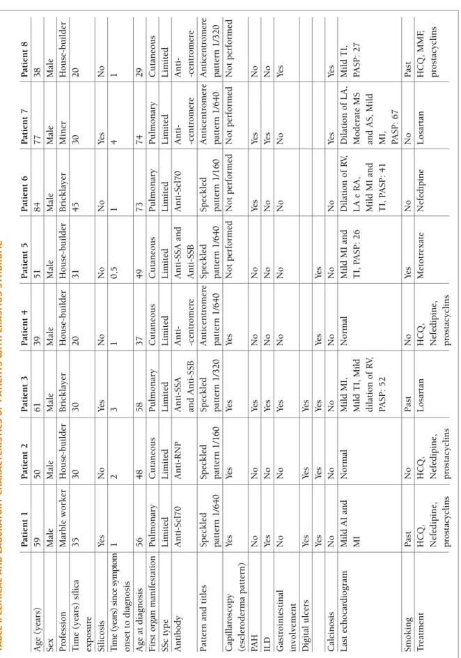
tA b le I. c lI n Ic A l A n d l A b o r At o ry c h A r A ct er Is tI cs o f p At Ie n ts w It h e r A sm u s sy n d r o m e P at ie n t 1 P at ie n t 2 P at ie n t 3 P at ie n t 4 P at ie n t 5 P at ie n t 6 P at ie n t 7 P at ie n t 8 A g e (y ea rs ) 5 9 5 0 6 1 3 9 5 1 8 4 7 7 3 8 S ex M al e M al e M al e M al e M al e M al e M al e M al e P ro fe ss io n M ar b le w o rk er H o u se -b u il d er B ri ck la y er H o u se -b u il d er H o u se -b u il d er B ri ck la y er M in er H o u se -b u il d er T im e (y ea rs ) sí li ca 3 5 3 0 3 0 2 0 3 1 4 5 3 0 2 0 ex p o su re S il ic o si s Ye s N o Ye s N o N o N o Ye s N o T im e (y ea rs ) si n ce s ym p to m 1 2 3 1 0 ,5 1 4 1 o n se t to d ia g n o si s A g e at d ia g n o si s 5 6 4 8 5 8 3 7 4 9 7 3 7 4 2 9 F ir st o rg an m an if es ta ti o n P u lm o n ar y C u ta n eo u s P u lm o n ar y C u ta n eo u s C u ta n eo u s P u lm o n ar y P u lm o n ar y C u ta n eo u s S S c ty p e L im it ed L im it ed L im it ed L im it ed L im it ed L im it ed L im it ed L im it ed A n ti b o d y A n ti -S cl 7 0 A n ti -R N P A n ti -S S A A n ti -A n ti -S S A a n d A n ti -S cl 7 0 A n ti -A n ti -an d A n ti -S S B -c en tr o m er e A n ti -S S B -c en tr o m er e -c en tr o m er e P at te rn a n d t it le s S p ec k le d S p ec k le d S p ec k le d A n ti ce n tr o m er e S p ec k le d S p ec k le d A n ti ce n tr o m er e A n ti ce n tr o m er e p at te rn 1 /6 4 0 p at te rn 1 /1 6 0 p at te rn 1 /3 2 0 p at te rn 1 /6 4 0 p at te rn 1 /6 4 0 p at te rn 1 /1 6 0 p at te rn 1 /6 4 0 p at te rn 1 /3 2 0 C ap il la ro sc o p y Ye s Ye s Ye s Ye s N o t p er fo rm ed N o t p er fo rm ed N o t p er fo rm ed N o t p er fo rm ed (e sc le ro d er m a p at te rn ) P A H N o N o Ye s N o N o Ye s Ye s N o IL D Ye s N o Ye s N o N o N o Ye s N o G as tr o in te st in al N o N o Ye s N o N o N o N o Ye s in v o lv em en t D ig it al u lc er s Ye s Ye s Ye s Ye s Ye s Ye s Ye s Ye s C al ci n o si s N o N o N o N o N o N o Ye s Ye s L as t ec h o ca rd io g ra m M il d A I an d N o rm al M il d M I, N o rm al M il d M I an d D il at io n o f R V, D il at io n o f L A , M il d T I, M I M il d T I, M il d T I, P A S P : 2 6 L A e R A , M o d er at e M S P A S P : 2 7 d il at io n o f R V, M il d M I an d an d A S , M il d P A S P : 5 2 T I, P A S P : 4 1 M I, P A S P : 6 7 S m o k in g P as t N o P as t N o Ye s N o N o P as t T re at m en t H C Q , H C Q , L o sa rt an H C Q , M et o tr ex at e N ef ed ip in e L o sa rt an H C Q , M M F, N ef ed ip in e, N ef ed ip in e, N ef ed ip in e, p ro st ac y cl in s p ro st ac y cl in s p ro st ac y cl in s p ro st ac y cl in s A I: A o rt ic i n su ff ic ie n cy ; A S : A o rt ic s te n o si s; I L D : In te rs ti ti al l u n g d is ea se ; H C Q : h y d ro x y ch lo ro q u in e; L A : L ef t at ri u m ; M I: M it ra l in su ff ic ie n cy ; M M F : M y co p h en o la te m o fe ti l; M S : M it ra l st en o si s; P A H : P u lm o n ar y a rt er ia l h y p er te n si o n ; P A S P : P u lm o n ar y a rt er y s y st o li c p re ss u re ( m m H g ); R A : R ig h t at ri u m ; R V : R ig h t v en tr ic le ; T I: T ri cu sp id I n su ff ic ie n cy ;
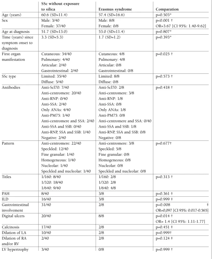
tAble II. clInIcAl And lAborAtory chArActerIstIcs of pAtIents wIth systemIc sclerosIs And systemIc sclerosIs wIthout exposure to sIlIcA, And theIr compArIson
SSc without exposure
to silica Erasmus syndrome Comparation
Age (years) 60.6 (SD=11.4) 57.4 (SD=16.6) p=0.503*
Sex Male: 3/40 Male: 8/8 p<0.001 †
Female: 37/40 Female: 0/8 OR=3.67 [CI 95%: 1.40-9.62]
Age at diagnosis 51.7 (SD=13.0) 53.0 (SD=11.4) p=0.807*
Time (years) since 3.3 (SD=5.3) 1.7 (SD=1.2) p=0.393*
symptom onset to diagnosis
First organ Cutaneous: 34/40 Cutaneous: 4/8 p=0.025 †
manifestation Pulmonary: 4/40 Pulmonary: 4/8
Articular: 2/40 Articular: 0/8
Gastrointestinal: 2/40 Gastrointestinal: 0/8
SSc type Limited: 35/40 Limited: 8/8 p=0.573 †
Diffuse: 5/40 Diffuse: 0/8
Antibodies Anti-Scl70: 7/40 Anti-Scl70: 2/8 p=0.418 †
Anti-centromere: 20/40 Anti-centromere: 3/8
Anti-RNP: 0/40 Anti-RNP: 1/8
Anti-SSA: 2/40 Anti-SSA: 0/8
Only ANAs: 4/40 Only ANAs: 1/8
Anti-PM75: 1/40 Anti-PM75: 0/8
Anti-centromere and SSA: 2/40 Anti-centromere and SSA: 0/40
Anti-SSA and SSB: 0/40 Anti-SSA and SSB: 1/8
Anti-RNP, SSA and SSB: 1/40 Anti-RNP, SSA and SSB: 0/8
Negative: 2/40 Negative: 0/8
Pattern Anti-centromere: 22/40 Anti-centromere: 3/8 p=0.677†
Speckled: 12/40 Speckled: 5/8
Fine granular: 1/40 Fine granular: 0/8
Homogeneous: 1/40 Homogeneous: 0/8
Nucleolar: 1/40 Nucleolar: 0/8
Speckled and nucleolar: 1/40 Speckled and nucleolar: 0/8
Titles 1/160: 8/40 1/160: 2/8 p=0.313 † 1/320: 18/40 1/320: 2/8 1/640: 9/40 1/640: 4/8 PAH 8/40 3/8 p=0.361 ‡ ILD 16/40 3/8 p=0.999 ‡ Gastrointestinal 31/40 2/8 p=0.008 ‡
involvement OR=0,097 [CI 95%: 0.017-0.565]
Digital ulcers 20/40 8/8 p=0.014 † OR= 1.4 [CI 95%: 1.11-1.77] Calcinosis 17/40 2/8 p=0.451 ‡ Dilation of LA 10/40 2/8 p=0.999† Dilation of RA 2/40 2/8 p=0.124 ‡ and/or RV LV hypertrophy 3/40 0/8 p=0.999 †
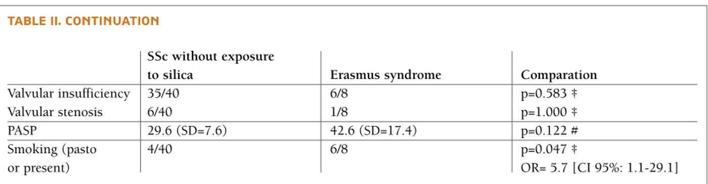
tAble II. contInuAtIon
SSc without exposure
to silica Erasmus syndrome Comparation
Valvular insufficiency 35/40 6/8 p=0.583 ‡
Valvular stenosis 6/40 1/8 p=1.000 ‡
PASP 29.6 (SD=7.6) 42.6 (SD=17.4) p=0.122 #
Smoking (pasto 4/40 6/8 p=0.047 ‡
or present) OR= 5.7 [CI 95%: 1.1-29.1]
* Statistically significant differences are underlined
CI: Confidence Interval; ILD: Interstitial lung disease; LA: Left atrium; LV: Left ventricle; OR: Odds Ratio; PAH: Pulmonary arterial hypertension; PASP: Pulmonary artery systolic pressure (mmHg); RA: Right atrium; RV: Right ventricle; SD= Standard deviation; * test T student; † Fisher’s exact test; ‡ Chi-square test; # Mann-Whitney U test
silica dust 25,26. However, our findings, consistent with
others studies, suggest that less intense exposures may
also play a role in ErS development7.
None of our female patients with SSc had a previous silica exposure and none developed ErS. Our findings can help to corroborate that the low prevalence of ErS in the female sex can be explained by occupational fac-tors and not by different susceptibility14,27.
The relevance of considering ErS as an occupatio nal disease is of paramount importance due to possible economic, social and professional implications.
We found an association between ErS and pul-monary involvement as the presenting manifestation of the disease, smoking exposition and history of di gital ulcers. The relevance of this association was proved in the EUSTAR registry, in a multivariable analysis adjus -ted for age, gender and all parameters considered po-tentially significant. In this registry, a history of digital ulcers was the strongest predictor of new digital ulcers, elevated PASP on heart echocardiogram,
cardiovascu-lar event and death28.
Magnant et al, in a study with 105 patients with SSc, reported that patients exposed to crystalline silica may have different characteristics when compared with
pa-tients with no exposure14. In that study, patients
ex-posed to silica more often exhibited: diffuse cutaneous SSc, presence of digital ulcers, interstitial lung disease,
myocardial dysfunction and cancer14. In our study, all
patients with ErS had limited cutaneous SSc and no statistically significant differences were found between SSc with or without silica exposition. In our SSc po -pulation (48 patients) only five (10.4%) had the dif-fuse type of disease, whilst literature reports an
inci-dence between 26 and 44.2%29. It was not possible to
evaluate cancer outcome due to the reduced sample.
We found an inverse association between silica ex-posure and gastrointestinal involvement. Although gas-trointestinal complications can be the most frequent
internal complications of SSc30, we did not find stu dies
evaluating the association of gastrointestinal involve-ment with exposure to silica. However, silica particles appear to be capable of inhibiting bacterial adhesion and are currently being studied in nanoparticles for the
treatment of infections31.
Rustin et al. report that 16 of 17 patients exposed to silica who developed SSc (ErS) had bibasilar
pul-monary fibrosis on chest radiographs12. In our study,
ILD was not more common in patients with exposure to silica than in those not exposed. This may be due to the fact that Rustin et al followed underground coal or uranium workers, therefore patients with very intense
exposure to silica crystals12. Studies in mice indicate
that intense silica exposures lead to the development of progressive pulmonary inflammation and ultimately fi-brosis, while inflammation caused by less intense
ex-posures may be reversible32.
Some studies have shown a relationship between sil-ica exposure and positivity of anti-Scl70 (anti-topoiso-merase I) antibodies, while others have shown a lower
prevalence of anti-centromere antibodies14,27,33. Ho
-wever, in our study, no relationship was found between positive anti-Scl70 or anti-centromere and toxic
expo-sure, as described in Czirjak and Kumánovic’s study34.
This study has some potential limitations. Firstly, our sample is relatively small and would be desirable to collect data from larger samples and other centers. Given the differences in mineral composition among Portuguese territory it would be advisable to include patients from different regions to obtain more reliable data. Secondly, silica exposition was self-reported and
not quantified in a standardized manner, so high/low exposure was not defined and its definitive role in SSc development is very hard to assure. Finally, this is a cross-sectional study with patients from different back-grounds and disease duration, so some patients’ fea-tures can change over the time.
conclusIon
The prevalence of ErS may be higher than previously described in silica-rich rocks regions. For a more curate ErS diagnosis it is necessary to be aware and ac-tively investigate silica exposures.
In our study, ErS patients presented pulmonary in-volvement as initial manifestation of the disease more frenquently than non-exposed patients, more digital ulcers and a higher exposure to tobacco. The gastroin-testinal involvement was found less frequently in ErS cases.
The fact that subjects’ exposure to silica dust could develop SSc, a rare but potencially severe disease, is a call for awareness regarding the identification of work-ers at risk, and should prompt the implementation of effective protection measures and screnning strategies. Further studies with bigger samples are warranted to understand if these differences may influence the
di-agnosis, treatment and prognosisof patients with SSc
with ErS and without silica expose.
correspondence to
Soraia Azevedo
Rheumatology Department, Unidade Local de Saúde do Alto Minho
Largo Conde Bertiandos, Ponte de Lima E-mail: [email protected]
references
1. Erasmus LD. Scleroderma in gold miners on the Witwatersrand whit particular reference to pulmonary manifestations. S Afr J Lab Clin Med 1957; 3:209-231.
2. Pollard KM, et al. Silica, Silicosis, and Autoimmunity. Front Im-munol. 2016; 7:97.
3. West SG, et al. Rheumatology Secrets. Third edition. Elsevier. Sytemic Sclerosis. 2015 chapter18, pg.141-153.
4. Elhai M, Meune C, Avouac J, Kahan A, Allanore Y. Trends in mortality in patients with systemic sclerosis over 40 years: a sys-tematic review and meta-analysis of cohort studies. Rheuma-tology (Oxford). 2012; 51:1017-1026.
5. Ferri C, Valentini G, Cozzi F, et al. Systemic Sclerosis Study Group of the Italian Society of Rheumatology (SIR-GSSSc). Sys-temic sclerosis: demographic, clinical, and serologic features and survival in 1,012 Italian patients. Medicine (Baltimore). 2002; 81:139-153.
6. Mora GF. Systemic Sclerosis: Environmental Factors. The
Jour-nal of Rheumatology. 2009, 36 (11) 2383-2396.
7. Haustein UH, Anderegg U, Rustin MHA. Silica induced sclero-derma — clinical and experimental aspects. J Rheumatol 1998; 25:1917-26.
8. Rosenman KD, Moore-Fuller M, Reilly MJ. Connective tissue disease and silicosis. Am J Ind Med 1999;25:375-81. 9. Van Loveren H, Vos JG, Germolec D, Yeseonova PP, Eijkemanns
G, McMichael AJ. Exploratory Meeting: Epidemiology of occu-pational and environmental factors associated with autoimmu-nity. Bilthoven, The Netherlands; 2000.
10. Rocha LF et al. Systemic sclerosis and silica exposure: a rare as-sociation in a large Brazilian cohort. Rhematol Int online 13 Jan 2016.
11. Sharma RK et al. Erasmus Syndrome: Association of Silicosis and Systemic Sclerosis. Indian Dermatol Online J. 2018; 9(3):185-187.
12. Rustin MH et al. Silica associated systemic sclerosis in clinical-ly, serologically and immunologically indistinguishable from id-iopathic systemic sclerosis. Br J Dermatol. 1990; 123:725-734. 13. Marie I. Association of occupational exposure with features of systemic sclerosis. J Am Acad Dermatol. 2015; 72(3):456-64. 14. Magnant J, de Monte M, Guilmot JL, et al. Relationship between
occupational risk factors and severity markers of systemic scle-rosis. J Rheumatol. 2005; 32:1713-1718.
15. Van den Hoogen F, Khanna D, Fransen J, et al. 2013 classifica-tion criteria for systemic sclerosis: an American College of Rheumatology/European League against Rheumatism collabo-rative initiative. Arthritis Rheum. 2013; 65: 2737-2747. 16. Sebastiani M, Manfredi A, Cassone G, Giuggioli D, Ghizzoni C,
Ferri C. Measuring microangiopathy abnormalities in systemic sclerosis patients: the role of capillaroscopy-based scoring mod-els. Am J Med Sci. 2014; 348(4):331-6.
17. Leroy EC, Black C, Fleischmajer R, et al. Scleroderma (systemic sclerosis): classification, subsets and pathogenesis. J Rheumatol. 1988; 15:202-5.
18. Marie I, Dominique S, Levesque H, et al. Esophageal involve-ment and pulmonary manifestations in systemic sclerosis. Arthritis Rheum. 2001; 45:346-354.
19. Elhai M, Meune C, Avouac J, Kahan A, Allanore Y. Trends in mortality in patients with systemic sclerosis over 40 years: a sys-tematic review and meta-analysis of cohort studies. Rheuma-tology (Oxford). 2012; 51:1017-1026.
20. Elhai M, Avouac J, Kahan A, Allanore Y. Systemic sclerosis: Re-cent insights. Joint Bone Spine. 2015; 82(3):148-53. 21. Makol A, Rosenman KD. Prevalence of connective tissue di sease
in silicosis (1985-2006): a report from the state of Michigan surveillance system for silicosis. Am J Ind Med. 2011; 54: 255-262.
22. Rodnan GP, Benedek TG, Medsger TA, Cammarata RJ. The as-sociation of progressive systemic sclerosis (scleroderma) with coal workers’ pneumoconiosis and other forms of silicosis. Ann Intern Med. 1967; 66: 323-34.
23. Jorge Carvalho, Recursos Minerais: o potencial de Portugal. Ini-ciativa Matérias Primas: rumo ao fornecimento seguro e à gestão dos recursos minerais europeus. 2010. Laboratório Nacional de Energia e Geologia
24. Comissão coordenadora de desenvolvimento regional do Norte, Norte Estrutura, 2018, ano II, nº5
25. Subrata Chakrabarti, Koushik Pan. Erasmus Syndrome in a 42-Year-Old Male: A Rare Case Report. J Clin Diagn Res. 2015 May; 9(5): OD01–OD03.
26. Bello S, Rinaldi A, Trabucco S, Serafino L, Bonali C, Lapadula G. Erasmus syndrome in a marble worker. Reumatismo. 2015 Dec 30; 67(3):116-22.
27. Burns CJ et al. The epidemiology of scleroderma among wom-en: assessment of risk from exposure to silicone and silica. J Rheumatol. 1996; 23:1904-1911.
28. Mihai C, Landewé R, van der Heijde D, et al. Digital ulcers pre-dict a worse disease course in patients with systemic sclerosis. Ann Rheum Dis. 2016;75(4):681-6.
29. Galluccio F, Walker UA, Nihtyanova S, et al. Registries in sys-temic sclerosis: a worldwide experience. Rheumatology Oxford 2011;50:60–8.
30. Forbes A, Marie I. Gastrointestinal complications: the most fre-quent internal complications of systemic sclerosis. Rheumatol-ogy (Oxford) 2009; 48(3):iii36–9.
31. Vallet-Regí M, González B, Izquierdo-Barba I. Nanomaterials as Promising Alternative in the Infection Treatment. Int J Mol Sci. 2019 Aug 4;20(15).
32. Velan GM, Kumar RK, Cohen DD. Pulmonary inflammation and fibrosis following subacute inhalational exposure to silica: determinants of progression. Pathology. 1993 Jul;25(3):282-90. 33. MacHugh NJ, Whyte J, Harvey G, Haustein UF. Antitopoiso-merase I antibodies in silica-associated systemic sclerosis. Arthritis Rheum 1994;37:1198-205.
34. Czirjak L, Kumánovics G. Exposure to solvents in female pa-tients with scleroderma. Clin Rheumatol 2002; 21:114-8.