Article
Printed in Brazil - ©2017 Sociedade Brasileira de Química0103 - 5053 $6.00+0.00*e-mail: [email protected]
Larvicidal Activity of
Beauveria bassiana
Extracts against
Aedes aegypti
and
Identification of Beauvericins
Juliana F. S. Daniel,*,a Andressa A. Silva,a Danielle H. Nakagawa,a
Lívia S. de Medeiros,b Mário G. Carvalho,c Lucineli J. Tavares,c Lucas M. Abreud and
Edson Rodrigues-Filhob
aDepartamento de Química, Universidade Tecnológica Federal do Paraná (UTFPR),
Avenida dos Pioneiros, 3131, 86036-370 Londrina-PR, Brazil
bDepartamento de Química, Universidade Federal de São Carlos (UFSCar),
CP 676, 13565-905 São Carlos-SP, Brazil
cDepartamento de Química, ICE, Universidade Federal Rural do Rio de Janeiro (UFRRJ),
Rodovia BR 465, Km 7, 23890-000 Seropédica-RJ, Brazil
dDepartamento de Fitopatologia, Universidade Federal de Viçosa (UFV),
36570-000 Viçosa-MG, Brazil
Beauveria bassiana is an entomopathogenic fungus that has been well known for its capacity to act as biopesticide on various disease vectors. The analysis of organic extracts of strains CG71 and UNI40 led to identification of cyclodepsipeptides beauvericin, beauvericin A or F, beauvericin E and bassianolide by ultra-high pressure liquid chromatography-high resolution mass spectrometry in tandem mode (UHPLC-HRMS/MS). The larvicidal activity on 3rd instar of Aedes aegypti
revealed LC50 0.9887 and 0.4653 ppm in 24 and 48 hours (CG71 methanolic extract), LC50 0.7834
ppm in 48 hours (CG71 ethyl acetate), LC50 0.7834 and 1.8149 ppm (UNI 40 for ethyl acetate and
methanolic extracts, respectively) in 48 hours. These findings highlight the potential of B. bassiana metabolites for controlling the vector of Dengue and Zika diseases.
Keywords: cyclohexadepsipeptides, Dengue, Zika, beauvericins, UHPLC-HRMS/MS
Introduction
The entomopathogenic Beauveria bassiana has a broad host range naturally attacking insects and variety of arthropod species.1-3 Due to this ability, it is used as a
biopesticide agent on diverse disease vectors, including crop pests, ticks and mosquitoes.4,5 The attack on insect
cuticle occurs after hyphae of B. bassiana penetrate the insect integument by enzymatic action.6
Species of entomopathogenic fungi have been described as producers of secondary metabolites, including bioactive non-ribosomal peptides and polyketides.7 B. bassiana
produces the cyclooligomer nonribosomal depsipeptides beauvericins and bassianolide,8,9 beauveriolides,10 tenellin,
bassianin,11 pyridovericin, pyridomacrolidin,12 oosporein13
and bassiacridin.14
In special, beauvericin is an ionophoric cyclodepsipeptide
detected in several fungi, mainly Beauveria, Paecilomyces
and Fusarium species.15-17 It forms complexes with
cations, causing an increase in permeability of natural and artificial membranes.18,19 Furthermore, beauvericin induces
programmed cell death similar to apoptosis.20 Initially
it was regarded as a toxin against brine shrimp,8 also
showing insecticidal and antibiotic activities.21-23 It contains
three residues each D-2-hydroxyisovaleric acid (Hiv) and L-N-methylphenylalanine linked alternately.
Recently, new analogues of beauvericin were described, including beauvericin A, B,24 D, E and F.25 Bassianolide
is another cyclooctadepsipeptide metabolite produced
B. bassiana and Lecanicillium lecanii.9 It is toxic to
insects and probably also acts as an ionophore, such as other cyclodepsipeptides.15 The structural diversity of
Dengue is a viral infection transmitted to humans by
Aedes aegypti mosquitoes that became a major concern of the public health sector.26 There were 96 million apparent
dengue infections globally in 2010, of which the Americas contributed 14% (13 million infections) of worldwide and over half developed in Brazil and Mexico.27 Due
to no effective antiviral agents and licensed vaccine for infection, the decrease in transmission depends only on vector control by elimination of artificial and disposable water inundated larvae breeding sites and application of insecticides.27,28 Furthermore, studies reveal resistance in
the vector population to various substances, in Brazil there is an irregular distribution of insecticide resistance.28
Furthermore, Aedes mosquitoes also transmit Zika virus; attracting worldwide attention in 2016, when the vector caused widespread epidemic cases in Brazil.29 This
disease has been recognized in Brazil since late 2014, but in 2015 coincided with an increase in the number of cases of microcephaly. The more affected Brazilian states were Bahia, Paraíba, and Pernambuco, where in the first trimester of pregnancy coincided with reports of cases of febrile and allergy compatible with Zika virus disease, suggesting an association between Zika virus infection during early pregnancy and the occurrence of microcephaly.30
Consequently, there is an urgent need to develop new methods for A. aegypti mosquitoes infections control. So, fungal extracts are an alternative because they constitute a rich source of bioactive metabolites.
The paper reports the activity of two strains of Beauveria bassiana extracts on mortality of Aedes aegypti larvae and identification of beauvericins and bassianolide using ultra-high pressure liquid chromatography-high resolution mass spectrometry in tandem mode (UHPLC-HRMS/MS). Additionally, the fragmentation data of beauvericin E and A of F is provided.
Experimental
Origin and preservation of B. bassiana
The entomopathogenic fungal strains B. bassiana
CG 71 and UNI 40 were kindly provided by Prof Dr Luis Francisco Angeli Alves from Universidade Estadual do Oeste do Paraná (Cascavel, PR, Brazil) and Prof Dr Pedro Manuel Oliveira Janeiro Neves from Universidade Estadual de Londrina (Londrina, PR, Brazil), respectively. The CG 71 and UNI 40 strains are preserved in the culture collection of Universidade Estadual do Oeste do Paraná and Universidade Estadual de Londrina, respectively.31
Both strains are also preserved at −80 °C in the Coleção
Micológica de Lavras, Universidade Federal de Lavras
(Lavras, MG, Brazil) under the accession numbers CML 2677 (CG 71) and CML 2678 (UNI 40).
Morphological characterization
The strains were preliminary identified as
Beauveria bassiana based on the macro and
micro-morphological characteristics of colonies, conidia and conidiophores after cultivation for 7 days at room temperature on malt extract agar (Himedia, Mumbai, India).32
DNA sequencing and phylogenetic analyses
Strains CG 71 and UNI 40 were cultivated on malt extract agar for five days at 24 °C. The mycelium was scrapped from the colonies, macerated under liquid nitrogen, and subjected to genomic DNA extraction with the Wizard Genomic Deoxyribonucleic Acid (DNA) Purification Kit (Promega, Madison, USA). A fragment of the second largest subunit of the ribonucleic acid (RNA) polymerase gene (RPB2) was amplified from the extracted DNA using the primers 5F and 7cR, and the polymerase chain reaction (PCR) conditions of Liu et al.33
The purified PCR products were sent for DNA sequencing by a commercial service.
Consensus sequences were assembled using SeqAssem ver. 07/2008 (SequentiX - Digital DNA Processing, Germany). Blast searches against the GenBank database suggested that both strains belonged to Beauveria bassiana. DNA sequences of RPB2 from reference strains of
Beauveria bassiana and related taxa34 were used to
compose a multiple sequence alignment using multiple sequence comparison by log-expectation (MUSCLE), as implemented by Mega 6 software.35 Maximum likelihood
(ML) phylogenetic analysis was conducted using Mega 6. 1000 bootstrap pseudo-replicates were employed for estimating node support. The general time reversible model with gamma distribution of rates across sites (GTR+G) model of evolution, estimated using JModeltest,36 was
used in the ML analysis. Isaria farinosa (ARSEF 4029) was used as the outgroup.
Culture of B. bassiana, extraction and chromatographic
fractionation
The B. bassiana CG71 and UNI 40 strains were
0.5 g MgSO4.7H2O; 0.5 g KH2PO4; 1 L water, as reported
by Bunyapaiboonsri et al.37 The flasks were incubated on
a rotary shaker (200 rpm) for 8 days. These seed cultures (240 mL of each strain) were used to inoculate 11 liters ML (55 Erlenmeyer flasks of 1 L with 200 mL of medium for each strain). After incubation at room temperature for 20 days in static mode, the mycelium was separated by reduced pressure filtration and the liquid phase was submitted to liquid-liquid fractionation with ethyl acetate (EtOAc). After lyophilization, the mycelial masses were combined and extracted totally with methanol (MeOH). The organic solvents were removed by vacuum distillation at 55 and 60 °C using a rotary evaporator.
The ethyl acetate extracts CG71a (4.7600 g) and UNI40a (4.9623 g), and the methanolic extracts CG71m (18.8054 g) and UNI40m (14.6802 g) were fractionated through Sephadex LH-20 (1.75 mm × 0.3 Ø) using MeOH as eluent. In both extracts of CG71 and UNI40 and some fractions were detected beauvericin, beauvericin A or F, beauvericin E and bassianolide.
Cyclodepsipeptides identification
The UHPLC-HRMS/MS analyzes were performed on an Accela High Speed LC coupled to a LTQ Orbitrap Velos FT-MS (ThermoFisher Scientific, San Jose, CA, USA), equipped with an electrospray source (HESI-II) and operated at a resolution of 60,000 FWHM, with positive ionization and 25 eV at HRMS/MS mode. The chromatographic system was fitted with an RP Luna C18
column (150 × 4.6 mm, 5 µm, Phenomenex) applying as mobile phase H2O/CH3OH (30:70 → 0:100, v/v, both
buffered with 0.1% of formic acid) in 15 min at a flow rate of 0.4 mL min-1. The software Thermo Xcalibur 2.1
(ThermoFisher Scientific) was used to control the full system. The sample injection (auto injector) volume was 0.5 µL.
Bioassay to control Aedes aegypti larvae Rockefeller strain
The bioassay was performed at the Laboratory of Biocontrol of Arthropods (LABIART), Veterinary Institute, Department of Animal Parasitology, Universidade Federal Rural do Rio de Janeiro (UFRRJ). The extracts tested were CG71a, CG71m, UNI40a and UNI40m.
Aedes aegypti of the Rockefeller strain were maintained in colonies at 27 ± 3 °C, and 80% humidity on a 12-h light/ dark cycle. Four replications of ten (10) third instar larvae (L-3) of Aedes aegypti susceptible in the insecticide were tested in five concentrations: 25, 50 100, 150, and 200 µL of each extract dissolved in dimethyl sulfoxide 10% (DMSO).
Two control groups were tested, one containing the solvent used in extracts and the other containing only pure water. The analyzes of the results of larval mortality were assessed at 15 minutes and at successive counts of 15, 30 minutes, 1, 2, 3 and 24 and 48 hours. The lethal concentrations LC50
and their respective confidence intervals were calculated through Probit analyses, using the R software (R Core Team).
Results and Discussion
The ethyl acetate and methanol extracts of entomopathogenic fungi Beauveria bassiana UNI40 and CG71 were analyzed by HRMS data analysis, obtained in an Orbitrap mass analyzer. The metabolites beauvericin (1), beauvericin A (2) or F (2a), beauvericin E (3) and bassianolide (4) were identified (Figure 1).
Cyclodepsipeptides fragmentation
Mass spectrometry has played essential role in the structure determination of cyclodepsipeptides originated from diverse fungal species, grains and foods. The fragmentation is a main step in sequencing and primary stage usually is bond rupture in the ester or amide group.38
Interpretation and elucidation of the amino acid sequence using collision-induced dissociation (CID) depends essentially on the sequence of specific ions present (an, bn,
cn and xn, yn, zn). The fragment ions produced in this process
can be divided in two series.39
Beauvericin is a cyclohexadepsipeptide isolated in 1969 from B. bassiana, derived from two dipeptidol monomers alternately, D-hydroxyisovalerate (D-HiV) and
N-methyl-L-phenylalanine (N-Me-Phe), while bassianolide isolated in 1977 from B. bassiana and Verticilliumlecanii
is a cyclooctadepsipeptide also formed by two dipeptidol monomers units of D-hydroxyisovalerate (D-HiV) and
N-methyl-L-leucine (N-Me-Leu).18
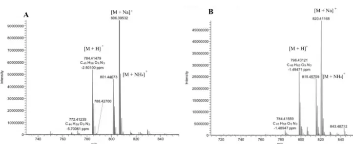
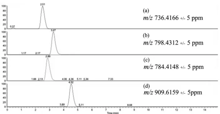
The cyclodepsipeptides beauvericin, beauvericin A or F, beauvericin E and bassianolide were detected in two strains of B. bassiana (CG71 and UNI 40) extracts and fractions semi-purified according to the observed accurate masses for protonated molecule and the corresponding cationized molecules, displayed at their full scan spectra (Figure 2) and selected ion chromatogram (Figure 3). Moreover, the product ion spectra allowed the comparison of the compounds fragmentation profile to the data found in the literature (Table 1).24,25,40-44
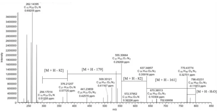
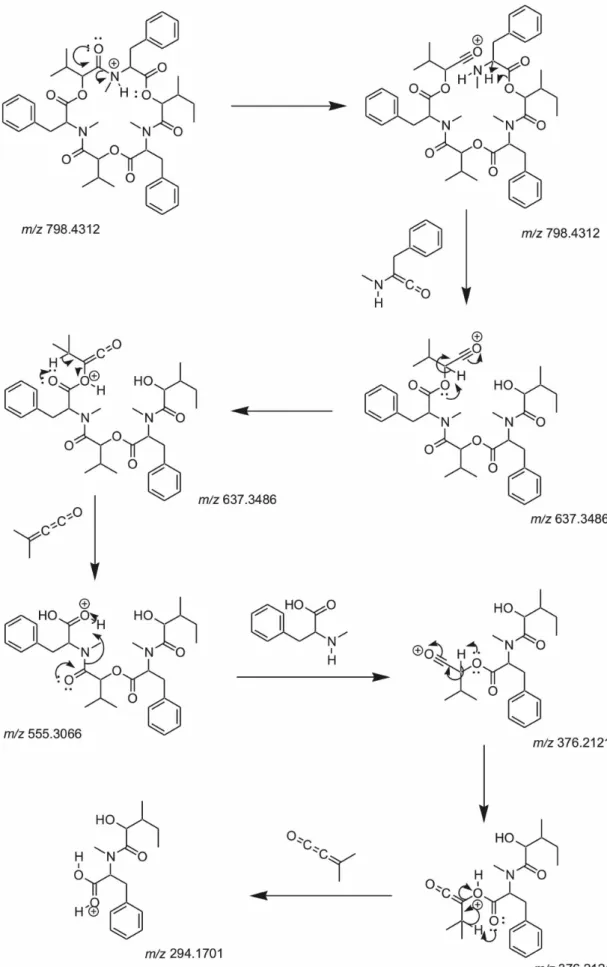
respectively. The N-methyl-L-phenylalanine (−161 Da)
loss gave rise to the fragment ion m/z 637.3486. While the peaks m/z 555.3066 and 294.1701 are detected due to the isopropylideneketene losses (82 Da), the ion m/z 376.2121 occurs through the rupture of the amide bond. It is worth to highlight that this new fragmentation proposal is fundamental for the confirmation of the beauvericin A or F presence in the organics extracts of entomopathogenic fungi.
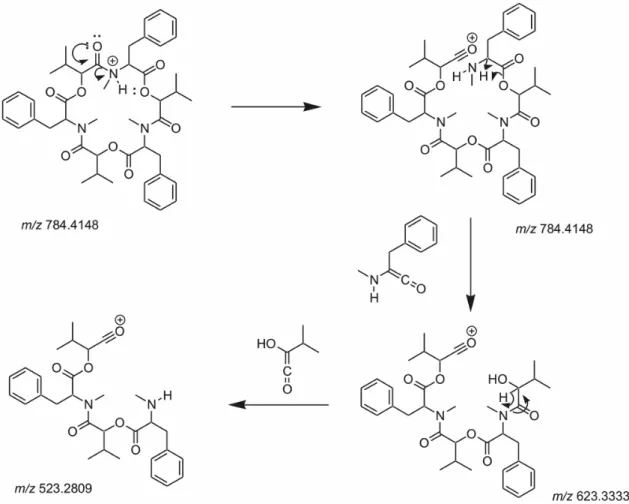
The inspection of the MS/MS data showed that the peaks at m/z 523.2809 in beauvericin and m/z 682.4630 in bassianolide spectra represent the loss of an
2-hydroxy-3-methyl-1-buten-1-one molecule (100 Da), the identical D-HiV monomer (Figures 6 and 7). Additionally, the peaks at m/z 623.3333 (Figure 6) and 782.5159 (Figure 7) show the loss of N-methyl-L-phenylalanine (−161 Da)
and N-methyl-L-leucine (−127 Da), respectively. Similar
fragments containing phenylalanine residues (m/z 262) are described for beauvericin and E, which then lost H2O to
give m/z 244 (Table 1).
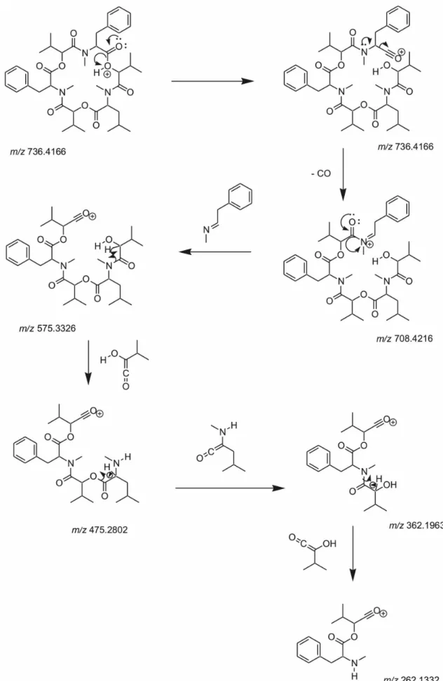
Figure 8 shows fragmentation mechanism proposal for beauvericin E (precursor ion m/z 736.4722). Differently from the mechanism of the other cyclodepsipeptides produced by the fungus, the MS/MS data from beauvericin E showed
Figure 1. Structures of compounds 1-4 identified from the extracts of B. bassiana.
CO loss, corresponding to the fragment ion m/z 708.4216 [M + H − CO]+. The other peaks m/z 575.3326, 475.2802,
362.1963 and 262.1438 occur due to the rupture of ester or amide bonds. The losses of N -(2-phenylethylidene)-methanamine (−133 Da),
2-hydroxy-3-methyl-1-buten-1-one (−100 Da), 2-amino-4-methyl-1-penten-1-one
(−113 Da) correspond to the peaks mentioned above. This
fragmentation mechanism proposal evidences the presence of beauvericins E in the organic extracts of B. bassiana
CG 71 and UNI 40 and may assist cyclodepsipeptide characterizations according to the additional structural information.
Table 1. Characterization (mass accuracy) of named ions cyclodepsipeptides and MS/MS parameters of Beauveria bassiana extracts
Beauvericin Error / ppm Beauvericin A
or F Error / ppm Beauvericin E Error / ppm Bassianolide Error / ppm Molecular formula C45H57N3O9 C46H59N3O9 C41H57N3O9 C48H84N4O12
Calcd. [M + H]+ 784.4175 798.4332 736.4175 909.6168
Calcd. [M + NH4]+ 801.4441 815.4598 753.4441 926.6433
Calcd. [M + Na]+ 806.3996 820.4153 758.3996 931.5988
Found [M + H]+ 784.4148 −2.50 798.4312 −1.49 736.4166 −0.26 909.6159 −0.85
Found [M + NH4]+ 801.4407 815.4171 753.4433 926.6425
Found [M + Na]+ 806.3953 820.4117 758.3986 932.6007
Precursor ion (m/z) 784.4174 0.84 798.4323 −0.12 736.4722 909.6159 −0.85
Product ions (m/z) 623.3333 1.09 637.3486 0.40 708.4216 782.5159 −1.25
523.2809 1.30 555.3066 0.29 575.3326 682.4630 2.76
362.1967 1.42 376.2121 0.57 475.2802 555.3640 3.37
262.1441 1.39 294.1701 0.51 362.1963 427.3168 −5.84
244.1336 (M-H2O)
1.45 262.1438
244.1332 (M-H2O)
328.2121 −7.41
Collision energy / eV 25 25 25
Tube lens offset / V 70 70 70
Calcd.: mass calculated.
Phylogenetic analyses
Strains CG 71 and UNI 40 were grouped in a well-supported clade with reference strains Beauveria bassiana
in the phylogenetic analyzes conducted with partial RPB2 DNA sequences (Figure 9), thus confirming their identity.
Larvicidal activity
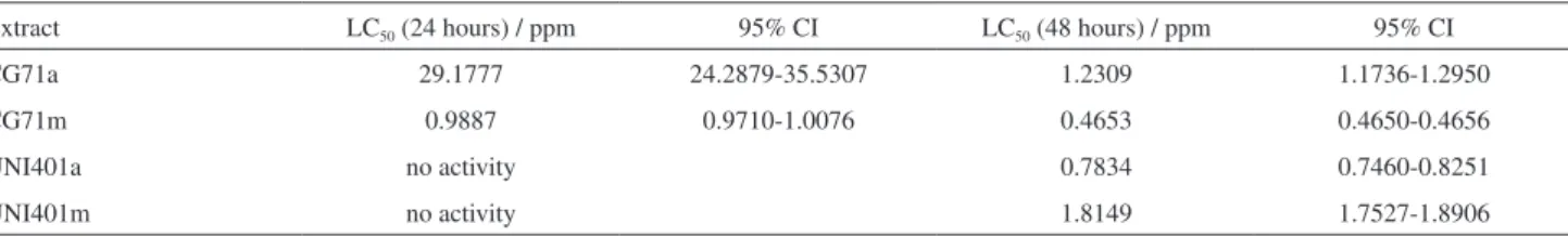
Table 2 shows larvicidal activity of the B. bassiana CG71 and UNI40 extracts against 3rd instar of Aedes aegypti. The
methanolic extract of strain CG71 revealed the highest activity with LC50 values of 0.9887 and 0.4653 ppm after
24 and 48 hours, respectively. The ethyl acetate extract of CG71 displayed LC50 of 29.1777 and 1.2309 ppm after 24
and 48 hours, respectively.
The UNI 40 strain showed LC50 values of 0.7834 and
1.8149 ppm for the ethyl acetate and methanolic extracts after 48 hours of incubation, respectively. These extracts showed no larvicidal activity in the first 24 hours of incubation. Besides the cyclodepsipeptides detected in both CG 71 and UNI 40 extracts, other peptides with peaks at m/z above 1000 were detected in the ethyl acetate and methanolic extracts of UNI 40.
New studies using fungal organic extracts aim to identify new bioactive secondary metabolites, such as
Fusarium sp. extract with trypanocidal activity, which revealed beauvericin as the responsible for the toxicity of
Fusarium sp. to Trypanosoma cruzi.45
Beauvericin has been reported to be toxic on Aedes aegypti
larvae with LC50 26 ppm,24 and 10 and 20 µg mL-1 showed
39 and 86% of mortality in 48 hours, respectively.46 The
presence of beauvericin, beauvericin A or F, beauvericin E and bassianolide in the extracts of two B. bassiana strains may explain the excellent larvicidal activity observed.
The cyclodepsipeptides have shown insecticidal activity against several species, such as fifth instar larvae of the silkworm Bombyx mori,9 Calliphora erythrocephala, Aedes aegypti, Lygus spp., Spodoptera frugiperda,
Schizaphis graminum47 and Sitophilus spp.48 However, the
mycelium and conidia of a B. bassiana strain, pathogenic to Tribolium castaneum and Sitophilus oryzae, showed no toxicity against rats and mice.49
Bassianolide was described as a virulence factor of
B. bassiana against Galleria mellonella, Spodoptera exigua,
Helicoverpa zea50 and Atta sexdens sexdens.51 Beauvericin
was cytotoxic (IC50 0.5 µM) on a lepidopteran Spodoptera frugiperda (SF-9) cell line52 and on the Colorado potato
beetle (LC50 633 ppm).15
Conclusions
The cyclodepsipeptides beauvericin, beauvericin A or F, beauvericin E and bassianolide were identified in the B. bassiana CG71 and UNI 40 extracts. The methanolic extract of CG71, ethyl acetate and methanolic extracts of UNI40 showed larvicidal activity against 3rd instar of Aedes aegypti
with LC50 values of 0.4653, 0.7834 and 1.8149 ppm after
48 hours of incubation, respectively. These results suggest
Figure 6. Plausible fragmentation mechanism for beauvericin in collision-induced dissociation (CID) spectra.
Table 2. Larvicidal activity Beauveria bassiana UNI40 and CG71 extracts against 3rd instar of Aedes aegypti
Extract LC50 (24 hours) / ppm 95% CI LC50 (48 hours) / ppm 95% CI
CG71a 29.1777 24.2879-35.5307 1.2309 1.1736-1.2950
CG71m 0.9887 0.9710-1.0076 0.4653 0.4650-0.4656
UNI401a no activity 0.7834 0.7460-0.8251
UNI401m no activity 1.8149 1.7527-1.8906
Figure 9. Maximum likelihood phylogenetic tree of partial RPB2 sequences of CG 71 and UNI 40, and reference strains of Beauveria bassiana and related species. Bootstrap values equal or higher than 70% are showed by the nodes. Isaria farinosa (ARSEF 4029) was used as the outgroup. T identifies ex-type strains.
that cyclodepsipeptides are probably the active principles responsible for the larvicidal action. Therefore, further studies must be undertaken in order to isolate and confirm the compounds responsible for this activity. Therefore, they may be considered as potential insecticidal components in the formulations for the Dengue and Zika vector. The liquid chromatography coupled with electrospray ionization tandem mass spectrometry (LC-ESI-MS/MS) methodology used proved to be an excellent tool for the identification of cyclodepsipeptides in fungal extracts, and contributed with new fragmentation studies for beauvericin A or F and E.
Acknowledgments
The authors would like to thank Dr Luis Francisco Angeli Alves and Prof Dr Pedro Manuel Oliveira Janeiro Neves for the fungal strains, and Prof Dr Ludwig H. Pfenning for infrastructure of DNA analyses. The authors thank the financial support from International Foundation of Science (IFS, grant 2010/F/4898-1) and DIRPPG (Diretoria de Pesquisa e Pós-Graduação of Universidade Tecnológica Federal do Paraná, Londrina Campus), Conselho Nacional de Desenvolvimento Científico e Tecnológico (CNPq) and Fundação Araucária for scholarships.
References
1. Clarkson, J. M.; Charnley, A. K.; Trends Microbiol. 1996, 4, 197.
2. Khachatourians, G. G.; J. Invertebr. Pathol.1992, 59, 212. 3. Goettel, M. S.; Eilenberg, J.; Glare, T. R. In Comprehensive
Molecular Insect Science; Gilbert, L. I.; Iatrou, K.; Gill, S., eds.; Elsevier: Boston, 2005, p. 361.
4. Leathers, T. D.; Gupta, S. C.; Alexander, N. J.; J. Ind. Microbiol.
1993, 12, 69.
5. De La Rosa, W.; Alatorre, R.; Barrera, J. F.; Toreillo, C.; J. Econ. Entomol. 2000, 93, 1409.
6. Leger, St. R. J.; Charnley, A. K.; Cooper, R. M.; J. Invertebr. Pathol.1986, 48, 85.
7. Von Döhren, H.; Adv. Biochem. Eng./Biotechnol. 2004, 88, 217.
8. Hamill, R. L.; Higgens, C. E.; Boaz, H. E.; Gorman, M.; Tetrahedron Lett.1969, 49, 4255.
9. Suzuki, A.; Kanaoka, M.; Isogai, A.; Murakoshi, S.; Ichinoe, M.; Tamura, S.; Tetrahedron Lett.1977, 25, 2167.
10. Kuzma, M.; Jegorov, A.; Kačer, P.; Havlíček, V.; J. Mass Spectrom.2001, 36, 1108.
11. McInnes, A. G.; Smith, D. G.; Wat, C. K.; Vining, L. C.; Wright, J. L. C.; J. Chem. Soc., Chem. Commun.1974, 8, 281. 12. Takahashi, S.; Kakinuma, N.; Uchida, K.; Hashimoto, R.;
Yanagisawa, T.; Nakagawa, A.; J. Antibiot.1998, 51, 596. 13. Vining, L. C.; Kelleher, W. J.; Schwarting, A. E.; Can. J.
Microbiol. 1962, 8, 931.
14. Quesada-Moraga, E.; Vey, A.; Mycol. Res. 2004, 108, 441. 15. Roberts, D. W.; Gupta, S.; Leger, R. J. St.; Pesq. Agropec. Bras.
1992, 27, 325.
16. Qinggui, W.; Lijian, X.; Molecules2012, 17, 2367.
17. Isaka, M.; Kittakoop, P.; Kirtikara, K.; Hywel-Jones, N. L.; Thebtaranonth, Y.; Acc. Chem. Res.2005, 38, 813.
18. Xu, Y.; Orozco, R.; Wijeratne, E. M. K.; Gunatilaka, A. A. L.; Stock, S. P.; Molnár, I.; Chem. Biol.2008, 15, 898.
19. Toman, E.; Makrlík, E.; Vaňura, P.; Monatsh. Chem. 2011, 142, 779.
20. Waetjen, W.; Debbab, A.; Hohlfeld, A.; Chovolou, Y.; Proksch, P.; Toxicol. Lett.2014, 231, 9.
21. Wu, Y.; Huang, X.; Deng, J.; J. Beijing Norm. Univ., Nat. Sci.
22. Gupta, S.; Krasnoff, S. B.; Underwood, N. L.; Renwick, J. A. A.; Roberts, D. W.; Mycopathologia1991, 115, 185.
23. Castlebury, L. A.; Sutherland, J. B.; Tanner, L. A.; Henderson, A. L.; Cerniglia, C. E.; World J. Microbiol. Biotechnol. 1999, 15, 119.
24. Gupta, S.; Montllor, C.; Wang, Y. S.; J. Nat. Prod. 1995, 58, 733.
25. Fukuda, T.; Arai, M.; Tomoda, H.; Omura, S.; J. Antibiot.2004, 57, 117.
26. WHO; Comprehensive Guidelines for Prevention and Control of Dengue and Dengue Hemorrhagic Fever; World Health Organization, Regional Office for South-East Asia, 2011, p. 1. 27. Bhatt, S.; Gething, P. W.; Brady, O. J.; Messina, J. P.; Farlow,
A. W.; Moyes, C. L.; Drake, J. M.; Brownstein, J. S.; Hoen, A. G.; Sankoh, O.; Myers, M. F.; George, D. B.; Jaenisch, T.; Wint, G. R. W.; Simmons, C. P.; Scott, T. W.; Farrar, J. J.; Hay, S. I.; Nature2013, 496, 504.
28. Linss, J. G. B.; Brito, L. P.; Garcia, G. A.; Araki, A. S.; Bruno, R. V.; Lima, J. B. P.; Valle, D.; Martins, A. J.; Parasites Vectors
2014, 7, 25.
29. Imperato, P. J.; J. Community Health2016, 41, 674.
30. Oliveira, W. K.; Cortez-Escalante, J.; De Oliveira, W. T. G. H.; Carmo, G. M. I.; Henriques, C. M. P.; Coelho, G. E.; França, G. V. A.; Morb. Mortal Wkly. Rep.2016, 65, 242.
31. Santoro, P. H.; Neves, P. M. O. J.; Alexandre, T. M.; Sartori, D.; Alves, L. F. A.; Fungaro, M. H. P.; J. Invertebr. Pathol. 2008, 97, 83.
32. Domsch, K. H.; Gams, W.; Anderson, T. H.; Compendium of Soil Fungi, 2nd ed.; IHW-Verlag: Eching, Germany, 2007.
33. Liu, Y. J.; Whelen, S.; Hall, B. D.; Mol. Biol. Evol. 1999, 16, 1799.
34. Rehner, S. A.; Minnis, A. M.; Sung, G. H.; Luangsa-ard, J. J.; Devotto, L.; Humber, R. A.; Mycologia2011, 103, 1055. 35. Tamura, K.; Stecher, G.; Peterson, D.; Filipski, A.; Kumar, S.;
Mol. Biol. Evol. 2013, 30, 2725.
36. Darriba, D.; Taboada, G. L.; Doallo, R.; Posada, D.; Nat. Methods2012, 9, 772.
37. Bunyapaiboonsri, T.; Yoiprommarat, S.; Srisanoh, U.; Choowong, W.; Tasanathai, K.; Hywel-Jones, N. L.; Luangsa-ard, J. J.; Isaka, M.; Phytochem. Lett.2011, 4, 283.
38. Wulfson, N. S.; Puchkov, V. A.; Rozinov, B. V.; Zyakoon, A. M.; Shemyakin, M. M.; Ovchinnikov, Y. A.; Kiryushkin, A. A.; Ivanov, V. T.; Tetrahedron Lett. 1965, 32, 2793.
39. Daniel, J. F. S.; Filho, E. R.; Nat. Prod. Rep.2007, 24, 1128. 40. Herebian, D.; Zühlke, S.; Lamshöft, M.; Spiteller, M.; J. Sep.
Sci. 2009, 32, 939.
41. Song, H. H.; Lee, H. S.; Lee, G. P.; Ha, S. D.; Lee, C.; Food Addit. Contam., Part A2009, 26, 518.
42. Gonzalezi, D. J.; Xu, Y.; Yang, Y. L.; Esquenazia, E.; Liu, W. T.; Edlund, A.; Duong, T.; Du, L.; Molnár, I.; Gerwick, W. H.; Jensen, P. R.; Fischbach, M.; Liaw, C. C.; Straight, P.; Nizet, V.; Dorrestein, P. C.; J. Proteomics2012, 75, 5069.
43. Devreese, M.; De Baere, S.; De Backer, P.; Croubels, S.; Talanta
2013, 106, 212.
44. El-Elimat, T.; Figueroa, M.; Ehrmann, B. M.; Cech, N. B.; Pearce, C. J.; Oberlies, N. H.; J. Nat. Prod.2013, 76, 1709. 45. Campos, F. F.; Junior, P. A. S.; Romanha, A. J.; Araújo, M. S.
S.; Siqueira, E. P.; Resende, J. M.; Alves, T. M. A.; Martins-Filho, O. A.; Santos, V. L.; Rosa, C. A.; Zani, C. L.; Cota, B. B.; Mem. Inst. Oswaldo Cruz2015, 110, 65.
46. Grove, J. F.; Pople, M.; Mycopathologia 1980, 70, 103. 47. Wang, Q.; Xu, L.; Molecules2012, 17, 2367.
48. Langenfeld, A.; Blond, A.; Gueye, S.; Herson, P.; Nay, B.; Dupont, J.; Prado, S.; J. Nat. Prod. 2011, 74, 825.
49. Dal Bello, G. M.; Padin, S. B.; Cagliada, P.; Carbone, C.; Vasicek, A.; Arcas, J.; Rev. Toxicol. 2000, 17, 36.
50. Xu, Y.; Orozco, R.; Wijeratne, E. M. K.; Espinosa-Artiles, P.; Gunatilaka, A. A. L.; Stock, S. P.; Molnár, I.; Fungal Genet. Biol.2009, 46, 353.
51. Loureiro, E. S.; Monteiro, A. C.; Arq. Inst. Biol. 2004, 71, 35. 52. Fornelli, F.; Minervini, F.; Logrieco, A.; J. Invertebr. Pathol.
2004, 85, 74.
Submitted: May 12, 2016
Published online: September 9, 2016