1 Graduate Program in Rehabilitation Sciences, School of Physical Education, Physical Therapy and Occupational Therapy (EEFFTO), Universidade Federal de Minas Gerais (UFMG), Belo Horizonte, MG, Brazil
2 Department of Physical Therapy, EEFFTO, UFMG, Belo Horizonte, MG, Brazil
3 Department of Elderly, Adult and Maternal and Child Physical Therapy, School of Physical Therapy, Universidade Federal de Juiz de Fora (UFJF), Juiz de Fora, MG, Brazil
4 Physical Therapist, Belo Horizonte, MG, Brazil
5 Department of Occupational Therapy, EEFFTO, UFMG, Belo Horizonte, MG, Brazil Received: 09/24/2012 Revised: 01/28/2013 Accepted: 02/01/2013.
a r t i c l e
Assessment of gait in toddlers with normal motor
development and in hemiplegic children with mild
motor impairment: a validity study
Priscilla R. P. Figueiredo1, Paula L. P. Silva1,2, Bruna S. Avelar1,
Paula S. C. Chagas3, Luísa C. P. Oliveira4, Marisa C. Mancini1,5
ABSTRACT | Background: The optimization of gait performance is an important goal in the rehabilitation of children with cerebral palsy (CP) who present a prognosis associated with locomotion. Gait analysis using videos captured by digital cameras requires validation. Objective: To evaluate the validity of a method that involves the analysis of videos captured using a digital camera for quantifying the temporal parameters of gait in toddlers with normal motor development and children with CP. Method: Eleven toddlers with normal motor development and eight children with spastic hemiplegia who were able to walk without assistive devices were asked to walk through a space contained in the visual ield of two instruments: a digital camera and a three-dimensional motion analysis system, Qualisys Pro-Relex. The duration of the stance and swing phases of gait and of the entire gait cycle were calculated by analyzing videos captured by a digital camera and compared to those obtained by Qualisys Pro-Relex, which is considered a highly accurate system. Results: The Intraclass Correlation Coeficient (ICC) demonstrated excellent agreement (ICC>0.90) between the two procedures for all measurements, except for the swing phase of the normal toddlers (ICC=0.35). The standard error of measurement was less than 0.02 seconds for all measures. Conclusions: The results reveal similarities between the two instruments, suggesting that digital cameras can be valid instruments for quantifying two temporal parameters of gait. This congruence is of clinical and scientiic relevance and validates the use of digital cameras as a resource for helping the assessment and documentation of the therapeutic effects of interventions targeted at the gait of children with CP.
Keywords: gait; toddlers; cerebral palsy; image processing; computer-assisted; rehabilitation.
HOW TO CITE THIS ARTICLE
Figueiredo PRP, Silva PLP, Avelar BS, Chagas PSC, Oliveira LCP, Mancini MC. Assessment of gait in toddlers with normal
motor development and in hemiplegic children with mild motor impairment: a validity study. Braz J Phys Ther. 2013 July-Aug; 17(4):359-366. http://dx.doi.org/10.1590/S1413-35552012005000105
Introduction
Gait is a key indicator of the functional capacity and independence of individuals1-3. Consequently,
the acquisition and improvement of this locomotor ability of patients have been a central objective in the rehabilitation process2. The implementation
of interventions with such focus requires reliable and valid evaluation methods to enable the use of gait analysis to help establish a precise functional diagnosis, with the subsequent deinition of therapeutic goals, and to document the effects of interventions1,2,4.
Rehabilitation professionals often evaluate locomotor skills of children with motor disabilities and gait prognosis, including of spastic hemiplegic children with mild motor impairment. The mobility
of these children is often observationally evaluated using standardized scales, including the Physicians Rating Scale(PRS)5-7and the Gross Motor Function
Measure (GMFM)6,8-11, or by documentation of
biomechanical variables, including spatiotemporal gait parameters10,12,13. Regardless of the method, the
digital camera serves a tool in this process, enabling the performance of patients to be recorded for subsequent analysis.
In the context of scientiic research, the evaluation of locomotion is typically based on the kinematic (i.e., the angular displacements of joints and spatiotemporal variables)14-17 and kinetic (i.e., the joint moments, joint
characterize the motor strategies used. Such analysis is performed using computerized systems for motion analysis and force platforms. In the clinical context, gait evaluation rarely uses kinetic parameters because motion analysis systems coupled to force platforms are not available in outpatient clinics, either public or private, with the exception of primary research centers. However, the determination of kinematic measures, particularly temporal parameters, has been increasingly possible in therapeutic settings through the analysis of videos recorded using digital cameras. Examples of those parameters are the duration of both the stance and swing phases of gait and of the entire gait cycle.
Temporal parameters are relevant indicators of gait performance19. For example, the onset of
and proiciency in gait development are marked by changes in biomechanical variables, including the temporal gait parameters20,21. Toddlers exhibit
unstable and short steps, a broad stance base, larger amplitudes of hip and knee lexion in the swing phase, and reduced gait speed at the onset of independent walking21,22. As gait pattern matures, swing phase
speed and time increases while double stance time decreases11,21,22.Additionally, the use of temporal
parameters enables the differentiation between gait patterns considered normal and those considered pathological, such as the gait patterns of children with cerebral palsy (CP)2,23. The gait performance of
children with spastic hemiplegia-type CP, compared to that of normal children, is characterized by lower speed and lower single stance time in the most compromised limb, as well as reduced step length and limb asymmetry2,24,25. Such changes are most
likely due to muscle weakness and impaired dexterity in the affected side of the body, which may hinder weight transfer to and weight bearing capacity of the affected limb during gait2. Lower-limb strengthening
and dexterity training, combined with the practice of functional ambulation are typical interventions prescribed for those children with the goal of eliciting changes in temporal gait parameters (for example, increasing the duration of the stance phase in the paralyzed limb to improve limb symmetry). Changes in temporal parameters are key indicators of the level of motor impairment in children with CP8,26. Therefore, the relation of those parameters
with the level of gait performance appears to be well established in the literature, and moreover, they are parameters that can be clinically documented.
Identiication of at least two reference events in the gait cycle, namely initial contact and toe-off, is required to determine the phase-stratiied temporal gait parameters. These events delimit the
stance and swing phases of a gait cycle27-29. The
use of force platforms27,28,30,31 and computerized
systems for motion analysis has been considered highly accurate methods of deining gait events and describing gait temporal parameters1. However, these
methods involve complex procedures that are usually restricted to the laboratory setting1. Both equipments
types have high costs and limitations with respect to clinical applications. Therefore, alternative low-cost, valid methods for assessing gait parameters to guide clinical practices would be relevant for motor rehabilitation30.
The visual inspection of videos recorded using digital cameras is an alternative for assessing gait events27. Such equipment is affordable and requires
no speciic location or special setup conditions1.
However, studies have reported the limitations of using digital cameras for gait analysis, including the long time period spent on analysis, human error and the imprecise nature of visual inspection, given the imperfect video capture procedures17,29. Some studies
show a signiicant increase in data consistency when the video analysis uses resources enabling frame-by-frame movement visualization in slow motion1,3.
Videos recorded using digital cameras are used both clinically and in research studies designed to validate scales for functional analysis of gait5,32,33. In
those studies, videos captured with digital cameras were used for scoring gait observational scales, and subsequently, the obtained scores were compared to results obtained using more sophisticated methods of three-dimensional gait analysis, including the Qualisys Pro-Relex®. However, the method of gait
analysis by digital camera video recording lacks evidence supporting its validity in speciic groups of individuals, despite its wide use.
The validity of measures of gait parameters must be known to enable the integration of those parameters into evaluation procedures. Differences in these parameters found before and after treatment may result from the therapeutic effects, measurement errors or both. Knowledge of the magnitude of the error rate of measurements may minimize the risk of over- or underestimating the results of an intervention. That is, such knowledge allows a clinician to determine whether the treatment effects outweigh the measurement errors34.
measurements by analyzing digital camera video recordings. The same measurements were quantiied using videos concomitantly recorded by a three-dimensional motion analysis system (Qualisys Pro-Relex, here after referred to as the laboratory method); these videos were used as the reference for the validity analysis of the clinical method. The aim of validating the proposed clinical method for populations with distinct gait patterns (toddlers in the maturation phase and hemiplegic children with a mature gait pattern, albeit adapted to the pathological condition) resulted from both being susceptible to signiicant changes. The former may change as a result of the natural process of development and the latter due to therapeutic interventions. As a result, the gait of toddlers and hemiplegic children has been evaluated using a digital camera in the clinical and scientiic contexts, which emphasizes the signiicance of validating this method.
Method
Sample
Eleven toddlers with normal motor development in the gait acquisition period (group 1) and eight children with spastic hemiplegia (group 2) participated in this study. The toddlers and hemiplegic children were selected based on convenience. The toddlers’ parents were informed of the study at the time of their child’s appointment with the pediatrician (a collaborator of the present study). The parents who expressed interest in participating in the study were contacted by the researchers in charge and informed of its objectives and procedures. Children with CP were recruited from rehabilitation services afiliated with the Universidade Federal de Minas Gerais (UFMG), Belo Horizonte, state of Minas Gerais (MG), Brazil; from the waiting lists of theses services; and from the local community. The children’s parents or guardians were informed of the objectives and procedures of the study. In both cases, the parents or guardians who agreed to the participation of their children in the study signed an informed consent form.
The inclusion criteria for group 1 were term birth, no complications in the pre-, peri- and post-natal periods and normal gross motor development according to the Alberta Infant Motor Scale (AIMS) test35. The inclusion criteria for group 2 were medical
diagnosis of spastic hemiplegic cerebral palsy, age from six to 12 years, ability to understand simple commands and walk without aid (classiied at levels I and II of the Gross Motor Function Classiication
System - GMFCS)36, suggesting mild motor
impairment. Additionally, the children with CP could not have received botulinum toxin injections, received serial casting treatment or undergone orthopedic surgery up to six months before the day of data collection.
The procedures used for data collection in the present study were approved by the Research Ethics Committee of UFMG, under the opinions ETIC No. 609/07 and ETIC No. 585/08.
Procedures and instrumentation
Data collection was held at the Motion Analysis Laboratory (Laboratório de Análise de Movimento - LAM) of the School of Physical Education, Physical Therapy and Occupational Therapy (Escola de EducaçãoFísica, Fisioterapia e Terapia Ocupacional - EEFFTO), UFMG. The children participants were accompanied by their parents or guardians. Initially, passive relexive markers were attached to the heads of the irst and ifth metatarsals and to the calcaneus to enable movement tracking of the forefoot and hindfoot, as required for delimiting the gait events. The markers were placed on the right lower limb for the toddlers and on the affected lower limb for the hemiplegic children.
After marker placement, the children were asked to walk barefoot, without support and/or orthoses, at a normal speed, for a distance of ive meters. The location deined for walking was within the visual ield of six cameras of Qualisys ProRelex MCU (QUALISYS MEDICAL AB®, 411 12 Gothenburg,
Sweden) motion analysis system and of the digital camera (Sony DCR/DVD 405 Recorder). The digital camera was placed on a tripod, at a perpendicular distance of two meters from the walking area, to record gait performance of the subjects in the sagittal plane. Each child walked ten to 20 times through the established area, depending on his or her willingness, to ensure that a minimum of three cycles suitable for analysis was obtained. The footage should show all of the markers on the foot of the child to be suitable for analysis.
A circuit of infrared light (light-emitting diode, LED), using a small bulb, was designed and placed within the visual ield of the digital camera and the Qualisys ProRelex® cameras. This procedure enabled
and reliable system that enables the accurate measurement of rapid movements37.
After data collection, two researchers, whose inter-rater reliability is shown in Table 1, independently analyzed the videos, recorded using either a digital camera or the Qualisys® system. To guarantee that the
same cycles would be analyzed with both clinical and laboratory systems their recording was synchronized using a LED circuit.
For the same gait cycle, the times (in seconds) in which the events of initial contact 1, toe-off and initial contact 2 occurred in both systems were manually recorded. For both groups, initial contact 1 was deined as the irst time-point at which any part of the foot touched the ground; toe-off was deined as the time-point before which the last part of the foot still in contact with the ground was raised from the ground; and initial contact 2 was deined as the time of new contact of any part of the foot with the ground.
Data reduction
The outcome variables of the present study were the duration of both stance and swing phases of gait, and the entire gait cycle. The stance phase was calculated by subtracting the time of toe-off from the time of initial contact 1. The swing phase, in turn, was calculated by subtracting the time of initial contact 2 from the time of toe-off. Finally, the entire gait cycle was calculated by subtracting the time of initial contact 2 from the time of initial contact 1.
On average, six gait cycles (a minimum of three and a maximum of nine) were analyzed for each of the 19 children, for a total of 123 cycles. That number corresponds to all cycles available for analysis. We excluded the videos in which children deviated from the sagittal plane and/or in which synchrony between the digital camera and the motion capturing system was not achieved due to failure in the LED circuit. The values of duration of the stance and swing phases of gait, and of the entire gait cycle were computed for each selected cycle. Subsequently, the mean of each variable was calculated for each child. This procedure was performed both for the video clips
using the digital camera (the clinical method) and the
Qualisys system (the laboratory method).
Statistical analysis
The agreement between the values found for the temporal gait variables using the clinical and laboratory methods was calculated using the intraclass correlation coeficient (ICC) for each group. The level of signiicance was set at α=0.05. The standard error of measurement (SEM) was calculated for each outcome variable (i.e., the duration of both the stance and swing phases and the entire gait cycle) obtained for each instrument.
Results
Group 1 consisted of 11 toddlers with normal motor development in the gait acquisition period, six boys (54.5%) and ive girls (45.5%), with a mean age of 1.03±0.07 years. Group 2 consisted of eight children with spastic hemiplegia, ive boys (62.5%) and three girls (37.5%), with a mean age of 7.5±1.30 years. In group 2, one child (12.5%) had the left side compromised, and seven (87.5%) had the right side compromised. Regarding the severity of motor impairment, ive children were classiied as level I (62.5%) and three as level II (37.5%) on the GMFCS.
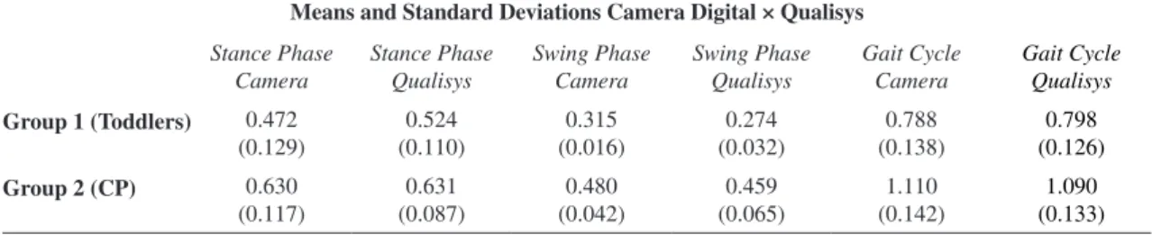
The mean and standard deviation values of each outcome variable are shown in Table 2.
Table 3 outlines the ICC values for the duration of the stance and swing phases of gait, the ICC values for the duration of the entire gait cycle, as well as the SEMs for each variable and instrument. The ICC reference values used in this study were proposed by Bartko38, where ICC magnitudes (1) smaller than
0.40 indicate poor to fair agreement; (2) from 0.41 to 0.60 indicate moderate agreement; (3) from 0.61 to 0.80 good agreement; and (4) from 0.81 to 1.00 indicate excellent agreement. Group 1 (toddlers) showed excellent agreement for the durations of the stance phase and the entire gait cycle and poor to fair agreement for the duration of the swing phase. Regarding group 2 (CP), the agreement was excellent
Table 1. Inter-examiner reliability for the duration of the temporal gait parameters in the analysis of videos obtained by a digital camera and by the Qualisys Pro-Relex system.
Inter-Examiner Reliability (ICC) Digital Camera × Qualisys
Stance Phase Camera
Stance Phase Qualisys
Swing Phase Camera
Swing Phase Qualisys
Gait Cycle Camera
Gait Cycle Qualisys
0.99* 0.99* 0.59** 0.88* 1.00* 1.00*
for all three variables. The magnitude of most of the ICCs shows excellent agreement between the measurements performed using the digital camera and the Qualisys system, except for the swing phase in the group of toddlers, which exhibited fair agreement.
Regarding the SEMs, values lower than 0.02 seconds were found for all outcome variables for both measuring instruments.
Discussion
In this study, excellent agreement (ICC>0.90) was observed between most temporal gait variables obtained using the digital camera and the Qualisys system for both toddlers with normal development and hemiplegic children. The results suggest that the digital camera is a valid instrument for the clinical quantiication of at least two temporal gait parameters in children with and without disabilities.
Speciically, in the group of toddlers, the coeficient was found to be weak (ICC=0.35) for the duration of the swing phase. This low correlation between the measurement of the duration of the swing phase assessed by clinical (digital camera) and laboratory (Qualisys system) methods may be explained if we consider the speciicities of the gait acquisition period. The duration of the swing phase is an indicator of stability. The lower stability of toddlers during the
gait acquisition period is indicated by their irregular and shorter steps, indexed by a shorter and more variable duration of the swing phase of gait2,20. The
digital camera video technology used in this study, which enables the delimitation of only 30 frames per second, most likely hindered the identiication of gait event times required for calculating the relatively shorter and more variable swing phase of toddlers. This phenomenon did not occur with the data from the Qualisys system, which yields 120 frames per second. Analyses using a digital video camera may be problematic in deining events of short duration because the temporal accuracy of this method is lower. The same rationale can be attributed to the lower inter-rater reliability achieved for the duration of the swing phase in the digital camera footage (ICC=0.59).
Other authors have used the same instruments as those used in this study, albeit for different purposes. Araújo et al.1 developed an observational scale to
assess the kinematic parameters representative of gait changes in spastic hemiplegic and diplegic children and tested its validity by comparing the results found using video analysis with those found with the
Qualisys system. The scale addressed the ankle/foot, knee, hip and pelvic joints. A very good correlation was found for the knee (r=0.64, p<0.05), and a good correlation was observed for the ankle/foot complex (r=0.59, p<0.05). The other items showed Table 2. The group mean and standard deviation of the duration of temporal gait parameters.
Means and Standard Deviations Camera Digital × Qualisys
Stance Phase Camera
Stance Phase Qualisys
Swing Phase Camera
Swing Phase Qualisys
Gait Cycle Camera
Gait Cycle Qualisys
Group 1 (Toddlers) 0.472
(0.129) (0.110)0.524 (0.016)0.315 (0.032)0.274 (0.138)0.788 (0.126)0.798
Group 2 (CP) 0.630
(0.117)
0.631 (0.087)
0.480 (0.042)
0.459 (0.065)
1.110 (0.142)
1.090 (0.133)
The numbers indicate mean and (standard deviation), in seconds. CP=Cerebral palsy.
Table 3. The Intraclass Correlation Coeficients (ICC) and standard errors of measurement (SEM) for temporal gait parameters obtained by the digital camera and Qualisys Pro-Relex system.
Stance Phase Duration Swing Phase Duration Gait Cycle Duration
Group 1 (Toddlers) 0.93* 0.35** 0.99*
Group 2 (CP) 0.93* 0.91* 0.96*
SEM† Digital
Camera
Qualisys System
Digital Camera
Qualisys System
Digital Camera
Qualisys System
0.02 0.01 0.01 0.01 0.01 0.01
a poor to fair correlation or no correlation with the measurements obtained using the Qualisys system.
Similarly, Dickens and Smith7 analyzed the
validity of a visual gait scale in children with spastic hemiplegia by comparing video analysis with three-dimensional analysis. The results showed a disagreement between both methods of analysis for the four kinematic parameters studied. The agreement for the position of the hip and knee in the swing phase was poor (kappa=-0.11 to 0.07) but ranged from fair to moderate (kappa=0.21 to 0.51) in the stance phase.
The agreement of the data analyzed using the digital camera recording and three-dimensional motion analysis systems of the abovementioned studies was not as high as in the present study. The differences in the magnitude of the coeficients may be explained by the different characteristics of the parameters analyzed. Whereas Araújo et al.1 and
Dickens and Smith7 compared visual and
three-dimensional analyses of the kinematics of individual joints, the present study used temporal gait variables, which were assessed based on the analysis of two-dimensional movements. Generally, the values of the kinematic parameters of individual joints do not show good agreement between both methods because these variables are more speciic, complex and require more sophisticated calculations. In contrast, the temporal parameters of the present study are more global variables, and simpler to assess, which may explain the good agreement found between the analyses by video recordings using the digital camera and using the Qualisys system.
The SEM values were equal to or less than 0.02 seconds for measures of duration of stance phase, swing phase and entire gait cycle obtained using both clinical and laboratory analyses. The SEM enables us to quantify the extent to which an instrument or test provides accurate measurements: low SEM values indicate a high level of accuracy and vice versa. In particular, the results found suggest that we can expect a maximum error of ±0.02 seconds when estimating the duration of the gait cycle and the stance and swing phases in both the clinical and laboratory methods. For example, we found a value of 0.47 seconds for the duration of the stance phase of toddlers using the digital camera. We may state with 95% conidence that the true average duration of that phase ranges between 0.45 and 0.49 seconds, indicating a relative error of only approximately 5% of the value found because the SEM of that measurement was 0.02 seconds. The same reasoning applies to the other measures. In summary, the SEM values found in this study suggest that measurements performed either
using a digital camera or the Qualisys system are stable and minimally error-prone.
A study limitation should be noted. Although our results support the use of a digital camera for gait evaluation of children in the clinical context, in this study, this instrument was used in a laboratory setting. This allowed the implementation of multiple procedures to ensure its validity, including training the examiners, having always the same examiner place the markers, and standardizing the data collection setting. The agreement values and SEMs may be less satisfactory in the clinical setting, where implementing the aforementioned procedures may not always be feasible. It is possible that obtaining valid measures of temporal parameters of gait with a digital camera depends on adaptation and standardization of the procedures used during gait recording.
The results of this study show that video analysis by a digital camera recording is a valid procedure for assessing the general phases of gait in normally developing toddlers and in hemiplegic children and, therefore, it is suitable for clinical use. The temporal parameters evaluated in this study would be dificult to measure and identify without some type of video recording resource. The digital camera enables frame-by-frame and slow motion visualization, allowing the footage to be rewound so that the same event can be watched multiple times. Rehabilitation professionals may responsibly use this method in therapeutic evaluation and follow up, after considering the rate of error in assessing short-duration phases.
Acknowledgments
References
1. Araújo PA, Kirkwood RN, Figueiredo EM. Validade
e confiabilidade intra e interexaminadores da Escala
Observacional da Marcha para crianças com paralisia
cerebral espástica. Rev Bras Fisioter. 2009;13(3):267-73. http://dx.doi.org/10.1590/S1413-35552009005000033 2. Dini PD, David AC. Repeatability of spatiotemporal gait
parameters: comparison between normal children and children with hemiplegic spastic cerebral palsy. Rev Bras
Fisioter. 2009;13(3):215-22. http://dx.doi.org/10.1590/ S1413-35552009005000031
3. Harris GF, Wertsch JJ. Procedures for gait analysis. Arch
Phys Med Rehabil. 1994;75(2):216-25. PMid:8311681. 4. Tanaka MS, Luppi A, Morya E, Fávero FM, Fontes SV,
Oliveira ASB. Principais instrumentos para a análise da
marcha de pacientes com distroia muscular de Duchenne. Rev Neurocienc. 2007;15(2):153-9.
5. Wren TAL, Rethlefsen SA, Healy BS, Do KP, Dennis SW, Kay RM. Reliability and validity of visual assessments
of gait using a modiied physician rating scale for crouch and foot contact. J Pediatr Orthop. 2005;25(5):646-50. PMid:16199948. http://dx.doi.org/10.1097/01. mph.0000165139.68615.e4
6. Curry VCR, Mancini MC, Melo AP, Fonseca ST, Sampaio RF, Tirado MGA. Efeitos do uso de órtese na mobilidade funcional de crianças com paralisia cerebral. Rev Bras
Fisioter. 2006;10(1):67-74. http://dx.doi.org/10.1590/ S1413-35552006000100009
7. Dickens WE, Smith MF. Validation of visual gait assessment scale for children with hemiplegic cerebral
palsy. Gait Posture. 2006;23(1):78-82 PMid:16311198. http://dx.doi.org/10.1016/j.gaitpost.2004.12.002 8. Damiano DL, Abel MF. Relation of gait analysis to
gross motor function in cerebral palsy. Dev Med Child
Neurol. 1996;38(5):389-96. PMid:8698147. http://dx.doi. org/10.1111/j.1469-8749.1996.tb15097.x
9. Provost B, Dieruf K, Burtner PA, Phillips JP,
Bernitsky-Beddingield A, Sullivan KJ, et al. Endurance and gait
in children with cerebral palsy after intensive body weight-supported treadmill training. Pediatr Phys
Ther. 2007;19:2-10. PMid:17304092. http://dx.doi. org/10.1097/01.pep.0000249418.25913.a3
10. Oeffinger D, Gorton G, Bagley A, Nicholson D, Barnes D, Calmes J, et al. Outcome assessments in children with cerebral palsy, Part I: descriptive characteristics of GMFCS Levels I to III. Dev Med Child
Neurol. 2007;49:172-180. PMid:17355472. http://dx.doi. org/10.1111/j.1469-8749.2007.00172.x
11. Delalić A, Kapidžić-Duraković S, Tahirović H. Assessment
of motor function score according to the GMFM-88
in children with cerebral palsy after postoperative
rehabilitation. Acta Med Acad. 2010;39:21-29.
12. Galli M, Cimolin V, Rigoldi C, Tenore N, Albertini G. Gait patterns in hemiplegic children with Cerebral Palsy:Comparison of right and left hemiplegia. Res Dev
Disabil. 2010;31(6):1340-5. PMid:20674265. http://
dx.doi.org/10.1016/j.ridd.2010.07.007
13. Zipp GP, Winning S. Effects of Constraint-Induced Movement Therapy on Gait, Balance and, Functional
Locomotor Mobility. Pediatr Phys Ther. 2012; 24(1):64-8. PMid:22207472. http://dx.doi.org/10.1097/ PEP.0b013e31823e0245
14. Öberg T, Karsznia A, Öberg K. Basic gait parameters:
reference data for normal subjects, 10-79 years of age. J Rehabil Res Dev. 1993;30(2):210-23. PMid:8035350. 15. Wall JC, Scarbrough J. Use of a multimemory stopwatch
to measure the temporal gait parameters. J Orthop Sports
Phys Ther. 1997;25(4):277-81. PMid:9083947.
16. Sutherland DH, Olshen R, Cooper L, Woo SL. The development of mature gait. J Bone Joint
Surg. 1980;62:336-53. PMid:7364807.
17. Chung TM. Avaliação cinética e cinemática da marcha de
adultos do sexo masculino. Acta Fisiátrica. 2000;7(2):61-7. 18. Russell S, Bennett B, Sheth P, Abel M. The gait of
children with and without cerebral palsy: work, energy,
and angular momentum. J Appl Biomech. 2011;27:99-107. PMid:21576717.
19. Lianza S, Gomes C, Consenso Nacional sobre Espasticidade. Diretrizes para diagnósticos e tratamentos. São Paulo: Sociedade Brasileira de Medicina Física e
Reabilitação; 2001.
20. Adolph KE. Motor/physical development: locomotion. In: Haith MM, Benson JB, editors. Encyclopedia of infant and early childhood development. San Diego: Academic
Press; 2008. p. 359-73. http://dx.doi.org/10.1016/ B978-012370877-9.00104-3
21. A d o l p h K , V e r e i j k e n B , S h r o u t P E . W h a t Changes in Infant Walking and Why. Child
Dev. 2003;74(2):475-497. PMid:12705568. http://dx.doi. org/10.1111/1467-8624.7402011
22. Ivanenko YP, Dominicini N, Lacquaniti F. Development
of Independent Walking in Toddlers. Exerc Sport Sci Rev. 2007;35(2):67-73. PMid:17417053. http://dx.doi. org/10.1249/JES.0b013e31803eafa8
23. Kirkwood RN, Franco RLLD, Furtado SC, Barela AMF, Deluzio KJ, Mancini MC. Frontal plane motion of the pélvis and hip during gait stance discriminates children with diplegia levels I and
II of the GMFCS. ISRN Pediatr. 2012;2012:1-10. PMid:22792478 PMCid:PMC3389695. http://dx.doi. org/10.5402/2012/163039
24. Norlin R, Odenrick P. Development of gait in spastic children
with cerebral palsy. J Pediatr Orthop. 1986;6(6):674-80. http://dx.doi.org/10.1097/01241398-198611000-00006 25. Wang X, Wang Y. Gait analysis of children with
spastic hemiplegic cerebral palsy. Neural Regen
Res. 2012;7(20):1578-1584.
26. O’Malley JM, Abel MF, Damiano LD. Fuzzy clustering of children with cerebral palsy based on temporal-distance
gait parameters. IEEE Trans Rehabil Eng. 1997;5(4):300-9. PMid:9422455. http://dx.doi.org/10.1109/86.650282 27. Hreljac A, Marshall RN. Algorithms to determine event
timing during normal walking using kinematic data. J
Biomech. 2000;33(6):783-6. http://dx.doi.org/10.1016/ S0021-9290(00)00014-2
Gait Posture. 2007;25(3):469-74. PMid:16876414. http://
dx.doi.org/10.1016/j.gaitpost.2006.05.016
29. Zeni JA Jr, Richards JG, Higginson JS. Two simple methods for determining gait events during treadmill and overground walking using kinematic data.
Gait Posture. 2008;27(4):710-4. PMid:17723303 PMCid:PMC2384115. http://dx.doi.org/10.1016/j. gaitpost.2007.07.007
30. Catalfamo P, Moser D, Ghoussayni S, Ewins D. Detection of gait events using F-Scan in-shoe pressure measurement
system. Gait Posture. 2008;28(3):420-26. PMid:18468441. http://dx.doi.org/10.1016/j.gaitpost.2008.01.019 31. Desailly E, Daniel Y, Sardain P, Lacouture P. Foot
contact event detection using kinematic data in cerebral palsy children and normal adults gait. Gait
Posture. 2009;29(1):76-80. PMid:18676147. http://dx.doi. org/10.1016/j.gaitpost.2008.06.009
32. Brown CR, Hillman SJ, Richardson AM, Herman JL, Robb JE. Reliability and validity of the Visual Gait Assessment Scale for children with hemiplegic cerebral palsy when
used by experienced and inexperienced observers. Gait Posture. 2008;27(4):648-52. PMid:17913500. http://
dx.doi.org/10.1016/j.gaitpost.2007.08.008
33. Wren TAL, Do KP, Hara R, Dorey FJ, Kay RM, Otsuka
NY. Gillette Gait Index as a gait analysis summary measure:
comparison with qualitative visual assessments of overall
gait. J Pediatr Orthop. 2007;27(7):765-8. PMid:17878782. http://dx.doi.org/10.1097/BPO.0b013e3181558ade
34. McGinley JL. The reliability of three-dimensional kinematic gait measurements: A systematic review. Gait
Posture. 2009;29(3):360-9. PMid:19013070. http://dx.doi. org/10.1016/j.gaitpost.2008.09.003
35. Piper MC, Darrah J. Motor assessment of the developing infant. Philadelphia: W.B.Saunders Company; 1994. 36. Palisano RJ, Rosenbaum P, Bartlett DJ, Livingston MH.
Content validity of the expanded and revised Gross Motor Function Classiication System. Dev Med Child Neurol. 2008;50(10):744-50. PMid:18834387. http://
dx.doi.org/10.1111/j.1469-8749.2008.03089.x
37. Qualisys Medical AB. Qualisys Track Manager User
Manual. Gothenburg; 2004.
38. Bartko JJ. The Intraclass Correlation Coefficient as
a measure of reliability. Psychol Rep. 1966;19:3-11. PMid:5942109. http://dx.doi.org/10.2466/pr0.1966.19.1.3
Correspondence
Marisa C. Mancini
Escola de Educação Física, Fisioterapia e Terapia Ocupacional Colegiado de Pós-graduação em Ciências da Reabilitação
Av. Antônio Carlos, 6627, Campus Pampulha CEP 31270-901, Belo Horizonte, MG, Brasil