Cephalometric study of
alterations induced by
maxillary slow expansion in
adults
Summary
Almiro José Machado Júnior1, Agrício NubiatoCrespo2
1 Dental surgeon specialized in maxillary functional orthopedics, MSc, Associate Professor at the Systemic Dentistry Society of São Paulo. 2 PhD in Medical Sciences, Head of the Ophthalmology-Otolaryngology Department at the Medical School of the Campinas State University - Unicamp.
Paper submitted to the ABORL-CCF SGP (Management Publications System) on April 9th, 2005 and accepted for publication on February 20th, 2006.
M
axilla expansion is a procedure that aims at increasing the maxillary dental arch to correct occlusal disharmony. Largely used in children, its efficacy in adults, when craniofacial growth has attained bone maturity, is controversial. Aim: The present study has the objective of evaluating cephalometric modifications resulting from maxilla expansion in adult patients, observing the following linear measurements: facial width, nasal width, nasal height, maxillary width, mandibular width and maxillary molar width. Material and methods: The sample was composed of 24 frontal teleradiographs, taken before and immediately after the expansions, from 12 male and female patients aged between 18 years and two months and 37 years and eight months. All patients were submitted to slow expansion of the maxillary bones by means of an appliance used in the technique named “dynamic and functional maxillary rehabilitation”. Wilcoxon paired statistical test was used for related samples with a 5% significance level. Results: There was a mean increase of 1.92 mm in nasal width and 2.5 mm in nasal height. As regards the linear measurements maxillary and mandibular width, the mean increase was 2.42 mm and 1.92 mm, respectively. A mean increase of 1.41 mm was found for facial width and 2.0 mm for maxillary molar width, alterations which were statistically significant, the mean time was 5.3 months. Conclusion: Based on the results obtained, it may be concluded that the use of maxillary expansion induces increase of the facial measurements studied in adults.Key words: Maxilla, Expansion, Cephalometry ORIGINAL ARTICLE
INTRODUCTION
At the beginning of the digestive process, mastica-tion grinds, humidifies and decreases the size of food particles, producing the food bolus which is then swal-lowed, ending the oral phase of digestion. This phase, represented by mastication and swallowing, is influenced by dental occlusion and plays a relevant role in human physiological equilibrium.
Dental occlusion is the physical relation between the dental and functional elements of the masticatory system components: superior and inferior dental arches, maxilla, jaw, hyoid bone, tongue, lips, cheek and muscles. It has a direct influence over mastication and swallowing, and indirect impact over respiration and speech1-3.
Odontology seeks the maintenance of this occlusal equilibrium, prevention and interception of deviations from normality of the stomatognatic system which may occur during an individual’s growth and development. When this equilibrium is not reached, deviations from the occlusal equilibrium set in, resulting in physical alterations named malocclusions.
Malocclusions are caused by hereditary and by ex-trinsic factors. If little can be done to avoid the hereditary factors, a lot can be done to prevent and treat the extrinsic factors. Among the maneuvers used in the treatment of malocclusions caused by extrinsic factors such as posterior cross bite, maxillary atresia and dental overlapping, maxil-lary expansion obtained through orthodontic, mechanical orthopedics, functional orthopedics, surgery and a combi-nation of all these resources can be used3,4.
Employed during growth, the maxilla expansion has been explained by the separation of the palate center and dental orientation associated with the augmentation of facial structures. There is controversy over the efficacy of maxilla expansion in adults, as soon as craniofacial growth has reached its bone maturity4-8.
Thus it is not well understood if there is maxillary expansion in adults and its local, regional effects as well as the way in which the growth of the superior dental arch occurs, if as a result of dental orientation or as an effect of palate center separation, or still bone structures growth adjacent to the maxilla.
The present study aims at evaluating possible ce-phalometric modifications caused by maxilla expansion in adult patients, observing the following linear measures: facial width, nasal width, nasal height, maxillary width, jaw width and molar-maxillary region width.
MATERIALS AND METHODS
The sample used in clinical, longitudinal, cohort prospective study was obtained at the dental clinic of the specialization course in orthodontics and functional maxil-lary orthopedics of the Society of Systemic Odontology of
the State of São Paulo.
A total of 12 patients, 11 females and 1 male were selected for sampling the patients that fit the following requirements: over 18 years of age, with maxillary atresia, uni or bilateral cross bite, dental line irregularity, and dental overlapping with maxillary atresia. Patients who had suffered head trauma, with periodontal disease and without the first superior molar were excluded from the sampling.
To expand the maxillary bone we used the tech-nique called “Dynamic and Functional Rehabilitation of the Maxilla” advocated by Vaz de Lima. We used a slow activation device for bilateral expansion of the maxillary bones9.
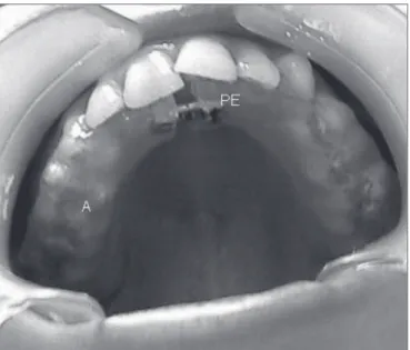
The dental piece for maxillary expansion has ex-panding screws in the palate center projection, wrapped in chemically activated acrylic, covering the whole exten-sion of the hard palate, palate surfaces, and occlusal third of the buccal surfaces of clinical crowns on the posterior teeth (Figure 1).
In placing the dental piece, we verified how the acrylic fits over the hard palate mucosa and posterior teeth surfaces. We asked the patient to remove and replace the dental piece many times, observing any difficulties to remove it, and instructing them to:
1. Use the dental piece constantly, even while sleep-ing, only removing it for eating and cleaning;
2. Opening the expansion screw: the patient acti-vates the dental piece, rotating ¼ towards the arrow indi-cating which way it should be done, every other day;
3. Hygiene: the dental piece should be cleaned periodically;
4. Maintenance: every fortnight the patient should return to check dental piece fit, presence of lesions or ulcers, and to make adjustments and polishing when necessary.
How long the patient should use the dental piece for maxilla expansion is yet to be determined. The end of the expansion was established by clinical criteria alone, when we confirmed the correction proposed for each case.
With the goal of evaluating possible cephalometric modifications, all patients were submitted to teleradio-graphic examinations before the beginning and the end of the treatment described above, with one only equipment, Siemens, Nanomobil model, regulated for exposures of 65 KVp, 10 mA, for 1.5 sec and focal distance of 1.52 meters. We used 18 x 24 cm Kodak x-omat XK1 films.
his/hers lips relaxed and in habitual occlusion10.
After obtaining all the teleradiography images prior and posterior to treatment we took the measurement of interest for the study. Over each teleradiography image we placed an acetate paper, attached with adhesive tape. Using a negatoscope in a dark room, we delimited the anatomic structures of interest to create the cephalogram: nostril opening, outer skull border, first superior right molar, zygomatic arches, jaw, tuberosity of the maxilla, nasal crest and anterior nasal spine.
The following linear measures were taken (Figure 2):
1. Facial width: distance between the bilateral points marked at the outermost tip of the zygomatic arches;
2. Nasal height: distance between the top of the nasal spine and the top of the nasal crest;
3. Nasal width: distance between the outermost external points in the nostril opening;
4. Maxillary width: distance between the two out-ermost points of the maxillary tuberosities;
5. Jaw width: distance between jaw angles; 6. Molar-maxillary width: distance between the lateralmost point of the first superior right molar crown to the intersection line between the jaw angle, right side, and maxillary tuberosity on, right side.
The observer who did all the measurements of the various distances was previously tested and calibrated, and did not know which patient it belonged to nor if the teleradiography was previous or posterior to the maxil-lary expansion, in order to avoid biasses. With the goal of minimizing the systematic error and establishing an intra-examiner agreement, each cephalometric measurement was done twice at a 10 day interval, obtaining a level of agreement close to 96%.
In order to compare initial and final measurements, we used the paired non-parametric test of Wilcoxon (for related samples). The level of significance adopted was of 5%. Descriptive statistics of the initial and final meas-urements (mm), p-value of the paired Wilcoxon are in corresponding tables.
To the patients we presented a signed consent form and the protocol for this research was previously approved by the Ethics Committee in Research of the Unicamp School of Medicine.
RESULTS
The results are presented in tables showing aver-ages, standard deviations, maximum values, medians and minimal values found for each cephalometric magnitude, time of expansion and age of patients.
Patient age varied between 18 years and two months of age and 37 years and eight months of age, with an average of 24.7, and median of 27 years of age. (Table 1). The time necessary to attain maxillary expansion was
Figure 1. Mouth piece used for maxillary expansion. A= acrylic cover; PE= expander screw.
Figure 2. Cephalometric exam by frontal teleradiography.
Cg-ENA=nasal height; ZY=facial width; Ma=maxillary width; NL=nasal width; GO=jaw width;
of 5.3 months in average, with a standard deviation of 2.57 months, minimum of 3 and maximum of 11 months (Table 2).
Tables 3 through 8 show the measurements evalu-ated before and after the maxillary expansion shown with their averages, Standard deviations, median, minimum and maximum values. All measurements taken show a significant increase after maxillary expansion of around 2.0 mm. The lower increase was observed in facial width, which was 1.41 mm in average (Table 3), and the high-est increases observed were on nasal height (2.50 mm
Table 1. Age of patients, in years, subject to maxillary expansion. N AVERAGE ST. DEV. MAX MEDIAN MIN 12 27.43 5.41 37.08 26.98 18.25 N: number of patients; St. Dev: Standard deviation; MAX: maximum age; MIN: minimum age
Table 2. Time for maxillary expansion, in months.
N AVERAGE ST. DEV. MAX MEDIAN MIN
12 5.33 2.57 11 5 3
N: number of patients; St. Dev: Standard deviation; MAX: maximum time; MIN: minimum time
Table 3. Cephalometric values of the face width obtained before and after maxillary expansion, in mm.
FACIAL WIDTH
N AVERAGE ST. DEV. MAX MEDIAN MIN INITIAL 12 128.67 7.41 144 129 115 FINAL 12 130.08 7.50 146 130 119 N: number of patients; St. Dev: Standard deviation; MAX: maximum value; MIN: minimum value
.Wilcoxon test:p-value=0.0078
Table 4. Cephalometric values of nasal width obtained before and after maxillary expansion, in mm.
NASAL WIDTH
N AVERAGE ST. DEV. MAX MEDIAN MIN INITIAL 12 30.00 2.52 34 29.0 26 FINAL 12 31.92 2.50 35 31.5 26 N: number of patients; St. Dev: Standard deviation; MAX: maximum value; MIN: minimum value
Wilcoxon test :p-value=0.0010
Table 5. Cephalometric values of nasal height obtained before and after maxillary expansion, in mm.
NASAL HEIGHT
N AVERAGE ST. DEV. MAX MEDIAN MIN INITIAL 12 59.83 4.53 67 59.0 54 FINAL 12 62.33 4.66 69 62.5 56 N: number of patients; St. Dev: Standard deviation; MAX: maximum value; MIN: minimum value
.Wilcoxon test :p-value =0.0020
Table 6. Cephalometric values of maxillary width obtained before and after maxillary expansion, in mm.
MAXILLARY WIDTH
N AVERAGE ST. DEV. MAX MEDIAN MIN INITIAL 12 65.25 3.70 70 66.5 57 FINAL 12 67.67 4.16 74 67.5 59 N: number of patients; St. Dev: Standard deviation; MAX: maximum value; MIN: minimum value
.Wilcoxon test :p-value =0.0034
in average) and maxillary width (2.42 mm in average) (Tables 5 and 6).
DISCUSSION
Maxillary expansion has been used to treat maloc-clusion growth in patients of various ages6,11-18.
Studies state that the post-natal growth reaches its peak in middle adolescence and is dramatically reduced at the end of this period. The average age for growth de-crease is around 14 years of age for women, and 16 years
Table 7. Cephalometric values of jaw width obtained before and after maxillary expansion, in mm.
JAW WIDTH
N AVERAGE ST. DEV. MAX MEDIAN MIN INITIAL 12 84.08 4.48 92 83.5 77 FINAL 12 86.00 4.68 95 86.5 79 N: number of patients; St. Dev: Standard deviation; MAX: maximum value; MIN: minimum value
.Wilcoxon test :p-value =0.0010
Table 8. Cephalometric values of molar-maxillary width obtained before and after maxillary expansion, in mm.
MOLAR-MAXILLARY WIDTH
N AVERAGE ST. DEV. MAX MEDIAN MIN INITIAL 12 5.08 1.62 8 5.0 3 FINAL 12 7.08 1.78 9 7.5 5 N: number of patients; St. Dev: Standard deviation; MAX: maximum value; MIN: minimum value
of age for men19-23.
Thus, using the biological concept defined for adult patients, we included in this study patients above 18 years of age, subject to slow maxillary expansion.
In researching the pertinent literature, no mention over the cephalometric observations caused by maxillary expansion in adult patients was found, such fact led to this study.
Cephalometry by frontal teleradiography, because of its ease of use and availability, has been used in anatomic studies and in diagnosing malocclusion10.
It has also been used in the observation of altera-tions caused by induced maxillary expansion8,24-33.
There is controversy over the types of mouth pieces used for rapid maxillary expansion. Various types of dia-grams are presented in the literature, but all are constituted basically of an expander screw placed transversally to the palate, differing only on the anchor type used26,33-36.
Some researchers support the use of an acrylic resin, covering the hard palate, offering the mouth piece a tooth-mucosa-supported anchor, to provide better sup-port, favoring the expansion and contention, mainly for the bone37-39.
For other authors, the mucosal support makes it harder to clean the resin-mucosa interface, and it also causes ulcers and eritematous lesions in the palate mucosa due to the contact and the pressure from the support, and these authors end up choosing a tooth-supported mouth piece34.
To attain slow maxillary expansion, we used a tooth-mucosa-supported mouth piece, expecting that the expansion forces would act not only over the poste-rior teeth, but also and mainly, over the maxillary bone structures37-40.
In this study we used the mouth piece for the slow maxillary expansion, with activation every other day, be-lieving that better results are obtained when the activation, and consequently, the liberation of the expansionary forces are applied intermittently over the maxillary bones4,39-41.
Even though we did not aim at observing the maintenance of linear measures in the long term after maxillary expansion, we believe the recurrence index, if any, would be lower in the cases where rapid maxillary expansion is used. Still, further studies are necessary to test this hypothesis.
Applied in children, maxillary expansion is contro-versial in adults, as the craniofacial growth has reached its bony maturity4,6-8,41.
As to the mechanism of maxillary expansion in adult patients, our results do not support the hypothesis that maxillary expansion occurred due to dental tilting, as suggested in the literature28,42-44.
If this hypothesis were right there should have been a decrease in molar-maxillary distance, which was not
observed in our cases where, on the contrary, there was an average increase of 2.0 mm (Table 8).
Some who use maxillary expansion attribute it to the separation of the palate center40,42,44-46.
With the methodology used in this study, we did not observe the behavior of palate suture, thus our find-ings do not allow us to define if the expansion occurred due to palate center separation.
On the other hand, the found average growth of 1.41 mm on facial width (table 3) and 1.92 mm on jaw width (table 7), measurements limited to the maxilla, have led us to suppose that if there is separation of palate center, it is not the only factor leading to maxillary expansion.
Despite the reduced sample size, the results of this study show that the use of maxillary expanders in adults allows for statistically significant expansion, observed by the average increase in the linear measures of facial width (1.41 mm), nasal width (1.92 mm), nasal height (2.5 mm), maxillary width (2.42 mm), jaw width (1.92 mm) and mo-lar-maxillary width (2.0 mm) (tables 3 through 8), in an average time frame of 5 months (table 2).
For the stomatognatic system the expansion move-ments might be considered fewer than two aspects: in-duced by mouth pieces which use mechanical forces and mouth pieces which use functional forces. The mechanical movements are a result of forces applied over the teeth and transmitted to the bones, aiming at changing growth direction. The functional movements use natural forces originated from the muscular movement, acting over the velocity and direction of growth and bone remodeling2,4.
The maxillary expansion mouth pieces used in this study produce mechanical forces, but their effects pro-duced by expansion such as increase in the mouth cavity, observed on the maxillary width and jaw width, allowing for extra space for the tongue functionality (swallowing, mastication, speech) and increase in the nasal measure-ments, nasal width and height, allowing for better anatomic functional improvements or nasal respiration, associated with the occlusive alteration, caused by the acrylic cover over the posterior teeth, showed factors which might fit the characteristics of functional movements, meaning, that maxillary expanders produce mechanical movements, al-lowing subsequent or concomitant functional movements, caused by the increase in mouth cavity size. Still, other studies are necessary to evaluate probable alterations in functionality (swallowing, mastication, and breathing) of the stomatognatic system after maxillary expansion.
the superior dental arch, bringing indisputable advantages in mechanotherapy for maxillary deficiencies.
The results found show the real possibility of max-illary expansion in adult patients, not being restricted to dental orientation or separation of palate center, but also playing a relevant role in the enlargement of facial struc-tureso by maxillary induced expansion.
CONCLUSIONS
The results obtained in this study allow us to con-clude:
• There is maxillary expansion in adults.
• Maxillary expansion leads to an increase in facial, nasal, maxillary, jaw and molar-maxillary widths, and nasal height.
REFERENCES
1. Moyers RE. Ortodontia. 4 ed. Rio de Janeiro: Ed. Guanabara Koogan; 1991. 483p.
2. Proffit WR. Ortodontia contemporânea. 2 ed. Rio de Janeiro: Ed. Guanabara Koogan; 1995. 596p.
3. Graber TM. Ortodontia - princípios e técnicas atuais. 2 ed. Rio de Janeiro: Ed. Guanabara Koogan; 1996. 897p.
4. Langlade M. Otimização transversal das oclusões cruzadas unilaterais posteriores. 1ª ed. São Paulo: Ed. Santos; 1998. 384 p.
5. Wertz RA. Skeletal and dental changes accompanying rapid midpalatal suture opening. Amer J Orthodont 1970;58(1):41-66.
6. Silva Filho OG, Valadares Neto J, Almeida RR. Early correction of posterior crossbite: Biomechanical characteristics of the appliances. J Pedod 1989;13(3):195-221.
7. Saadia M, Torres E. Sagittal changes after maxillary protraction with expansion in class III patients in the primary, mixed, and late mixed dentitions: a longitudinal retrospective study. Am J Orthod Dentofacial Orthop 2000;117(6):669-80.
8. Cross DL, McDonald JP. Effect of rapid maxillary expansion on skeletal, dental and nasal structures: a postero-anterior cephalometric study. Eur J Ortho 2000;22(5):519-28.
9. Vaz De Lima M, Soliva H. Reabilitação dinâmica e funcional dos maxilares sem extração. 3ª ed. Rio de Janeiro: Pedro Primeiro; 1999. 176p.
10. Ricketts RM. Cephalometric synthesis. Am J Orthod 1960;46:647. 11. Badcock JH. The screw expansion plate. Dent Rec 1911;31:588-90. 12. Krebs A. Expansion of the midpalatal suture, studied by means of
metallic implants. Acta Odont Scand 1959;92(5):491-501.
13. Hass AJ. Rapid expansion of the maxillary dental arch and nasal cavity by opening the midpalatal suture. Angle Othodont 1961;31(2):73-90.
14. Hass AJ. Palatal expansion: Just the bigining of the dentofacial or-thopedics. Amer J Orthodont 1965;35(3):200-17.
15. Gray LP. Rsults of 310 cases of rapid maxillary expansion selected for medical reasons. J Laryngol Otolaryngol 1975;89(6):601-9.
16. Sato K. Avaliação cefalométrica da disjunção palatina mediana, através da telerradiografia em norma frontal. Rev Odont Metodista 1985;6(1):123-36.
17. Rizzato SD, Costa NP, Marchioro EM, Saffer M. Avaliação do efeito da expansão rápida da maxila na resistência nasal por rinomanometria ativa anterior em crianças. Ortodontia Gaúcha 1998;2(2):79-93. 18. Paiva JB. Estudo rinomanométrico e nasofibroendoscópico da
cavidade nasal dos pacientes submetidos à expansão rápida da maxila. São Paulo, 1999. (Tese de Doutorado - Universidade de São Paulo).
19. Andrew R. The anatomy of aging in man and animals. New York:
Grune e Stratton; 1971.
20. Finch CE, Hayflick L. Handbook of the biology of aging. New York: Van Nostrand Reinhold; 1977.
21. Sinclair D. Human growth after birth. 3rd Ed. New York: Oxford University Press; 1978.
22. Kohn RR. Principles of mammalian aging. 2nd ed. Englewood Cliffs: Prentice-Hall; 1978.
23. Enlow DH. Crescimento facial. 3ª ed. São Paulo: Artes Médicas; 1993. 553p.
24. Linder-Aronson S, Lindgren J. The skeletal and dental effects of rapid maxillary expansion. Brit J Orthodont 1979;6(1):25-9.
25. Santos-Pinto CCM, Henriques JFC. Expansão rápida da maxila: pre-ceitos clínicos e radiográficos. Rev Odont USP 1990;4(2):164-6. 26. Mazzieiro ET. Estudo cefalométrico, em norma frontal, das alterações
dentoesqueléticas após a expansão rápida da maxila, em pacientes na faixa etária de 10 a 16 anos e 2 meses. Bauru, 1994. (Dissertação - Mestrado - Universidade de São Paulo).
27. Almeida GA, Capelozza Filho L, Trindade Junior AS. Expansão rápida da maxila: estudo cefalométrico prospectivo. Ortodontia 1999;32(1):45-56.
28. Pearson LE, Pearson BL. Rapid maxillary expansion with incisor in-trusion: a study of vertical control. Am J Orthod Dentofacial Orthop 1999;115(5):576-82.
29. Memikoglu TU, Iseri H. Effects of a bonded rapid maxillary ex-pansion appliance during orthodontic treatment. Angle Orthod 1999;69(3):251-6.
30. Trenouth MJ. Cephalometric evaluation of the Twin-block ap-pliance in the treatment of Class II Division 1 malocclusion with matched normative growth data. Am J Orthod Dentofacial Orthop 2000;117(1):54-9.
31. Garib DG, Henriques JFC, Janson GRP. Avaliação cefalométrica lon-gitudinal das alterações produzidas pela expansão rápida da maxila. Rev Det Press Ortodon Ortoped Facial 2001;6(5):17-30.
32. Ursi WJS, Dale RCXS, Claro CA, Chagas RV, Almeida G. Alterações transversais produzidas pelo aparelho de expansão maxilar com cobertura oclusal, avaliada pelas telerradiografias póstero-anteriores. Ortodontia 2001;34(3):43-55.
33. Siqueira DF, Almeida RR, Henriques JFC. Estudo comparativo, por meio de análise cefalométrica em norma frontal, dos efeitos den-toesqueléticos produzidos por três tipos de expansores palatinos. Rev Dent Press Ortodon Ortoped Facial 2002;7(6):27-47.
34. Biederman W. Rapid correction of class III maloclusion by midpalatal expansion. Amer J Orthodont 1973;63(1):47-55.
35. Bramate FS. Estudo cefalométrico em norma lateral das alterações dentoesqueléticas produzidas por três tipos de expansores: colado, tipo Haas e Hyrax. Bauru, 2000. (Dissertação - Mestrado - Universi-dade de São Paulo).
36. Silva Filho OG, Ferrari Junior FM, Aiello CA, Zoponi N. Correção da mordida cruzada posterior nas dentaduras decídua e mista. Rev Assoc Paul Cir Dent 2000;54(2):142-7.
37. Hass AJ. Palatal expansion: Just the bigining of the dentofacial or-thopedics. Amer J Orthodont 1970;57(3):219-55.
38. Hass AJ. Long-term posttreatment evolution of rapid palatal expan-sion. Angle Orthodont 1980;50(3):189-217.
39. Soliva H. Expansão superior e inferior em pacientes adultos: uma possibilidade real de tratamento. J Bras Ortodon Ortop Facial 1998;3(15):41-4.
40. Medau V. Expansor do Dr. Maurício Vaz de Lima pode fazer disjunção da sutura palatina. J Bras Orthodont Ortop Facial 2001;6(31):42-51. 41. Zimring JF, Isaacson RJ. Forces produced by rapid maxillary
ex-pansion - III. - Forces present during retention. Angle Orthodont 1965;35(3):178-86.
42. Timms DJ. A study of basal movement with rapid maxillary expan-sion. Amer J Orthodont 1968;77(5):500-7.
43. Riccioli GA. La disgiunzione rapida della sutura palatina in ortodonzia. Mondo Odontostomat 1973;15:356-7.
45. Angell ML. Treatment of irregularity of the permanent or adult teeth. Dent Cosmos 1860;1:540-4 e 599-600.