Recurrent Invasive Pneumococcal Disease in
Children: Underlying Clinical Conditions, and
Immunological and Microbiological
Characteristics
Laia Alsina1,5, Maria G. Basteiro2, Hector D. de Paz3, Melania Iñigo3, Mariona F. de Sevilla2, Miriam Triviño2, Manel Juan4,5, Carmen Muñoz-Almagro3*
1Allergy and Clinical Immunology Department, Hospital Sant Joan de Déu, University of Barcelona, Barcelona, Spain,2Department of Pediatrics, Hospital Sant Joan de Déu, University of Barcelona, Barcelona, Spain,3Department of Molecular Microbiology, Hospital Sant Joan de Déu, University of Barcelona, Barcelona, Spain,4Immunology Service, Hospital Clinic and Instituto de Investigaciones Biomédicas August Pi y Sunyer (IDIBAPS), Barcelona, Spain,5Functional Unit of Immunology, Hospital Sant Joan de Déu and Hospital Clinic. Barcelona, Spain
*cma@hsjdbcn.org
Abstract
Purpose
Clinical, immunological and microbiological characteristics of recurrent invasive pneumo-coccal disease (IPD) in children were evaluated, differentiating relapse from reinfection, in order to identify specific risk factors for both conditions.
Methods
All patients<18 years-old with recurrent IPD admitted to a tertiary-care pediatric center
from January 2004 to December 2011 were evaluated. An episode of IPD was defined as the presence of clinical findings of infection together with isolation and/or pneumococcal DNA detection by Real-Time PCR in any sterile body fluid. Recurrent IPD was defined as 2 or more episodes in the same individual at least 1 month apart. Among recurrent IPD, we differentiated relapse (same pneumococcal isolate) from reinfection.
Results
593 patients were diagnosed with IPD and 10 patients died. Among survivors, 23 episodes of recurrent IPD were identified in 10 patients (1.7%). Meningitis was the most frequent form of recurrent IPD (10 episodes/4 children) followed by recurrent empyema (8 episodes/4 chil-dren). Three patients with recurrent empyema caused by the same pneumococcal clone ST306 were considered relapses and showed high bacterial load in their first episode. In contrast, all other episodes of recurrent IPD were considered reinfections. Overall, the rate of relapse of IPD was 0.5% and the rate of reinfection 1.2%. Five out of 7 patients with rein-fection had an underlying risk factor: cerebrospinal fluid leak (n = 3), chemotherapy a11111
OPEN ACCESS
Citation:Alsina L, Basteiro MG, de Paz HD, Iñigo M, de Sevilla MF, Triviño M, et al. (2015) Recurrent Invasive Pneumococcal Disease in Children: Underlying Clinical Conditions, and Immunological and Microbiological Characteristics. PLoS ONE 10(3): e0118848. doi:10.1371/journal.pone.0118848
Academic Editor:Bernard Beall, Centers for Disease Control & Prevention, UNITED STATES
Received:August 19, 2014
Accepted:January 6, 2015
Published:March 4, 2015
Copyright:© 2015 Alsina et al. This is an open access article distributed under the terms of the Creative Commons Attribution License, which permits unrestricted use, distribution, and reproduction in any medium, provided the original author and source are credited.
Data Availability Statement:All relevant data are within the paper.
treatment (n = 1) and a homozygous mutation in MyD88 gene (n = 1). No predisposing risk factors were found in the remainder.
Conclusions
recurrent IPD in children is a rare condition associated with an identifiable risk factor in case of reinfection in almost 80% of cases. In contrast, recurrent IPD with pleuropneumonia is usually a relapse of infection.
Introduction
Streptococcus pneumoniae(pneumococcus) is a commensal organism of the human nasopharynx [1] and a major cause of morbidity and mortality worldwide. Although all age groups may be af-fected, the highest rate of pneumococcal disease occurs in young children and in the elderly [2]. In 2000, it was estimated that about 14.5 million episodes of serious pneumococcal disease and more than 800,000 deaths in children less than 5 years of age occurred [3]. In addition, pneumo-coccus is the leading cause of mild infections of the upper respiratory tract (otitis media, sinusi-tis) [1]. Despite this high burden, the occurrence of 2 or more episodes of invasive pneumococcal disease (IPD) in the same individual one month apart, called recurrent IPD [4], is much less fre-quent, with an estimated rate between 2 and 4% [4]. The event of a recurrent IPD must then pose the question of an underlying medical condition predisposing to this rare phenomenon.
The best-known acquired factors that determine susceptibility to IPD, other than young age, are co-infection by the human immunodeficiency virus (HIV), splenectomy, malignancies, chronic cardiopulmonary diseases, traumatic cerebrospinal fluid leaks and cochlear implant [5]. Inherited factors have also long been known to predispose patients to IPD, notably sickle-cell disease and primary immunodeficiency diseases (PIDs). Classically, the PIDs most related to IPD were congenital asplenia, defects in the classical pathway of complement activation and defect in the antibody response to polysaccharides [6]. All these PIDs share a common patho-physiological mechanism, which is a high level of interference with the ability of opsonization and phagocytosis of the encapsulated bacteria by the splenic macrophages. Patients suffering from these PIDs are not only prone to IPD, but also to serious infections by other encapsulated
bacteria (Streptococcusgroup B,Haemophilus influenzaetype B,Salmonella typhi,Neisseria
meningitidis). Recently a new group of PIDs was identified, affecting innate immunity and
pre-disposing to infection by Gram-positive bacteria, pneumococcus in particular [7–12]. They
in-clude anhidrotic ectodermal dysplasia with immunodeficiency [8] [of which there are two
forms, X-linked and autosomal dominant, caused by defects inNF-kappa-B essential
modula-tor(NEMO) or gain-of-function inalpha inhibitor ofNF-kappa-B(IKBA)], and the IL-1
recep-tor-associated kinase type 4 (IRAK-4) deficiency [7–12] and myeloid differentiation primary
response 88 (MyD88) deficiency [11–12], both autosomal recessive. These are monogenic
dis-eases in which there is a mutation in the NEMO gene, IRAK-4 or MyD88, respectively, whose products are crucial in the signaling pathways of Toll-like receptors (TLR) and IL-1 receptors (the Toll-IL1R or TIR pathway), the most relevant sensors of pathogens and inflammation in the innate immune system [13].
In recent years there have appeared several publications analyzing the underlying factors in IPD, but only a few have focused on patients with recurrent IPD in children [4,14,15]. Their re-sults demonstrated that underlying risk conditions were present in 80% of patients with recur-rent IPD, both children and adults, and which might not have been identified before the IPD
study design, data collection and analysis, decision to publish, or preparation of the manuscript.
recurrence [15]. Of note, most patients with recurrent IPD without underlying risk conditions were young children less than 12 months old [4].
The aim of this study was to evaluate the clinical features of a pediatric population display-ing recurrent IPD, as well as to identify the underlydisplay-ing microbiological characteristics and risk factors associated with this condition. We performed a broad immunological evaluation of the patients with no apparent underlying factors, including evaluation of the TIR pathway that has only been performed in one recent study [16].
Patients and Methods
Patients and Setting
We identified all children and adolescents with IPD who were admitted to Sant Joan de Déu Hospital in Barcelona (January 2004 to December 2011). Our hospital is located in the south-ern area of Barcelona, Catalonia, Spain, and serves a paediatric referral population of 200,000
children<18 years, around 19% of the Catalan paediatric population (data obtained from
Cat-alonian Department of Statistics,www.idescat.net). Conjugate vaccines are not subsidized by
the Spanish Health Service but the uptake of the first one available, heptavalent conjugate vac-cine (PCV7), has increased since its introduction in June 2001. It has been estimated to have provided PCV7 coverage of around 50% in the year 2009 [17]. In 2010 PCV7 was replaced by high-valent conjugate vaccines (PCV10 and PCV13).
An episode of IPD was defined as the presence of clinical findings of infection together with
isolation and/or DNA detection ofpneumolysine (ply)gene and an additional capsular gene
(wzg) ofS. pneumoniaeby Real-Time PCR in any sterile body fluid such as blood, cerebrospi-nal fluid, pleural fluid or articular fluid. A recurrent IPD was defined as 2 or more episodes of IPD in a same individual at least one month apart [17]. Among recurrent IPDs, we differentiat-ed relapse IPD if the same pneumococcal isolate was identifidifferentiat-ed (defindifferentiat-ed as 2 strains with the same clonal type and/or serotype) and reinfection IPD if it was different (defined as 2 strains with different serotype and/or clonal type).
The Clinical Microbiology Laboratory monitored all confirmed pneumococcal infections and several variables were routinely recorded including demographic data, identification hos-pital number, type of infection, antimicrobial susceptibility, serotype and clonal type or se-quence type. IPD was classified according to the International Classification of Disease Ninth
Revision (ICD-9) specific for diseases caused byS. pneumoniaeas follows: meningitis,
pneu-monia, parapneumonic empyema, occult bacteremia, sepsis, arthritis, peritonitis, and endophthalmitis. Identification number of all isolates from sterile fluids was used to review electronic medical records and recorded demographic, epidemiological and clinical variables including age, gender, date of birth, date of admission, clinical data, treatment, outcome, and pneumococcal polysaccharide-protein conjugate vaccination status. A thorough medical chart review and interview with patients with recurrent IPD and family was performed in order to identify underlying medical conditions that are known to predispose to IPD [5]. Brain magnet-ic resonance imaging (MRI) was performed in patients who experienced one or more episodes of meningitis, to rule out CSF leak.
Immunological studies
B/NK lymphocyte phenotyping, antibodies against polysaccharide antigens (isohemagglutinins and pneumococcus, post-infection), and protein antigens (diphtheria, tetanus, pneumococcus post-Prevenar vaccination); 2) evaluation of innate immunity: CH50 (assay for evaluation of classical pathway by 50% of hemolysis), C3 and C4 levels, Howell-Jolly bodies in blood smear and/or abdominal ultrasound (both to rule out asplenia), and evaluation of TIR pathway through CD62L shedding [18] and whole blood stimulation with TLR ligands and cytokine
measurement (defined in the paragraph that follows) [7–11]. In patients suffering from
pneu-monia or parapneumonic pleural effusion as the only form of recurrent IPD, with no other in-fections, the TIR pathway was not evaluated, since isolated pneumonia is not a common presentation of TIR deficiencies [18,19].
IgG, A and M and complement C3, C4 were evaluated using immunoturbidimetric specific protein methods (Architect ci8200). CH50 was determined by ELISA (Binding Site). IgG sub-classes were evaluated by nephelometric technique. Specific polysaccharide antibody deficiency was defined as normal serum IgG, A and M and IgG subclass levels and a defect of the antibody
responses toS. pneumoniae(or other polysaccharide vaccine) either after documented invasive
infection or after test immunization and/or absent isohemagglutinins above 1 year of age. Immunodeficiences were defined according criteria of ESID (European Society
Immunodefi-ciences).http://esid.org/Working-Parties/Registry/Diagnosis-criteria. IgA + IgG subclass
deficien-cies were defined in patients aged>4years with marked decrease in IgA [ie<0.05g/l] and at least
one of IgG 1–3 subclasses less than the 5th percentile for age along with poor response to vaccines
and/or negative isohemagglutinins (<1/8). IgG subclass deficiencies were defined in patients
aged>4years with normal levels of IgM and IgA at least two of IgG 1–3 subclasses less than the 5th
percentile for age along with poor response to vaccines and/or negative isohemaglutinins (<1/8).
Specific polysaccharide deficiency was defined in patients>4 years with normal levels of IgG
sub-classes and IgA, and poor polysaccharide response (negative isohemaglutinins and/or absent pneu-mococcal antibodies after natural infection or less than 2-fold increase in pneupneu-mococcal antibodies after polysaccharide vaccination with Pneumovax23). Hypogammaglobulinemia was defined as marked decrease of at least one of Ig,IgG subclass(es), IgA or IgM levels (measured at least twice), secondary causes of hypogammaglobulinaemia having been excluded, with normal isohaemaggluti-nins and/or antibody response to vaccines. Complement deficiencies required genetic confirmation of the defect after detection of low CH50 in two different determinations 6 months apart.
Evaluation of TIR pathway of whole blood (WB) stimulation with TLR
ligands through CD62L shedding and cytokine measurement [
7
–
11
]
WB was diluted with an equal amount of RPMI before adding different stimuli. Diluted WB was activated for 24 hours with 10μg/ml of polymyxin B to clear LPS contamination plus TLR1/2 ago-nist Pam3CSK4 0.5μg/ml (Invivogen, Cayla, France); TLR2/6 agoago-nist FSL1 1μg/ml (Invivogen); TLR4 agonist LPS 100ng/ml (Invivogen); 1μg/ml TLR5 agonist Flagellin-BS (Invivogen); 1μg/ml
TLR7/8 agonist Imiquimod (Invivogen); TNFα0.2μg/ml (Miltenyi Biotech, Bergisch Gladbach,
Germany); PMA 2μg/ml (Sigma Aldrich, St Louis, MO, USA) or left unstimulated, at 37°C and 5% CO2. The expression of CD62L on neutrophils was measured by flow cytometry (FACScalibur, BD Bioscience, NJ, USA), at baseline and after 1 h TLR stimulation as published before [20]. Cytokine
production (GM-CSF, IFN-γ, IL-1β, IL-2, IL-4, IL-5, IL-6, IL-8, IL-10 and TNF-α) was measured
at 24 h after stimulation through Luminex assay (LifeTechnologies, Carlsbad, NM, USA).
Microbiological studies
(defined as the lowest concentration of an antibiotic needed to inhibit visible bacterial growth) of several antibiotics, including penicillin and cefotaxime. American Type Culture Collection (ATCC) 49619 (serotype 19) was used as a control. Antibiotic susceptibilities were defined ac-cording to the 2013 breakpoints of the European Committee on Antimicrobial Susceptibility
Testing (EUCAST)http://www.eucast.org/clinical_breakpoints/
Isolates with resistance to 3 or more antimicrobial classes were considered multiresistant.
Detection ofplygene ofS. pneumoniaewas performed with quantitative Real-Time PCR
ac-cording to a published assay [21]. Serotyping of strains isolated by culture was carried out with the Quellung reaction, using antisera provided by the Statens Serum Institut (Copenhagen, Denmark), or with Dot-Blot serotyping. MICs and serotyping of the strains were performed at the National Center for Microbiology (Majadahonda, Madrid). Detection of pneumococcal
se-rotypes in negative culture clinical samples that wereplypneumococcal gene positive was
per-formed according to a published Multiplex Real-Time PCR methodology [22]. This procedure
includes the DNA detection of conservedwzgcapsular gene ofS. pneumoniaeand other genes
selected to distinguish 24 serotypes (1, 3, 4, 5, 6A, 6B, 7F⁄A, 8, 9V⁄A⁄N⁄L, 14, 15B⁄C, 18C⁄
B, 19A, 19F⁄B⁄C, 23A, and 23F). Clonal type of strains was analyzed using Multi-Locus
Se-quence Typing (MLST) as reported elsewhere [23]. The assignment of alleles and seSe-quence
types (ST) was carried out using the software at the pneumococcal web pagewww.mlst.net
Statistical analysis
We used theχ2test or Fisher’s exact test to compare proportions, and student’s t-test to
com-pare means. Statistical analyses were performed using SPSS for Windows, version 17.0 (SPSS), and Epi Info, version 6.0 (Centers for Disease Control and Prevention). We calculated 95%
CIs, and 2-sidedPvalues0.05 were considered to be statistically significant.
Ethical issues
Written informed consent was obtained from parents or legal guardians of patients included in the study. Data and informed consent were recorded following the guidelines of the Sant Joan de Deu Hospital Clinical Research Ethical Committee, which approved the study.
Results
During the study period a total of 606 episodes (588 hospital admission episodes) of IPD oc-curred in 593 patients. Three-hundred sixty-three episodes were identified only by positive de-tection of pneumococcal DNA by Real-Time PCR with negative bacterial culture. Among 593 patients, bacteremic pneumonia occurred most frequently in the first episode of this cohort (n = 439; 74%, 311 of them with empyema), followed by bacteremia/sepsis (n = 75; 13%), men-ingitis (n = 64; 11%), arthritis (n = 12; 2%), appendicitis (n = 2) and endophthalmitis (n = 1). The median age of patients was 36 months, range 18 days-18 years, and they included 339 males (57.2%) and 254 females (42.8%); 10 patients (7 males and age range 4 months-15 years) died, yielding a mortality rate of 1.7%. Metabolic disease (n = 3) cerebral palsy (n = 1) and lym-phoblastic leukaemia (n = 1) were identified as underlying risk factor in 5 of these 10 patients. No risk factors were identified in the other patients despite two of them having no appropriate elevation of C-reactive protein during the acute-phase of infection.
Rate and type of recurrent IPD
infections and 1 child experienced 4 infections (n = 23 episodes). Sixteen out of 23 episodes (69.5%) were caused by serotypes included in the 13-valent conjugate vaccine. The most fre-quently detected serotypes were 1 (6/23, 26.1%), 6A (3/23, 13%), 6C (1/23, 4.3%) and 19A (3, 13.0%). Six out of 23 episodes (26%) were identified only by real-Time PCR, and antimicrobial susceptibility and sequence type were not available. Among the 17 strains, 15 different clonal
types or sequence types were identified and 4 of them were multiresistant strains. Tables1and
2show the main clinical and microbiological characteristics of children with recurrent IPD.
None of the children who suffered recurrent IPD died during the study period. In terms of age, recurrent IPD was detected in 0.9% of patients younger than 5 years (4 of 444 children)
and in 4.3% of children5 years (6 of 139) P = 0.01.
Meningitis was the most frequent form of recurrent IPD, with 10 episodes (43.5%) in 4 children, followed by pulmonary infections, with 8 episodes (34.8%) in 4 children. Seven out of 8 episodes were pneumonia complicated with empyema, and one episode of non-complicated pneumonia. Other infections were occult bacteremia (3 episodes; 13.0%) and arthritis (2 episodes; 8.7%).
Three patients (2 episodes each) with recurrent empyema caused byS.pneumoniaeST306
were considered as having a relapse of infection. Of note, these 3 patients had an extremely
high bacterial load (>10,000,000 copies/ml) in their first episode of empyema. The other
epi-sodes (n = 17) of recurrent IPD suffered by the remaining 7 patients were considered reinfec-tion. Different serotypes and clonal types were detected in each child, except in two of them in whom the same serotype but different clonal type was found. The mean of days between
epi-sodes was 39 in relapses, and 314 in reinfection P<0.02. Overall, the rate of relapse of IPD was
0.5% and the rate of reinfection 1.2%. No significant differences were found in the rate of
rein-fection in children younger than 5 years (0.9%; 4 of 444) and5 years (2.1%; 3 of 139) p = 0.2.
Risk factors and immunological characteristics in children with recurrent
IPD
Among the 10 patients with recurrent IPD, 4 patients had a well-recognized underlying risk factor: Cerebrospinal fluid (CSF) leak (3 patients) and chemotherapy treatment (1 patient). Five of the remaining 6 patients were then evaluated for immunodeficiency (Table 3).
For patient 4, a severe defect in the Toll-IL1R pathway was detected: a nearly absent CD62L shed-ding was observed on granulocytes upon activation with agonists of TLRs 1/2, 2/6, 5, 7/8. These
re-sults were confirmed with evaluation of cytokine production (GM-CSF, IFN-γ, IL-1β, 2, 4,
IL-5, IL-6, IL-8, IL-10 and TNF-α) after 24h of whole blood stimulation with the same TLR agonists;
this was also severely impaired (data not shown). A homozygous mutation in MyD88 gene (E65del/ E65del) was identified, thus confirming the primary immune defect of the TIR pathway [11].
For patient 6, the first episode of IPD was meningitis and the second an occult bacteremia, 16 weeks later with the same serotype but different clonal type. The child had previously suf-fered meningococcal meningitis by serotype B. However, all the immune work-up turned out to be normal. Moreover, we detected an appropriate antibody response to pneumococcus after the first IPD episode, highlighting the absence of a defect in humoral immunity to polysaccha-ride antigens, such as pneumococcus capsule. Due to the association of pneumococcal and me-ningococcal infection in the patient, mannose binding lectin genotype, AH50 and Properdin levels were also determined; all were normal. After the second episode of IPD, and despite the normality of the immune work-up, the child was given amoxicillin prophylaxis, for suspected innate immune deficiency. No new episodes of invasive bacterial disease were recorded in the subsequent 2 years.
recurrent IPD of all these patients was respiratory tract IPD, and in 3 of them, the recurrent IPD was identified as a relapse of empyema episodes.
Vaccination status
Only 3 children had received at least one dose of PCV7 vaccine before the first IPD episode, and all of their IPDs were caused by non-PCV7 serotypes. After the first IPD episode, 5 out of 10 patients received at least one dose of another pneumococcal vaccine: four PPV23 and one PCV13. Three out of 4 patients who received PPV23 vaccine after the first episode had a new infectious episode caused by serotypes included in this vaccine (1, 10A and 19A). CSF leak was detected in all of them. Also, the only patient who had received 2 doses of the PCV13 after the first episode suffered a reinfection caused by a serotype included in PCV13 (serotype 6A). In this patient no risk factors were detected.
Discussion
The present work aims at analyzing the underlying medical conditions and microbiological data of children with recurrent IPD in Barcelona, among a representative population in the north of Spain. There are two main conclusions that may be drawn from the results.
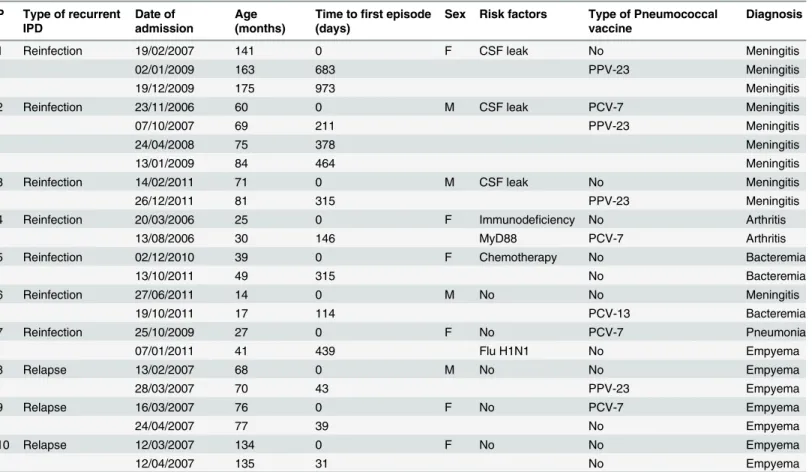
Table 1. Clinical characteristics of patients with recurrent invasive pneumococcal disease. P Type of recurrent
IPD
Date of admission
Age (months)
Time tofirst episode (days)
Sex Risk factors Type of Pneumococcal vaccine
Diagnosis
1 Reinfection 19/02/2007 141 0 F CSF leak No Meningitis
02/01/2009 163 683 PPV-23 Meningitis
19/12/2009 175 973 Meningitis
2 Reinfection 23/11/2006 60 0 M CSF leak PCV-7 Meningitis
07/10/2007 69 211 PPV-23 Meningitis
24/04/2008 75 378 Meningitis
13/01/2009 84 464 Meningitis
3 Reinfection 14/02/2011 71 0 M CSF leak No Meningitis
26/12/2011 81 315 PPV-23 Meningitis
4 Reinfection 20/03/2006 25 0 F Immunodeficiency No Arthritis
13/08/2006 30 146 MyD88 PCV-7 Arthritis
5 Reinfection 02/12/2010 39 0 F Chemotherapy No Bacteremia
13/10/2011 49 315 No Bacteremia
6 Reinfection 27/06/2011 14 0 M No No Meningitis
19/10/2011 17 114 PCV-13 Bacteremia
7 Reinfection 25/10/2009 27 0 F No PCV-7 Pneumonia
07/01/2011 41 439 Flu H1N1 No Empyema
8 Relapse 13/02/2007 68 0 M No No Empyema
28/03/2007 70 43 PPV-23 Empyema
9 Relapse 16/03/2007 76 0 F No PCV-7 Empyema
24/04/2007 77 39 No Empyema
10 Relapse 12/03/2007 134 0 F No No Empyema
12/04/2007 135 31 No Empyema
P, patient identification; PCV-7, 7-valent polysaccharide-protein conjugate vaccine; PCV 13, 13-valent polysaccharide-protein conjugate vaccine; PPV-23, 23-valent pneumococcal polysaccharide vaccine.
First, as expected, the frequency of recurrent IPD in this pediatric population is low (1.7%),
even lower than other reported studies [4,15,16,24]. It is well known that the rate of
recur-rence in children is lower than in adults [4,25–27]. No patient died of recurrent IPD during
the study period, which is in agreement with previous reports comparing IPD in children and
adult population [24,27].
Second, except for recurrent complicated pneumonia, all except one child with recurrent IPD had an underlying condition that should be identified by the practitioner in order to re-duce future recurrences and related sequelae. In our study, the most frequent underlying dis-ease was a local anatomical factor (CSF leak in recurrent meningitis). Indeed, meningitis was the most prevalent form of recurrence, and more than 2 episodes occurred despite vaccination. Thus, in cases of recurrent meningitis, there is a clear need to perform CNS imaging for the di-agnosis of CSF leak. These data contrast with those reported by Gaschinard et al [16] and
Einarsdóttiret al. [24], who described immunoglobulin disorders as the main risk factor for
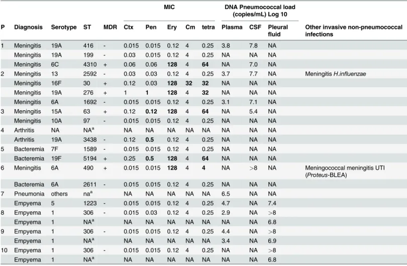
Table 2. Microbiological characteristics of patients with recurrent invasive pneumococcal disease.
MIC DNA Pneumococcal load
(copies/mL) Log 10 P Diagnosis Serotype ST MDR Ctx Pen Ery Cm tetra Plasma CSF Pleural
fluid
Other invasive non-pneumococcal infections
1 Meningitis 19A 416 - 0.015 0.015 0.12 4 0.25 3.8 7.8 NA
Meningitis 19A 199 - 0.03 0.015 0.12 4 0.25 NA NA NA
Meningitis 6C 4310 + 0.06 0.06 128 4 64 NA 7.0 NA
2 Meningitis 13 2592 - 0.03 0.03 0.12 4 0.25 3.7 7.7 NA MeningitisH.influenzae
Meningitis 16F 30 + 0.12 0.03 128 32 32 NA NA NA
Meningitis 19A 276 + 1 1 128 4 32 NA NA NA
Meningitis 6A 1692 - 0.015 0.015 0.12 4 0.25 3.1 7.1 NA
3 Meningitis 15A 63 + 0.12 0.12 128 4 64 NA 5.4 NA
Meningitis 10A 97 - 0.015 0.015 0.12 4 0.25 NA NA NA
4 Arthritis NA NAa NA NA NA NA NA NA NA NA
Arthritis 19A 3438 - 0.12 0.5 0.12 4 0.25 NA NA NA
5 Bacteremia 7F 1589 - 0.015 0.015 0.12 4 0.25 NA NA NA
Bacteremia 19F 5194 + 0.25 0.5 128 4 64 NA NA NA
6 Meningitis 6A 490 + 0.015 0.015 128 4 4 NA >8 NA Meningococcal meningitis UTI
(Proteus-BLEA)
Bacteremia 6A 2611 - 0.015 0.015 0.12 4 0.25 NA NA NA
7 Pneumonia others naa NA NA NA NA NA 6.5 NA NA
Empyema 5 1223 - 0.015 0.015 0.12 4 0.25 4.7 NA 7.4
8 Empyema 1 306 - 0.015 0.03 0.12 4 0.25 2.9 NA >8
Empyema 1 NAa NA NA NA NA NA NA NA 6.8
9 Empyema 1 306 - 0.015 0.015 0.12 4 0.25 4.4 NA >8
Empyema 1 NAa NA NA NA NA NA 3.4 NA 6.9
10 Empyema 1 306 - 0.015 0.015 0.12 4 0.25 NA NA >8
Empyema 1 NAa NA NA NA NA NA NA NA 6.8
P, patient identification; ST, sequence type by Multi-locus sequence typing; MDR, Multi-drug resistant strain; PCV-7, 7-valent polysaccharide-protein conjugate vaccine; PCV 13, 13-valent polysaccharide-protein conjugate vaccine; PPV-23, 23-valent pneumococcal polysaccharide vaccine; CSF, cerebrospinalfluid; NA, Not available; MIC, minimal inhibitory concentration; Pen, penicillin; Ery, erythromycin; Cm, chloramphenicol; Tet, tetracycline; Ctx, Cefotaxime; Bold text, resistant strain according to MICs forStreptococcus pneumonia. EUCAST Clinical Meningeal Breakpoints.
aNA, ST were not available in episodes identi
fied only by PCR.
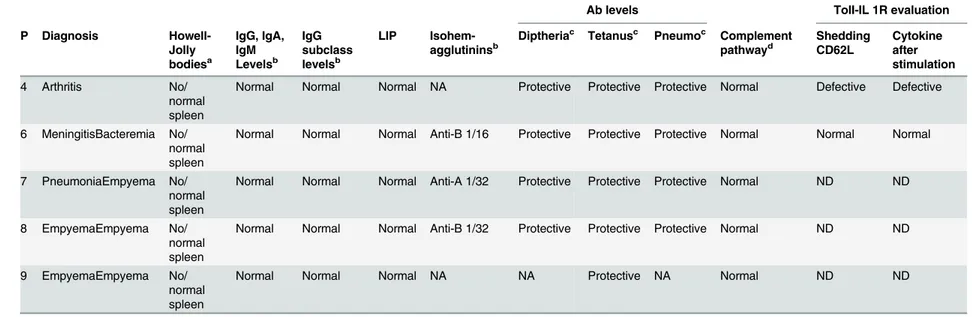
Table 3. Immunological work-up of patients with recurrent invasive pneumococcal disease with unknown underlying risk factors.
Ab levels TolI-IL 1R evaluation
P Diagnosis Howell-Jolly bodiesa
IgG, IgA, IgM Levelsb
IgG subclass levelsb
LIP Isohem-agglutininsb
Diptheriac Tetanusc Pneumoc Complement pathwayd
Shedding CD62L
Cytokine after stimulation
4 Arthritis No/ normal spleen
Normal Normal Normal NA Protective Protective Protective Normal Defective Defective
6 MeningitisBacteremia No/ normal spleen
Normal Normal Normal Anti-B 1/16 Protective Protective Protective Normal Normal Normal
7 PneumoniaEmpyema No/ normal spleen
Normal Normal Normal Anti-A 1/32 Protective Protective Protective Normal ND ND
8 EmpyemaEmpyema No/ normal spleen
Normal Normal Normal Anti-B 1/32 Protective Protective Protective Normal ND ND
9 EmpyemaEmpyema No/ normal spleen
Normal Normal Normal NA NA Protective NA Normal ND ND
P, Patient identification; LIP, Lymphocyte immune-phenotype (T/B/NK); NA, Not available; ND, Not determined; Data for patient 10 were not available. aIn blood smear and/or abdominal ultrasound study.
bRange for age.
cCriteria of positive isohemagglutinins: antiA>1/16, antiB>1/8; Criteria of positive antibody (ab) levels of diphtheria and tetanus:>0.1 UI/mL. Protective antibody (ab) levels>
250 UI/mL. Measurement of antibodies to 23 pneumococcal capsular polysaccharides through enzyme-linked immunosorbent assay. dComplement pathway includes CH50 (normal between 42
–95 UI/mL), C3, C4 for all patients. AH50, properdin and MBL for patient 6.
doi:10.1371/journal.pone.0118848.t003
Recurrent
Invasive
Pneumoc
occal
Disease
in
|DOI:10.137
1/journal.p
one.0118848
March
4,
recurrent IPD in children. The levels of immunoglobulins IgG, IgA and IgM were determined in all patients with recurrent meningitis before the identification of the CSF leak and in all pa-tients with no identified risk factor, and these levels were normal. In our population both pri-mary and secondary immunodeficiencies were detected in 2 patients (and suspected in another): one patient was receiving chemotherapy and the other suffered an identified PID (MyD88 deficiency). The incidence of PID among patients with IPD might be higher than identified here where only recurrent IPD was studied. Indeed, 10 children died of IPD during the study period and 5 of them had underlying risk factors. Of note, 2 patients who died with-out underlying risk factors showed no C-reactive elevation that might be suggestive of a defect in the TIR pathway [19].
Our low rate of PID in children compared with that reported for Gashingnard et al [16] could be explained by the distribution of circulating serotypes in our geographical area. Temporal trends of serotypes causing IPD could be related with associated risk factors for IPD and for re-current IPD. There are more than 94 pneumococcal serotypes that cause varying rates of IPD.
Some of these serotypes are called“opportunistic serotypes”; they are frequently detected in
car-riers and they are more prevalent causing IPD in patients with comorbidities [28]. In contrast,
se-rotypes with“high-attack-rate”, also called“high-invasive potential serotypes”, are seldom
detected in carriers and often cause IPD particularly in older children and adults without
comor-bidities [28,29]. Moreover, we recently reported that young children with genetically-determined
low-mannose binding lectine (MBL) production are at a higher risk of developing IPD, particu-larly that caused by opportunistic or low-attack-rate pneumococcal serotypes [30]. Serotype 1, a well-known high attack serotype, was highly prevalent during the study period mainly associated to pneumonia [17,29], so this could explain a high proportion of disease in healthy children and consequently a low rate of recurrent IPD related with immunological disorders. Of note, in the study of Gaschingnard et al bacteremic pneumonia was not included as IPD. However, if bacter-emic pneumonia episodes were not consider, the recurrent IPD of our study raise to 3,2%, that is
still significantly lower in comparison with the 10% of Gaschingnard’s study.
Based on our results, recurrent pneumococcal pneumonia seems to be pathophysiologically different from other forms of recurrent IPD. Recurrent pneumonia would be more related to microbiological factors than to host factors. The three patients suffering complicated pneumo-nia without underlying predisposing conditions corresponded to episodes of relapse caused by the virulent clone ST306 expressing serotype 1. The interval between relapses was much lower (39 days) than between episodes of reinfection (314 days). Also, a much higher bacterial load in pleural fluid was detected despite the antibiotic course usually being standardized in the same way. This suggests that the main cause of this recurrence is insufficient treatment or clearance of the infection in the first episode of empyema. One limitation of our study was the inability to determine the clonal composition of strains detected in the second episode of the 3 cases of relapses detected in patients suffering complicated pneumonia. However, the high
homogeneity of serotype 1 as previously described [29,31] and the short time interval between
episodes suggests that these were relapses rather than reinfections.
explained by the limitation in evaluating Ab responses to pneumococcus through the pool of 23 serotypes (ELIZEN Pneumoccocus IgG assay, ZenTech) instead of the quantification of the 13-serotype-specific Ab. As previous studies have shown, nonfunctional antibodies to the cell wall polysaccharides can interfere with the determination of serotype-specific antibodies in these ELISAs. In addition, because of the variable immunogenicity among different serotypes, the measurement of overall antibody responses can give rise to the observation of high levels of antibodies all induced by a single serotype, and may mask a possible deficiency in antibody
re-sponses to serotypes that are less immunogenic [32,33].
Finally, a PID was diagnosed in a patient due to the broad immune work-up established in our hospital for recurrent IPD. This evaluation must include specific antibody responses to polysaccharides, complement and spleen function, since these three deficiencies are the most
commonly described [16,34]. The recommendation of including TIR evaluation in the
im-mune evaluation of recurrent IPD in children was made in 2007 [35]. Recently, Gaschinard
et al[16] published a systematic evaluation of PID, including TIR pathway, in a prospective study on 163 children hospitalized in France for IPD, of which 10% were recurrent. They iden-tified 11% of patients with PID, with primary antibody and complement deficiencies being the most common. They also identified a patient with MyD88 deficiency after a single episode of pneumococcal meningitis and a previous episode of ethmoiditis. The identification of MyD88 deficiency in our patient enabled us to optimize her treatment and antibiotic prophylaxis, with an improved outcome. PIDs affecting TIR pathway are rare and represent less than 1% of all forms of PIDs [19], but due to their recent description, and to the need for performing highly
specific tests for their diagnosis [11,20], they are probably underdiagnosed. The main clinical
suspicion sign for defects in innate immunity, TIR pathway in particular, is recurrent IPD [12,19]. So in children with recurrent IPD in whom no risk factor has been identified, we strongly recommend a stepwise immune work-up which can begin with the evaluation of antibody responses (since humoral immunodeficiencies are the most common forms of PID
representing over 60–70% of all forms), followed by innate immunity evaluation (CH50,
Howell-Jolly and, finally, TIR defects, if the above-mentioned are normal).
In conclusion, recurrent IPD in children is a rare condition. Whenever we face it, a predis-posing condition needs to be searched for. In the absence of underlying diseases known to pre-dispose to recurrent IPD (which currently indicates pneumococcal vaccination),
immunological status of patients should be studied in depth, including TIR pathway, and also followed-up periodically, enabling the implementation of preventive measures, such as pneu-mococcal vaccination, antibiotic prophylaxis and other specific treatments, if needed. However, if recurrent IPD is a relapse of pneumonia, microbiological factors, rather than immune fac-tors, seem to play a pivotal role.
Author Contributions
Conceived and designed the experiments: LA CMA. Performed the experiments: HDP MI MJ CMA. Analyzed the data: LA CMA. Contributed reagents/materials/analysis tools: LA MGB MFS MT. Wrote the paper: LA HDP CMA.
References
1. Bogaert D, de Groot R, Hermans PW. Streptococcus pneumoniae colonisation: the key to pneumococ-cal disease. Lancet Infect Dis. 2004; 4: 144–54. PMID:14998500
2. Obaro S, Adegbola R. The pneumococcus: carriage, disease and conjugate vaccines. J. Med Micro-biol. 2002; 51: 98–104. PMID:11863272
4. King MD, Whitney CG, Parekh F, Farley MM. Recurrent invasive pneumococcal disease: a population-based assessment. Clin Infect Dis. 2003; 37: 1029–36. PMID:14523766
5. CDC. Prevention of pneumococcal disease among infants and children—use of 13-valent
pneumococ-cal conjugate vaccine and 23-valent pneumococpneumococ-cal polysaccharide vaccine: recommendations of the Advisory Committee on Immunization Practices (ACIP) MMWR 59 (No. RR-11).2010.
6. Picard C, Puel A, Bustamante J, Ku CL, Casanova JL. Primary immunodeficiencies associated with pneumococcal disease. Curr Opin Allergy Clin Immunol. 2003; 3: 451–9. PMID:14612669
7. Picard C, Puel A, Bonnet M, Ku CL, Bustamante J, Yang K, et al. Pyogenic bacterial infections in hu-mans with IRAK-4 deficiency. Science. 2003; 299: 2076–9. PMID:12637671
8. Ku CL, Picard C, Erdös M, Jeurissen A, Bustamante J, Puesl A, et al. IRAK4 and NEMO mutations in otherwise healthy children with recurrent invasive pneumococcal disease. J Med Genet. 2007; 44: 16–23. PMID:16950813
9. Ku CL, Yang K, Bustamante J, Puel A, von Bernuth H, Santos OF, et al. Inherited disorders of human Toll-like receptor signaling: immunological implications. Immunol Rev. 2005; 203: 10–20. PMID: 15661018
10. Currie AJ, Davidson DJ, Reid GS, Bharya S, MacDonald KL, Devon RS, et al. Primary immunodeficien-cy to pneumococcal infection due to a defect in Toll-like receptor signaling. J Pediatr. 2004; 144: 512–8.
PMID:15069402
11. von Bernuth H, Picard C, Jin Z, Pankla R, Xiao H, Ku CL, et al. Pyogenic bacterial infections in humans with MyD88 deficiency. Science. 2008; 321: 691–6. doi:10.1126/science.1158298PMID:18669862 12. Picard C, Casanova JL, Puel A. Infectious diseases in patients with IRAK-4, MyD88, NEMO, or IκBα
deficiency. Clin Microbiol Rev. 2011; 24: 490–7. doi:10.1128/CMR.00001-11PMID:21734245 13. Beutler BA. TLRs and innate immunity. Blood. 2009; 113: 1399–407. doi:
10.1182/blood-2008-07-019307PMID:18757776
14. Mufson MA, Hao JB, Stanek RJ, Norton NB. Clinical features of patients with recurrent invasive Strepto-coccus pneumoniae disease. Am J Med Sci. 2012; 343: 303–9. doi:10.1097/MAJ.0b013e31822d9860
PMID:21934596
15. Mason EO Jr, Wald ER, Tan TQ, Schutze GE, Bradley JS, Barson WJ, et al. Recurrent systemic pneu-mococcal disease in children. Pediatr Infect Dis J. 2007; 26: 480–4. PMID:17529863
16. Gaschignard J, Levy C, Chrabieh M, Boisson B, Bost-Bru C, Dauger S, et al. Invasive pneumococcal disease in children can reveal a primary immunodeficiency. Clin Infect Dis. 2014; 59: 244–51. doi:10. 1093/cid/ciu274PMID:24759830
17. de Sevilla MF, García-García JJ, Esteva C, Moraga F, Hernández S, Selva L, et al. Clinical presentation of invasive pneumococcal disease in Spain in the era of heptavalent conjugate vaccine. Pediatr Infect Dis J. 2012; 31: 124–8. doi:10.1097/INF.0b013e318241d09ePMID:22173137
18. Mahlaoui N, Minard-Colin V, Picard C, Bolze A, Ku CL, Tournilhac O, et al. Isolated congenital asplenia: a French nationwide retrospective survey of 20 cases. J Pediatr. 2011; 158: 142–8. doi:10.1016/j. jpeds.2010.07.027PMID:20846672
19. Picard C, von Bernuth H, Ghandil P. Clinical features and outcome of patients with IRAK-4 and MyD88 deficiency. Medicine. 2010; 89: 403–425. doi:10.1097/MD.0b013e3181fd8ec3PMID:21057262 20. von Bernuth H, Ku CL, Rodriguez-Gallego C, Zhang S, Garty BZ, Maródi L, et al. A fast procedure for
the detection of defects in Toll-like receptor signaling. Pediatrics. 2006; 118: 2498–503. PMID: 17142536
21. Muñoz-Almagro C, Gala S, Selva L, Jordan I, Tarragó D, Pallares R, et al. DNA bacterial load in
chil-dren and adolescents with pneumococcal pneumonia an empyema. Eur J Clin Microbiol Infect Dis. 2011; 30: 327–35. doi:10.1007/s10096-010-1086-9PMID:20972810
22. Tarragó D, Fenoll A, Sánchez-Tatay D, Arroyo LA, Muñoz-Almagro C, Esteva C, et al. Identification of
pneumococcal serotypes from culture-negative clinical specimens by novel real-time PCR. Clin Micro-biol Infect. 2008; 14: 828–834. doi:10.1111/j.1469-0691.2008.02028.xPMID:18844683
23. Enright MC, Spratt BG. A multilocus sequence typing scheme for Streptococcus pneumoniae: identifi-cation of clones associated with serious invasive disease. Microbiology. 1998; 144: 3049–3060. PMID: 9846740
24. Einarsdóttir HM, Erlensdóttir H, Kristinsson KG, Gottfredsson M. Nationwide study of recurrent invasive pneumococcal infections in a population with a low prevalence of human immunodeficiency virus infec-tion. Clin Microbiol Infect. 2005; 11: 744–9. PMID:16104990
25. Rodríguez-Créixems M, Muñoz P, Miranda E, Peláez T, Alonso R, Bouza E. Recurrent pneumococcal
26. Coccia MR, Facklam RR, Saravolatz LD, Manzor O. Recurrent pneumococcal bacteremia: 34 episodes in 15 patients. Clin Infect Dis. 1998; 26: 982–5. PMID:9564486
27. McEllistrem MC, Mendelsohn AB, Pass MA, Elliott JA, Whitney CG, Kolano JA, et al. Recurrent inva-sive pneumococcal disease in individuals with human immunodeficiency virus infection. J Infect Dis. 2002; 185: 1364–8. PMID:12001059
28. Brueggemann AB, Griffiths DT, Meats E, Peto T, Crook DW, Spratt BG. Clonal relationships between invasive and carriage Streptococcus pneumonia and serotype- and clone-specific differences in inva-sive disease potential. J Infect Dis. 2003; 187:1424–32. PMID:12717624
29. Esteva C, Selva L, de Sevilla MF, Garcia-Garcia JJ, Pallares R, Muñoz-Almagro C.Streptococcus
pneumoniaeserotype 1 causing invasive disease among children in Barcelona over a 20-year period
(1989–2008). Clin Microbiol Infect. 2011; 17: 1441–4. doi:10.1111/j.1469-0691.2011.03526.xPMID: 21729192
30. Muñoz-Almagro C, Bautista C, Arias MT, Boixeda R, Del Amo E, Borrás C, et al. High prevalence of
ge-netically-determined mannose binding lectin deficiency in young children with invasive pneumococcal disease. Clin Microbiol Infect. 2014; 20: O745–52. doi:10.1111/1469-0691.12615PMID:24602163 31. Muñoz-Almagro C, Ciruela P, Esteva C, Marco F, Navarro M, Bartolome R, et al. Catalan study group
of invasive pneumococcal disease. Serotypes and clones causing invasive pneumococcal disease be-fore the use of new conjugate vaccines in Catalonia, Spain. J Infect. 2011; 63: 151–62. doi:10.1016/j. jinf.2011.06.002PMID:21679725
32. Jeurissen A, Moens L, Raes M, Wuyts G, Willebrords L, Sauer K, et al. Laboratory diagnosis of specific antibody deficiency to pneumococcal capsular polysaccharide antigens. Clin Chem. 2007; 53: 505–10.
PMID:17259230
33. Balmer P, North J, Baxter D. Measurement and interpretation of pneumococcal IgG levels for clinical management. Clin Exp Immunol. 2003; 133: 364–9. PMID:12930362
34. Ingels H, Lambertsen L, Harboe ZB, Marquart HV, Konradsen H, Christensen JJ, et al. Recurrent inva-sive pneumococcal disease in children: epidemiological, microbiological, and clinical aspects from a Danish 33-year nationwide survey (1980–2013). Scand J Infect Dis. 2014; 46: 265–71. doi:10.3109/ 00365548.2013.877156PMID:24628485