Development of Ss-NIE-1 Recombinant
Antigen Based Assays for Immunodiagnosis
of Strongyloidiasis
Lisa N. Rascoe1, Courtney Price1, Sun Hee Shin2, Isabel McAuliffe1, Jeffrey W. Priest3, Sukwan Handali1*
1Division of Parasitic Diseases and Malaria, Centers for Disease Control and Prevention, Atlanta, Georgia, United States of America,2Emory College, Emory University, Atlanta, Georgia, United States of America,
3Division of Foodborne, Waterborne, and Environmental Diseases, Centers for Disease Controls and Prevention, Atlanta, Georgia, United States of America
Abstract
Strongyloides stercoralisis a widely distributed parasite that infects 30 to 100 million people worldwide. In the United States strongyloidiasis is recognized as an important infection in immigrants and refugees. Public health and commercial reference laboratories need a sim-ple and reliable method for diagnosis of strongyloidiasis to identify and treat cases and to prevent transmission. The recognized laboratory test of choice for diagnosis of strongyloidi-asis is detection of disease specific antibodies, most commonly using a crude parasite ex-tract for detection of IgG antibodies. Recently, a luciferase tagged recombinant protein ofS. stercoralis, Ss-NIE-1, has been used in a luciferase immunoprecipitation system (LIPS) to detect IgG and IgG4specific antibodies. To promote wider adoption of immunoassays for
strongyloidiasis, we used the Ss-NIE-1 recombinant antigen without the luciferase tag and developed ELISA and fluorescent bead (Luminex) assays to detectS.stercoralisspecific IgG4. We evaluated the assays using well-characterized sera from persons with or without
presumed strongyloidiasis. The sensitivity and specificity of Ss-NIE-1 IgG4ELISA were
95% and 93%, respectively. For the IgG4Luminex assay, the sensitivity and specificity
were 93% and 95%, respectively. Specific IgG4 antibody decreased after treatment in a manner that was similar to the decrease of specific IgG measured in the crude IgG ELISA. The sensitivities of the Ss-NIE-1 IgG4ELISA and Luminex assays were comparable to the
crude IgG ELISA but with improved specificities. However, the Ss-NIE-1 based assays are not dependent on native parasite materials and can be performed using widely available laboratory equipment. In conclusion, these newly developed Ss-NIE-1 based immunoas-says can be readily adopted by public health and commercial reference laboratories for rou-tine screening and clinical diagnosis ofS.stercoralisinfection in refugees and immigrants in the United States.
OPEN ACCESS
Citation:Rascoe LN, Price C, Shin SH, McAuliffe I, Priest JW, Handali S (2015) Development of Ss-NIE-1 Recombinant Antigen Based Assays for Immunodiagnosis of Strongyloidiasis. PLoS Negl Trop Dis 9(4): e0003694. doi:10.1371/journal. pntd.0003694
Editor:Aysegul Taylan Ozkan, Hitit University, TURKEY
Received:September 10, 2014
Accepted:March 10, 2015
Published:April 10, 2015
Copyright:This is an open access article, free of all copyright, and may be freely reproduced, distributed, transmitted, modified, built upon, or otherwise used by anyone for any lawful purpose. The work is made available under theCreative Commons CC0public domain dedication.
Data Availability Statement:All relevant data are within the paper.
Funding:The authors received no specific funding for this work.
Author Summary
Strongyloidiasis is a neglected tropical disease that affects millions worldwide and needs more attention and better diagnostic methods.Strongyloides stercoraliscan undergo an au-toinfection cycle and can cause hyperinfection involving the pulmonary and gastrointesti-nal systems and disseminated infection in other organs. Although endemic areas are mostly developing countries in tropical and subtropical regions with only sporadic trans-mission in temperate areas, the disease is a threat to developed world populations through immigrants, refugees, travelers, and military personnel. The disease can have catastrophic effects when a patient is immunocompromised or when an infected organ is transplanted into a vulnerable recipient. Due to the threat to public health, the intricate life cycle ofS.
stercoralis, the need to perform multiple follow-up diagnostics to ensure treatment success, and the necessity to rule out multiple co-endemic parasitic infections, it is imperative to develop new diagnostic assays that are simple and efficient while retaining maximal sensi-tivity and specificity. In this study, we use a well-known recombinant protein, Ss-NIE-1, to optimize assays using both an ELISA format and a multiplex platform to meet these needs.
Introduction
Strongyloides stercoralis, an intestinal nematode that migrates through the skin and lung, is a widely distributed disease that infects 30 to 100 million people worldwide [1]. Unlike other hel-minthic parasites,S.stercoraliscan complete its entire life cycle within a single human host through autoinfection and can cause an asymptomatic chronic infection that may go undetect-ed for decades in immunocompetent hosts [2,3]. In the United States,S.stercoraliscauses more deaths than any other soil-transmitted helminth, with mortality rates as high as 87% in cases of hyper-infection in immunocompromised hosts [3].
The standard diagnosis of strongyloidiasis relies on the detection of larvae in the stool [4], but a single stool sample analysis will identify no more than 70% of positive cases [5]. Due to the low sensitivity of the stool assay, immunodiagnosis using a crude antigen-based enzyme-linked immunosorbent assay (ELISA) has been developed as the laboratory test of choice for clinical diagnosis of strongyloidiasis. The Immunoglobulin G (IgG) ELISA utilizes crude ex-tract prepared from L3S.stercoralislarvae obtained from infected dogs. Reliance on native par-asite materials and the canine infection model are major disadvantages of this test. As a result, a number of recombinant antigen-based ELISAs have recently been developed. Recombinant antigens can be purified easily and can be reproducibly generated in large amounts [6–8]. Anti-body detection assays utilizing recombinant protein Ss-NIE-1, a 31-kDa antigen derived from
S.stercoralisL3 parasites [8], have reported sensitivities and specificities of 84–98% and 95– 100%, respectively, and are comparable in performance to the crude antigen-based ELISA [6–13].
treated with a single course of therapy [18], we also used the fluorescent bead assay to deter-mine if a decrease in antibody was measureable after treatment using a select set of sera.
Materials and Methods
Serum Specimens
Although some samples were exhausted during the initial ELISA development and some new samples were added during Luminex assay development, the same sets of sera were used for testing the Ss-NIE-1 ELISA and Ss-NIE-1 Luminex assays and many samples were assayed using both techniques. The sets of human sera used were: (1) samples proven positive forS.
stercoralisbased on the presence of larvae in the stool or sputum (ELISAN= 258, Luminex
N= 175); (2) presumed negative samples from U.S. residents with no history of foreign travel (ELISAN= 182, LuminexN= 207); (3) a convenience panel of samples from patients with var-ious diseases other thanS.stercoralisfocusing mainly on worm infections and including 63 sera from proven cases of lymphatic filariasis from Haiti (ELISAN= 143, LuminexN= 159) [19]; (4) and sera from patients withS.stercoralisinfections, before and after treatment (ELISA
N= 48, LuminexN= 25) [18]. All sera were anonymous and were used in accordance with ap-proved human subjects’protocols.
Recombinant Protein Preparation
Ss-NIE-1 ELISA antigen. Ss-NIE-1 with a 6x His tag was expressed inE.colifrom a clone in pET30b (kindly provided by T. Nutman, NIAID, NIH, Bethesda, MD) by Genscript (Piscat-away, NJ). Expression was analyzed and confirmed by Western Blot using anti- 6xHis antibod-ies andS.stercoralispositive serum. The protein was purified in a one-step affinity purification using a Nickel metal affinity column and concentrations were measured with the Bradford pro-tein assay (Bio-Rad Laboratories, Hercules, CA).
linear NaCl gradients: 0–400 mM NaCl in 4 min, 400–600 mM NaCl in 10 min, and 600–1000 mM in 4 min. A total of 4.2 mg of protein was collected in three 1-ml fractions at approximate-ly 0.44 M NaCl in the gradient profile. Purity was estimated to be>98% by SDS polyacryl-amide gel electrophoresis. The recombinant GST/Ss-NIE-1 fusion protein was used in all multiplex assays. Protein concentrations were measured with the BCA microassay (Pierce, Rockford, IL).
Ss-NIE-1 ELISA Development
The development of an IgG4standard reference curve for the Ss-NIE-1 ELISA was performed as described by Scheel et al [22]. Immunoglobulin G4, human myeloma plasma was purchased and stored frozen in 20 mM phosphate, pH 7.4, with 150 mM NaCl and 0.05% Sodium azide (NaN3) (Athens Research & Technology, Athens, GA). IgG4was diluted into antigen sensitiz-ing buffer (ASB) (0.05 M Tris/HCl, pH 8.0 + 1 M KCl + 2 mM EDTA) to create standard curve points. IgG4concentrations were chosen based on previous experiments in standard curve de-velopment and adjusted to produce the highest OD value, ~ 2.0.
Checkerboard titrations for antigen concentration, serum dilution, conjugate dilution, and substrate 3, 3’, 5, 5’-Tetramethylbenzidine (TMB) time were carried out on Immulon 2HB Microwell plate (Thermo Scientific, Cat. Number 6506). For optimization of the Ss-NIE-1 ELISA, optimal conditions were chosen based on the signal to noise ratio between defined strong
S.stercoralispositive and normal human serum samples. The optimized Ss-NIE-1 ELISA steps are as follows: the micro-well plate was sensitized with 100μL/well of Ss-NIE-1 antigen at a
con-centration of 0.3μg/mL in antigen sensitizing buffer (0.05 M Tris/HCl, pH 8.0 + 1 M KCl + 2
mM EDTA) for 2 hours at room temperature on a plate shaker. Following antigen sensitization, the plate was washed 4 times with PBS/0.3% Tween. The plate was blocked for 30 minutes with 100μL/well of 10 mM Nickel Chloride (Aldrich, Cat. Number 339350) in PBS/0.3% Tween/5%
Instant Nonfat Dry Milk (Nestle, Glendale, CA), and then washed as before. StabilCoat Immu-noassay Stabilizer (SurModics, East Prairie, MN) was then added 100μL to each well and
incu-bated for 30 minutes on a plate shaker at room temperature. After discarding the blocking solution, the plate was dried for 4 hours at 30°C in a vacuum oven chamber. The sensitized and blocked plate was stored at 4°C in sealed aluminum foil with desiccator.
Ss-NIE-1 ELISA Protocol
Human serum samples were tested in 100μL/well at 1:50 dilution in PBS/0.3% Tween/5%
In-stant Nonfat Dry Milk. Following 30 minutes incubation at room temperature on a plate shak-er (speed ~ 800 rpm), the plate was washed 4 times with PBS/0.3% Tween. Propshak-er conjugate concentration of mouse anti-human IgG4 (clone HP6025), affinity purified, horseradish perox-idase labeled (Southern Biotech, Birmingham, AL; Cat. Number 9200–05) was added to each well at 100μL/well at 1:1,000 dilution in PBS/0.3% Tween and incubated 30 minutes at room
temperature on a plate shaker with the plate being washed 4 times following incubation with PBS/0.3% Tween. The substrate used was SureBlue, 3, 3’, 5, 5’-Tetramethylbenzidine (TMB) Microwell Peroxidase Substrate (KPL, Gaithersburg, Maryland). We used 100μL/well of TMB
Ss-NIE-1 Luminex Assay Development
Protein coupling to MagPlex magnetic beads. Coupling of protein to MagPlex Magnetic Microspheres (Luminex, Austin, TX) was carried out using EDC-Sulfo NHS protocol [23,24]. Briefly, beads were washed and activated in buffer containing 50mM MES, pH 5, 0.85% NaCl, and 0.05% Tween-20. After 40 minutes of incubation using end-over-end mixing in the dark with Sulfo-NHS and EDC, beads were washed 2 times with MES buffered saline. The activated beads were then transferred to a new tube and washed once more. Beads were resuspended in the MES buffer without Tween-20 and 0.3μg of GST-Ss-NIE-1/1.25 x 10E6 beads were added.
The total volume of reaction was brought to 500μL with MES buffer without Tween-20. The
coupling was performed for 3 hours in the dark at room temperature by end-over-end mixing. Beads were blocked with blocking buffer (PBS + 1% BSA + 0.05% sodium azide (NaN3), pH 7.4) for 30 minutes. The coupled beads were stored at 4°C in PBS + 1% BSA + 0.05% NaN3+ 0.05% Tween-20 + PMSF (1:500), Pepstatin (1:1000), Leupeptin (1:1000) until used. The concentration of the beads was determined by viewing them in a hemocytometer using a 20X objective.
Luminex immunoassay. FiftyμL of the working MagPlex microsphere mixture (50 beads/ μL in PBS/0.3% Tween-20/5%—Instant Nonfat Dry Milk) and 50μL of diluted sera (1:100 in
PBS/0.3% Tween-20/5% Instant Nonfat Dry Milk) were added into each well of Costar 96-well black, round-bottom plate (Fisher Scientific, Cat.# 3792). After 30 minutes incubation at room temperature with shaking at speed 6 (~800 rpm), the beads were washed using the Biotek Mag-netic Washer ELx50 (2 minutes magMag-netic separation followed by 2 cycles of dispensing 100μL
of PBS/Tween-20 0.3% and a 40 second soak before aspiration). The complex of antibody and coupled beads was detected using 50μL of biotinylated mouse anti-human IgG4(clone HP6025, affinity purified, Southern Biotech, Birmingham, AL, Cat. Number 9200–08) diluted 1:200 in PBS-1% BSA, 0.05% NaN3.After 30 minute incubation, the beads were washed as pre-viously described. As a detector, 50μL/well ofR- phycoerythrin-labeled streptavidin conjugate
(Invitrogen, Cat. # S866) at a 1:250 dilution in PBS-1% BSA, 0.05% NaN3was added to the well and incubated for 30 minutes. After washing, the beads were resuspended using 100μL/well of
PBS-1% BSA, 0.05% NaN3. The mean fluorescence intensity from each well was determined by using BioPlex manager software, version 6.02 (BioRad) and a Luminex 100 platform.
Data Analysis
Data were tabulated and analyzed using Microsoft Excel. Determination of the cut-off value and assay performance was calculated using SAS version 9.0. The concentration of Ss-NIE-1 ELISA was measured in in ng/mL; Luminex results are reported as mean fluorescence intensity minus background blank (MFI). The J-index, a single measurement of assay performance, was calculated as described previously ([25,26].
Results
S
.
stercoralis
NIE-1 IgG
4ELISA
The recombinant Ss-NIE-1-His protein expressed at Genscript was successfully used to devel-op an ELISA (Table 1). Using a cutoff value of 0.80 ng IgG4/mL, the assay correctly identified 245 of 258 parasitologically confirmed strongyloidiasis cases for a sensitivity of 95% (Table 1). The overall specificity of the ELISA was determined to be 93% using a panel of non-endemic US controls and sera from patients with other (mainly parasitic worm) diseases (Tables1and
among sera from echinococcosis, gnathostomiasis, and hookworm patients.Table 2shows the cross-reactivity of the various presumed negative sera. Given the likelihood of polyparasitism among many of these serum donors (i.e., 63 lymphatic filariasis patients fromS.stercoralis -en-demic Haiti), the 99% specificity observed among US negative controls may be a reasonable upper bound to the value. The J-index, a single measure of assay performance, was 0.88.
Positive sera with low and medium level reactivity were used to measure inter-assay tion as previously described in the Materials and Methods. The inter-assay coefficient of varia-tion was determined to be 22% for the low positive control serum and 10% for the medium positive control serum.
Table 1. Performance of SS-NIE-1 ELISA and Luminex.
Assay Characteristics SS-NIE-1 ELISA 95% CI* Ss-NIE-1 Luminex 95% CI
Cut-off point 0.8 ng IgG4/ mL 8 MFI units
J-index 0.88 0.88
Sensitivity:NPositive/NTested (%) 245/258 (95) 92–97 162/175 (93) 88–96
Specificity:NNegative/NTested (%) 303/325 (93) 90–96 349/366 (95) 93–97
Note:*CI = confidence interval
doi:10.1371/journal.pntd.0003694.t001
Table 2. Cross-reactivity of SS-NIE-1 ELISA and Luminex.
SS-NIE-1 ELISA SS-NIE-1 Luminex
Cross reactivity (%)
No. of positives
No. of sera tested
Conditions represented by sera
No. of sera tested
No. of positives
Cross reactivity (%)
1 2 182 U.S. Negatives 207 3 1
NT NT NT Amebiasis 2 1 50
7 1 14 Ascariasis 8 1 13
0 0 1 Baylisascariasis 1 0 0
50 1 2 Cancer 2 0 0
0 0 2 Cysticercosis 4 0 0
NT NT NT Dirofilariasis 1 0 0
50 1 2 Echinococcosis 5 0 0
0 0 2 Eosinophilic myalgia 1 0 0
10 6 63 Filiariasis 63 6 10
50 2 4 Gnathostomiasis NT NT NT
33 2 6 Hookworm(N.americanus) 14 2 14
NT NT NT Hymenolepiaisis 3 1 33
NT NT NT Paragonimiasis 6 0 0
0 0 4 Schistosomiasis 5 0 0
NT NT NT Taeniasis 4 0 0
12 4 33 Toxocariasis 28 1 4
0 0 4 Toxoplasmosis 3 0 0
25 1 4 Trichinellosis 4 1 25
100 2 2 Trichuriasis 4 1 25
NT NT NT Tuberculosis 1 0 0
NT = Not tested
Fluorescent Bead Ss- NIE-1 IgG
4Assay
The His tagged Ss-NIE-1 recombinant protein could not be coupled to magnetic beads. Thus, we proceeded using a GST-tagged Ss-NIE-1 protein and successfully coupled the Ss-NIE-1 pro-tein to the MagPlex microspheres. The intra- and inter-assay coefficients of variation were de-termined to be 4.2% and 13.9%, respectively, for average values of 641 MFI and 585. Using a cutoff value of 8 MFI, the sensitivity and specificity of the IgG4Luminex assay were 93% and 95% (Table 1), respectively. As with the ELISA described above, the specificity among US nega-tive controls was much higher (99%) than among donors with defined diseases or parasitic in-fections (91%) (Table 2). High reactivities (33%) were only observed among sera from the two amebiasis and three hymenolepiasis donors. The J-index was identical to that of the ELISA at 0.88.
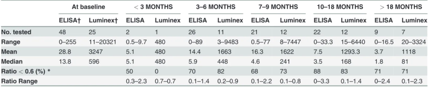
Post Treatment Analysis
Antibody longevity in subjects infected with strongyloidiasis following treatment with thiaben-dazole [18] can be seen inTable 3. Peak and median antibody responses decreased over time using both assay formats, but the antibody levels remained above the cut-off point for most of the subjects even 18 months post treatment. Using the Kobayashi criteria of cure, which con-siders a patient to be cured if the ratio of serological results post-treatment compared to pre-treatment is less than 0.6, 70% of the subjects would be reported as cured 3–6 months following treatment [27].
Discussion
Strongyloidiasis is an increasingly important health problem in the US among immigrants and refugees. Patients with occult strongyloidiasis are at risk of disease if they become immunosup-pressed, and organ donors with unrecognizedS.stercoralisinfection pose a risk to recipients if their infected organs are transplanted [2]. Identification of the parasite in stool specimens is in-sensitive, and, because parasitological examination requires collection of multiple stool speci-mens over 3 days, serological testing would be preferred if available. We elected to develop novel immunodiagnostic assays to meet this need. We employed a well described recombinant protein with proven performance as an immunodiagnostic antigen, the Ss-NIE-1 protein [6–
8]. Based on the potential importance of IgG4antibody responses in filarial infections, we de-cided to develop methods that detectS.stercoralis-specific IgG4antibodies. The Ss-NIE-1 IgG4
Table 3. Change in antibody response following treatment.
At baseline <3 MONTHS 3–6 MONTHS 7–9 MONTHS 10–18 MONTHS >18 MONTHS
ELISA† Luminex† ELISA Luminex ELISA Luminex ELISA Luminex ELISA Luminex ELISA Luminex
No. tested 48 25 2 1 26 11 21 12 22 12 9 7
Range 0–255 11–20321 0.5–9.7 480 0–89 3–9483 0.5–77 8–7447 0–33.3 15–6440 0–16.5 20–3324
Mean 28.8 3247 5.1 480 14.4 1663 16.3 1622 7.5 1293.3 3.7 1118
Median 13.8 596 5.1 480 5.9 448 4.6 241 3.5 168 1.8 81
Ratio<0.6 (%)* 50 0 70 82 68 73 88 83 71 71
Ratio Range 0.3–2.3 0.7–0.7 0.1–1.4 0.2–0.9 0.1–2.2 0.1–0.8 0–3.3 0.1–1.4 0–2.4 0.1–2.3
Note:
†For SS-NIE-1 ELISA, units are expressed in ng/mL and for SS-NIE-1 Luminex, the unit is a ratio of meanfluorescent intensity minus the background of the test/control.
*Follow-up serologic result divided by the initial result
Luminex bead assay achieved a sensitivity and a specificity comparable to those reported for other strongyloidiasis assays such as the crude antigen ELISA, and 26-kDA ELISA, and Ss-NIE-1 ELISAs and the Ss-Ss-NIE-1 LIPS [6,7,9,10,12,18]. Compared to the CDCS.stercoralis
crude antigen ELISA, the Ss-NIE-1 IgG4ELISA and the Ss-NIE-1 IgG4Luminex assay achieved similar sensitivity without compromising specificity. The possible factors contributing to im-proved specificity could be the use of recombinant antigen, assay optimization, or detection of IgG4versus IgG. During Ss-NIE-1 ELISA optimization, we found that non-specific antibody binding in the normal human sera could be decreased by adding a pre-blocking step with 10 mM nickel chloride in PBS/0.3% Tween/5% milk. The decrease in background noise allows the assay to have a higher specificity (93%).
ELISA based tests can only be used to detect antibody responses against one antigen of in-terest at a time. Because the differential diagnosis ofS.stercoralisoften includes multiple hel-minths, a submitted serum sample must frequently be tested using several parasite-specific ELISAs to determine the possible cause(s) of infection. xMAP Luminex technology offers an assay platform that can simultaneously detect antibodies to multiple diseases/infections, and multiplex bead-based antibody assays are generally as sensitive as conventional ELISA, have a wide dynamic range, and are highly reproducible from assay to assay [28]. For these reasons, we elected to transfer the Ss-NIE-1 assay to the Luminex platform. In our hands, the Ss-NIE-1 IgG4Luminex bead assay was slightly less sensitive and slightly more specific than the ELISA, but had comparable overall performance as measured by the J-index.
With the exception of toxocariasis and lymphatic filariasis, our cross-reactivity data must be interpreted with some caution due to the small numbers of samples available for testing. When sera from echinococcosis, hookworm, and trichuriasis patients were tested with Ss-NIE-1 by ELISA, 33% or more of the sera reacted, some quite strongly. A portion of this reactivity likely resulted from ELISA-specific background as many of these same sera did not react with the Ss-NIE-1 antigen in our newly developed Luminex bead assay. Bisoffi et al. [9] found no ELISA or LIPS assay cross-reactions to Ss-NIE-1 among theirEchinococcus-or hookworm-infected do-nors. Although true cross-reactivity between these parasites may exist, polyparasitism cannot be ruled out in the remaining Luminex-positive subjects. An analysis of Ss-NIE-1 LIPS results from a hookworm/Ascaris lumbricoides/H.nana/S.stercoralisco-endemic region of Argen-tina failed to demonstrate an association between infection with other parasites and an anti-body response to the Ss-NIE-1 antigen [10].
The Ss-NIE-1 ELISA and Luminex had 10% cross-reactivity against the subjects with lym-phatic filariasis. Again, this problem could be a true cross-reactivity or could also be explained by polyparasitism with soil-transmitted helminthes in Haiti. Unfortunately, we have no data about the presence of other parasitic infections in the individuals from whom these serum sam-ples were obtained [29]. Norsyahida [11] reported that although the use of IgG4conjugate did decrease the cross-reactivity to filariasis compared to the total IgG responses, some cross-reac-tivity was found with the IgG4based assay. However, the Norsyahida group did not use a recom-binant antigen [8], and the observed cross-reactivity could be due to the use of crude extract antigen in their assay. Of note, the group mentioned the importance of testing for filariasis in subjects with strongyloidiasis. Such testing could most easily be accomplished on a multiplexing platform such as Luminex that can detect antibodies against filariasis and strongyloidiasis simultaneously.
considered as cured based on the SS-NIE-1 ELISA and Luminex assay. We have no definitive explanation for these observed differences, except the fact that the crude ELISA uses a complex antigen versus a single antigen which was used in our studies.
Overall, excellent assays for detectingS.stercoralisspecific antibodies have been developed. Because these assays use recombinant proteins, negating the need for native parasite materials these assays can be adopted for use in public health laboratories for refugee screening or in commercial laboratories for diagnosis of clinical strongyloidiasis and for screening possible transplant donors with occult disease. As both ELISA and Luminex based assays performed similarly, studies in low infrastructure, endemic setting could use the ELISA format. Although polyparasitism is a potential problem, with a strong specificity of 93%, cross-reactivity should not be an issue. For a country-wide study to determine the prevalence of strongyloidiasis, a multiplexing capability of Luminex will be more cost efficient.
Acknowledgments
We thank T. Nutman and F. Neva from NIAID, NIH, Bethesda, MD for providing the Ss-NIE-1 clones. The use of trade names is for identification only and does not imply endorsement by the Public Health Service or by the U.S. Department of Health and Human Services. The find-ings and conclusions in this report are those of the authors and do not necessarily represent the official position of the Centers for Disease Control and Prevention.
Author Contributions
Conceived and designed the experiments: SH. Performed the experiments: LNR CP SHS IM JWP. Analyzed the data: CP SHS SH. Contributed reagents/materials/analysis tools: IM JWP SH. Wrote the paper: LNR CP SHS IM JWP SH. Contacted NIH for NIE clones: SH JWP.
References
1. Gilman R.H., Intestinal Nematodes that migrate through skin and lung, in Hunter's Tropical Medicine and Emerging Infectious Diseases, Strickland G.T., Editor. 2000, W.B. Saunders: Philadelphia. p. 736–740.
2. Centers for Disease, C. and Prevention, Transmission of Strongyloides stercoralis through transplanta-tion of solid organs—Pennsylvania, 2012. MMWR Morb Mortal Wkly Rep, 2013. 62(14): p. 264–6. PMID:23575239
3. Roxby A.C., Gottlieb G.S., and Limaye A.P., Strongyloidiasis in transplant patients. Clin Infect Dis, 2009. 49(9): p. 1411–23. doi:10.1086/630201PMID:19807271
4. Sato Y., et al., Efficacy of stool examination for detection of Strongyloides infection. Am J Trop Med Hyg, 1995. 53(3): p. 248–50. PMID:7573706
5. Hirata T., et al., Increased detection rate of Strongyloides stercoralis by repeated stool examinations using the agar plate culture method. Am J Trop Med Hyg, 2007. 77(4): p. 683–4. PMID:17978071
6. Ramanathan R., et al., A luciferase immunoprecipitation systems assay enhances the sensitivity and specificity of diagnosis of Strongyloides stercoralis infection. J Infect Dis, 2008. 198(3): p. 444–51. doi: 10.1086/589718PMID:18558872
7. Ravi V., et al., Strongyloides stercoralis recombinant NIE antigen shares epitope with recombinant Ves v 5 and Pol a 5 allergens of insects. Am J Trop Med Hyg, 2005. 72(5): p. 549–53. PMID:15891128
8. Ravi V., et al., Characterization of a recombinant immunodiagnostic antigen (NIE) from Strongyloides stercoralis L3-stage larvae. Mol Biochem Parasitol, 2002. 125(1–2): p. 73–81. PMID:12467989
9. Bisoffi Z., et al., Diagnostic accuracy of five serologic tests for Strongyloides stercoralis infection. PLoS Negl Trop Dis, 2014. 8(1): p. e2640. doi:10.1371/journal.pntd.0002640PMID:24427320
11. Norsyahida A., et al., Laboratory detection of strongyloidiasis: IgG-, IgG4—and IgE-ELISAs and cross-reactivity with lymphatic filariasis. Parasite Immunol, 2013. 35(5–6): p. 174–9. doi:10.1111/pim.12035 PMID:23521712
12. Sudre A.P., et al., Identification of a 26-kDa protein fraction as an important antigen for application in the immunodiagnosis of strongyloidiasis. Parasitol Res, 2007. 101(4): p. 1117–23. PMID:17569087
13. van Doorn H.R., et al., Use of enzyme-linked immunosorbent assay and dipstick assay for detection of Strongyloides stercoralis infection in humans. J Clin Microbiol, 2007. 45(2): p. 438–42. PMID: 17151215
14. Arnold B.F., et al., Serological measures of malaria transmission in Haiti: comparison of longitudinal and cross-sectional methods. PLoS One, 2014. 9(4): p. e93684. doi:10.1371/journal.pone.0093684 PMID:24691467
15. Hamlin K.L., et al., Longitudinal monitoring of the development of antifilarial antibodies and acquisition of Wuchereria bancrofti in a highly endemic area of Haiti. PLoS Negl Trop Dis, 2012. 6(12): p. e1941. doi:10.1371/journal.pntd.0001941PMID:23236534
16. Moss D.M., et al., Longitudinal evaluation of enteric protozoa in Haitian children by stool exam and mul-tiplex serologic assay. Am J Trop Med Hyg, 2014. 90(4): p. 653–60. doi:10.4269/ajtmh.13-0545PMID: 24591430
17. Priest, J.W.,Seroepidemiology of Toxoplasma in a coastal region of Haiti:Multiplex bead assay detec-tion of immunoglobulin G antibodies that recognize the SAG2A antigen. Epidemiology and Infection, 2014: p.http://dx.doi.org/10.1017/S0950268814001216.
18. Loutfy M.R., et al., Serology and eosinophil count in the diagnosis and management of strongyloidiasis in a non-endemic area. Am J Trop Med Hyg, 2002. 66(6): p. 749–52. PMID:12224585
19. Weil G.J., et al., A multicenter evaluation of a new antibody test kit for lymphatic filariasis employing re-combinant Brugia malayi antigen Bm-14. Acta Trop, 2011. 120 Suppl 1: p. S19–22. doi:10.1016/j. actatropica.2010.04.010PMID:20430004
20. Priest J.W., et al., Detection by enzyme immunoassay of serum immunoglobulin G antibodies that rec-ognize specific Cryptosporidium parvum antigens. J Clin Microbiol, 1999. 37(5): p. 1385–92. PMID: 10203492
21. Priest J.W., et al., Multiplex assay detection of immunoglobulin G antibodies that recognize Giardia intestinalis and Cryptosporidium parvum antigens. Clin Vaccine Immunol, 2010. 17(11): p. 1695–707. doi:10.1128/CVI.00160-10PMID:20876825
22. Scheel C.M., et al., Development of a normal human immunoglobulin G standard curve for enzyme-linked immunosorbent assay: use for comparison of antigen efficacy. J Immunoassay Immunochem, 2006. 27(2): p. 173–81. PMID:16711254
23. Handali S., et al., Development and evaluation of a magnetic immunochromatographic test to detect Taenia solium, which causes taeniasis and neurocysticercosis in humans. Clin Vaccine Immunol, 2010. 17(4): p. 631–7. doi:10.1128/CVI.00511-09PMID:20181766
24. Hermanson G.T., Bioconjugate Techniques. 2 ed. 2008, Burlington, MA: Academic Press.
25. Handali S., et al., Porcine antibody responses to taenia solium antigens rGp50 and sTs18var1. Am J Trop Med Hyg, 2004. 71(3): p. 322–6. PMID:15381814
26. Youden W.J., Index for rating diagnostic tests. Cancer, 1950. 3(1): p. 32–5. PMID:15405679
27. Kobayashi J., et al., Application of enzyme immunoassay for postchemotherapy evaluation of human strongyloidiasis. Diagn Microbiol Infect Dis, 1994. 18(1): p. 19–23. PMID:8026153
28. Lammie P.J., et al., Development of a new platform for neglected tropical disease surveillance. Int J Parasitol, 2012. 42(9): p. 797–800. doi:10.1016/j.ijpara.2012.07.002PMID:22846784
29. Adjobimey T. and Hoerauf A., Induction of immunoglobulin G4 in human filariasis: an indicator of immu-noregulation. Ann Trop Med Parasitol, 2010. 104(6): p. 455–64. doi:10.1179/
136485910X12786389891407PMID:20863434
30. Satoh M., et al., Association of a sex-related difference of Strongyloides stercoralis-specific IgG4 anti-body titer with the efficacy of treatment of strongyloidiasis. Am J Trop Med Hyg, 2004. 71(1): p. 107–