Class II Division 2 subdivision left malocclusion
associated with anterior deep overbite in an adult
patient with temporomandibular disorder
Ivan Toshio Maruo1
The orthodontic treatment of patients with chief complaint of temporomandibular disorders (TMD) presents doubtful prognosis, due to the poor correlation between malocclusions and TMDs. The present case report describes the treatment of an adult patient with Angle Class II Division 2 subdivision left malocclusion associated with anterior deep overbite and TMD. This case was presented to the Brazilian Board of Orthodontics and Dentofacial Orthopedics (BBO), as part of the requirements to obtain the title of BBO Diplomate.
Keywords: Malocclusion, Angle Class II. Deep overbite. Temporomandibular joint disorders.
» Patients displayed in this article previously approved the use of their facial and in-traoral photographs.
» The author reports no commercial, proprietary or financial interest in the products or companies described in this article.
Submitted: March 15, 2017 - Revised and accepted: April 20, 2017
Contact address: Ivan Toshio Maruo E-mail: [email protected]
How to cite: Maruo IT. Class II Division 2 subdivision left malocclusion associated with anterior deep overbite in an adult patient with temporomandibular disorder. Dental Press J Orthod. 2017 July-Aug;22(4):102-12.
DOI: https://doi.org/10.1590/2177-6709.22.4.102-112.bbo
1 Professor, Pontifícia Universidade Católica do Paraná, Especialização em
Ortodontia (Curitiba/PR, Brasil). Professor, Associação Brasileira de Odontologia do Paraná, Especialização em Ortodontia (Curitiba/PR, Brasil).
DOI: https://doi.org/10.1590/2177-6709.22.4.102-112.bbo
INTRODUCTION
The patient, a 24-year-old man, attended to the ini-tial appointment, referred by a general dentist. His chief complaints were clicking and occasional pain in tem-poromandibular joints (TMJs).
He was healthy and presented no signiicant infor-mation in his medical records. In his dental records,
biannual attendance to the general dentist, diurnal and nocturnal clenching habits, as well as clicking and occa-sional pain in his TMJs were noted. Attempts of occlu-sal adjustment, third molars extractions and the use of interocclusal device to sleep, during approximately two years, were made to lower the symptoms of TMJs pain. However, none of these procedures were successful. O tratamento ortodôntico de pacientes com queixa principal de disfunção nas articulações temporomandibulares (DTM) apresenta prognóstico duvidoso, devido à baixa correlação entre as más oclusões e as DTMs. O presente relato de caso descreve o tratamento de um paciente adulto com má oclusão de Classe II, divisão 2, subdivisão esquerda, de Angle e sobremordida pro-funda, associadas à DTM. Esse caso foi apresentado à Diretoria do Board Brasileiro de Ortodontia e Ortopedia Facial (BBO), como parte dos requisitos para a obtenção do título de Diplomado pelo BBO.
Figure 1 - Initial facial and intraoral photographs. In functional assessment, mild clicking in both TMJs, during mouth opening and closing movements, indicated a possible “anterior articular disc displacement with reduction”.
Before any dental intervention, due to the chief com-plaints, medical assessments by an otorhinolaryngologist, an endocrinologist and a rheumatologist were required, in order to diagnose eventual medical pathologies related to pain in the TMJs region. Nothing was found.
DIAGNOSIS
In facial examination (Fig 1), passive lip seal, facial balance and harmony and good proile were noted. The patient presented slight facial asymmetry, with right side more rounded than let side. Nasolabial
an-gle and mentolabial anan-gle were normal. Smile analysis showed adequate maxillary incisors exposure and there was an 1.5-mm maxillary dental midline deviation to the right. As the patient presented nose deviation to the let, there was an impression that the maxillary midline deviation to the right was greater.
Figure 2 - Initial dental casts.
Figure 3 - Initial panoramic radiograph.
maxillary irst molar was reconstructed with prosthesis. In the panoramic radiograph (Fig 3) and in the periapi-cal radiographs (Fig 4), third molars absence, endodon-tic treatment and porcelain-fused-to-metal crown on the right maxillary irst molar, and restoration on the let mandibular irst molar were noted.
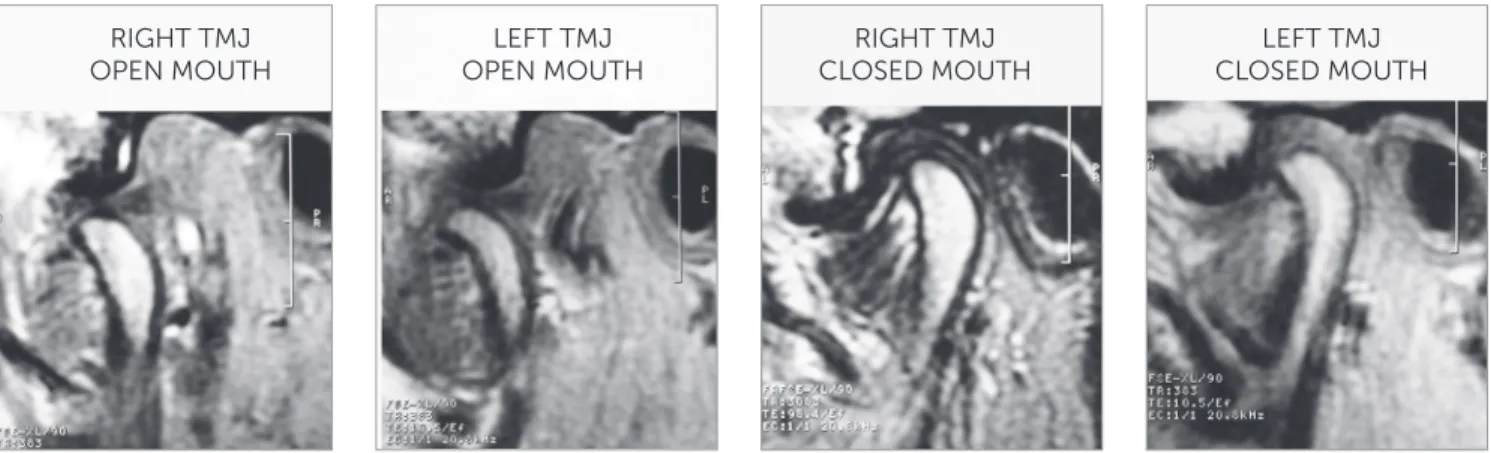
In order to confirm the clinical diagnosis and to create a baseline for comparison at the end of
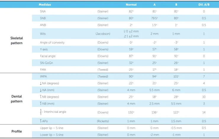
treat-ment, magnetic resonance of TMJs was requested. It revealed “anterior articular disc displacement with reduction” in both TMJs. Left displacement was greater than the right one (Fig 5). Cephalomet-ric analysis (Fig 6 and Table 1) showed a brachy-facial skeletal pattern (SN-GoGn = 25o; FMA = 17o; and Y-Axis = 57o) and balanced anteroposterior re-lationship between the maxilla and the mandible
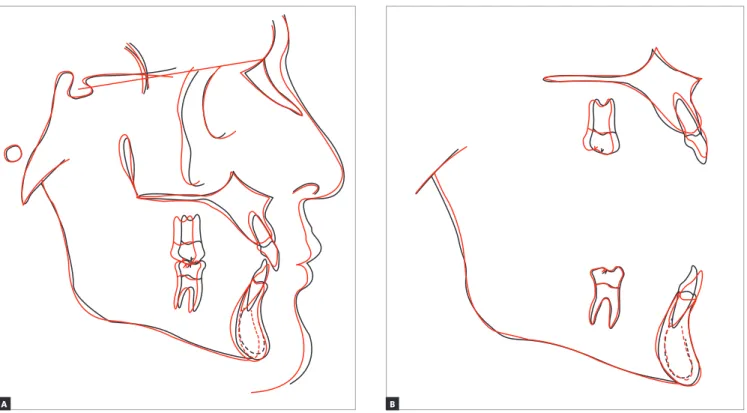
Figure 6 - Initial lateral cephalometric radiograph (A) and cephalometric tracing (B).
Figure 5 - Initial magnetic resonance of right and left TMJs, with opened and closed mouth.
B A
RIGHT TMJ OPEN MOUTH
LEFT TMJ OPEN MOUTH
RIGHT TMJ CLOSED MOUTH
(SNA = 81o; SNB = 79.5o; and ANB = 1,5o), as well as well-positioned maxillary incisors (1.NA = 21o, 1-NA = 5.5mm), and retruded and retroclined man-dibular incisors (1.NB = 18o, and 1-NB = 2.5mm), in relation to their supporting bone bases.
TREATMENT PLAN
The treatment objectives were the correction of Class II Division 2 subdivision let, anterior deep over-bite, dental midline deviations and crowding.
When explaining to the patient what his orth-odontic problems were, it was pointed out that, ac-cording to the current literature evidence, there was no guarantee that the correction of his malocclusion would solve his chief complaints of clicking and pain in his TMJs. However, it was also explained that Class II Division 2 subdivision left and anterior deep overbite are contributing factors to increase the pain and discomfort in TMJs.
With these clariications, the patient decided to un-dergo orthodontic treatment to improve his dental oc-clusion, and airmed that he was fully aware that orth-odontic treatment could have no efect in his clicking and occasional pain in the TMJs.
Considering the skeletal bases balance (brachy-facial and Class I skeletal pattern) and the pleasant facial esthetics, the following treatment plans were discussed:
1) Distalization of let maxillary posterior teeth using asymmetric Kloehn low-pull extraoral headgear (HG), until Class II correction, followed by corrective orth-odontic treatment, with mandibular incisors and ca-nines intrusion.
2) Aligning and leveling of mandibular and max-illary teeth, followed by the correction of let Class II with temporary anchorage devices (TADs).
3) Extraction of let maxillary irst premolar, right maxillary second premolar and mandibular second pre-molars, followed by corrective orthodontic treatment using Tweed-Merriield technique, with high-pull “J-hook” extraoral headgear.
Orthodontic treatment involving dental extractions was discarded, because it could latten the proile and worse the deep overbite, in addition to increasing TMJs pain (TMJs pain was occasional) caused by the “ante-rior articular disc displacement with reduction” in both TMJs and clenching habits.
After understanding that the orthodontic treat-ment to correct the left Class II utilizing Kloehn HG would promote more extrusion of posterior teeth than utilizing TADs, and that this extrusion could contribute to decrease the TMD pain, the patient opted for the treatment plan #1, i.e., using asymmet-ric Kloehn low-pull extraoral HG, followed by cor-rective orthodontic treatment, with mandibular inci-sors and canines intrusion.
TREATMENT PROGRESS
Treatment was initiated with asymmetric HG. Al-though the let maxillary posterior teeth distalization was taking time, the patient insisted to continue utiliz-ing HG and did not want to use TADs.
When the let Class II was corrected, 0.022 x 0.028-in edgewise standard brackets were bonded on the remain-ing maxillary teeth, until the correction of the midline deviation.
Once aligning and leveling of maxillary incisors permitted, 0.022 x 0.028-in edgewise standard brack-ets were bonded on the mandibular teeth. So, it was possible to align and level mandibular teeth, not only intruding incisors and canines, but also correcting the midline deviation.
As maxillary lateral incisors presented size anomaly, ater the brackets and bands removal, the general dentist reshaped these teeth with composite resin.
Fixed mandibular canine-to-canine lingual arch and Hawley maxillary removable appliance were utilized as retention.
RESULTS
In the end of orthodontic treatment, the patient re-ported that he no longer felt pain in the TMJs. So, a new magnetic resonance exam was requested.
The magnetic resonance (Fig. 7) showed that “anterior articular disc displacement with reduction” in both TMJs persisted. This inding was explained to the patient, who understood the need of monitoring his clenching habit.
preserved, and the maxillary midline right devia-tion was corrected. As the discreet nose deviadevia-tion to the left persisted, it caused the impression that slight maxillary and mandibular midline deviations in relation to the midsagittal plane were present.
With the performed orthodontic treatment, the an-teroposterior relationship between let posterior teeth, the overbite as well as the maxillary and mandibular midline deviations were corrected, and the normal overjet was maintained (Figs 8 and 9).
In the panoramic radiograph (Fig 10) and in the peri-apical radiographs (Fig 11), no alterations were veriied. Dental and periodontal health were maintained.
Final cephalometric analysis (Fig 12, Table 1) showed a slight mandibular plane opening (SN-GoGn = 26o; FMA = 18o; and Y-axis = 58o), and maintenance of a balanced anteroposterior relationship between maxilla and mandible (SNA = 81o; SNB = 80o; and ANB = 1o). Besides, mandibular incisors were buccally inclined
(1.NB = 28o and 1-NB = 5.5mm), and maxillary incisors presented with a discreet buccal inclination (1.NA = 25o and 1-NA = 6mm).
Total cephalometric superimposition (Fig 13) dem-onstrated a slight mandibular plane opening, due to the let maxillary posterior teeth distalization, and a discreet improvement of facial lower proile, caused by the im-proved balance of upper and lower lips.
Maxillary cephalometric superimposition (Fig 13) showed let maxillary molar distalization, without ex-trusion, and intrusion of maxillary incisors, with lingual root torque and with crowns maintaining their antero-posterior position.
Mandibular cephalometric superimposition (Fig 13) evinced that anteroposterior and vertical position of let mandibular molar was maintained. Besides, man-dibular incisors were intruded and buccaly inclined, with their crowns moving buccally and their roots moving lingually.
Figure 7 - Final magnetic resonance of right and left TMJs, with opened and closed mouth. RIGHT TMJ
OPEN MOUTH
LEFT TMJ OPEN MOUTH
RIGHT TMJ CLOSED MOUTH
Figure 8 - Final facial and intraoral photographs.
Figure 10 - Final panoramic radiograph.
Figure 11 - Final periapical radiographs.
Figure 12 - Final lateral cephalometric radiograph (A) and cephalometric tracing (B).
Figure 13 - Total superimposition (A), maxillary and mandibular superimpositions (B) of initial (black) and final (red) cephalometric tracing.
B A
FINAL CONSIDERATIONS
Orthodontic treatment of patients with chief complaint of temporomandibular disorders (TMD) is complex, because this dysfunction has many eti-ological factors and the literature in this subject is controversial. According to some authors, it is not possible to affirm that malocclusions are an etiologi-cal factor of TMD1-3, while other authors claim the opposite.6 Additionally, it is not possible to affirm that orthodontics plays a relevant etiological or ther-apeutic role in TMDs.4,5
Because of that, it is important that, before any dental intervention, differential diagnosis is made, re-quiring medical assessment to rule out the possibility of presence of systemic pathologies with similar TMD symptoms, as gout,7 osteosarcoma8 and pseudotumor9 in TMJs, Eagle’s syndrome,10 fibromyalgia,11 rheu-matoid arthritis12 and trigeminal neuralgia.13
Following this principle, in this case, any other medical pathology which could cause similar TMDs symptoms was excluded.
Considering both the clicking in the end of mouth opening and closing movements, and the magnetic resonance images, the diagnosis of “anterior articu-lar disc displacement with reduction” was reached. This is an intracapsular disorder, in which the disc is in an anterior position relative to the condylar head, in the closed mouth position, and the disc reduces upon opening of the mouth.14
Table 1 - Baseline (A) and final (B) cephalometric values
Medidas Normal A B Dif. A/B
Skeletal pattern
SNA (Steiner) 82o 81o 81o 0
SNB (Steiner) 80o 79.5o 80o 0.5
ANB (Steiner) 2o 1.5o 1o 0.5
Wits (Jacobson) ♀ 0 ±2 mm
♂ 1 ±2 mm 2 mm 1 mm 1
Angle of convexity (Downs) 0o -2o -3o 1
Y-axis (Downs) 59o 57o 58o 1
Facial angle (Downs) 87o 91o 91o 0
SN-GoGn (Steiner) 32o 25o 26o 1
FMA (Tweed) 25o 17o 18o 1
Dental pattern
IMPA (Tweed) 90o 94o 101o 7
1.NA (degrees) (Steiner) 22o 21o 25o 4
1-NA (mm) (Steiner) 4 mm 5.5 mm 6 mm 0.5
1.NB (degrees) (Steiner) 25o 18o 28o 10
1-NB (mm) (Steiner) 4 mm 2.5 mm 5.5 mm 3
1
1- Interincisal angle (Downs) 130o 136o 122o 14
1-APo (Ricketts) 1 mm 1 mm 1.5 mm 0.5
Profile
Upper lip — S-line (Steiner) 0 mm 0 mm -0.5 mm 0.5
Lower lip — S-line (Steiner) 0 mm -2 mm -1 mm 1
et al15 verified, there is correlation among TMD, deep overbite and Class II malocclusion, when these vari-ables are associated with clenching habit, although is not possible to affirm if occlusal factors are predispos-ing, precipitating or perpetuating the disease.
Considering the malocclusion, the orthodontic treatment could be with or without extractions, uti-lizing either HG or TADs. However, as there was the possibility of overbite increase, that would accentuate TMD symptoms, and because of the pleasant profile, the chosen treatment plan was without extractions.
The choice for HG utilization, instead of TADs, in the treatment, aimed to act not only in the maloc-clusion but also in the TMD.
There is not enough evidence to measure the efects of orthodontic treatment in the signs and symptoms of TMD.16 However, there are studies that point out that orthodontic appliances are as efective as interoclusal
devices, in the treatment of pain in “anterior articular disc displacement with reduction” cases.17
In this case, the utilization of Kloehn HG aimed to take advantage of its potential to distalize and ex-trude molars.18 If both of these movements occurred, not only the left Class II malocclusion would be cor-rected, but also the TMD symptoms would be re-lieved, through the seven common features of con-ventional interocclusal devices, described by Oke-son19: 1) change of occlusal conditions; 2) change of condylar position; 3) increase in vertical dimension; 4) cognitive sensitization; 5) placebo effect; 6) in-crease in peripheral stimuli to central nervous system; and 7) symptoms regression to the mean.
REFERENCES
1. Iodice G, Danzi G, Cimino R, Paduano S, Michelotti A. Association between posterior crossbite, masticatory muscle pain, and disc displacement: a systematic review. Eur J Orthod. 2013 Dec;35(6):737-44.
2. Greene CS. Relationship between occlusion and temporomandibular
disorders: Implications for the orthodontist. Am J Orthod Dentofacial Orthop. 2011 Jan;139(1):11, 13, 15.
3. Kanavakis G, Mehta N. The role of occlusal curvatures and maxillary arch dimensions in patients with signs and symptoms of temporomandibular disorders. Angle Orthod. 2014 Jan;84(1):96-101.
4. Leite RA, Rodrigues JF, Sakima MT, Sakima T. Relationship between temporomandibular disorders and orthodontic treatment: a literature review. Dental Press J Orthod. 2013 Jan-Feb;18(1):150-7.
5. Manfredini D, Stellini E, Gracco A, Lombardo L, Nardini LG, Siciliani G. Orthodontics is temporomandibular disorder-neutral. Angle Orthod. 2016 July;86(4):649-54.
6. Slavicek R. Relationship between occlusion and temporomandibular
disorders: implications for the gnathologist. Am J Orthod Dentofacial Orthop. 2011 Jan;139(1):10, 12, 14 passim.
7. Oliveira INF, Gomes RCF, Santos RR, Oliveira TP, Pereira LLC, Mainenti P. Gout of the temporomandibular joint: report of a case. Int Arch Otorhinolaryngol. 2014 July;18(3):316-8.
8. Uchiyama Y, Matsumoto K, Murakami S, Kanesaki T, Matsumoto A,
Kishino M, et al. MRI in a case of osteosarcoma in the temporomandibular joint. Dentomaxillofac Radiol. 2014;43(2):20130280.
9. Yoshitake H, Kayamori K, Nakamura R, Wake S, Harada K. Pseudotumor
in the temporomandibular joint: a case report. Int J Surg Case Rep. 2015;15:5-9.
10. Thoenissen P, Bittermann G, Schmelzeisen R, Oshima T, Fretwurst T. Eagle’s syndrome: a non-perceived diferential diagnosis of temporomandibular disorder. Int J Surg Case Rep. 2015;15:123-6.
11. Salvetti G, Manfredini D, Bazzichi L, Bosco M. Clinical features of the stomatognathic involvement in ibromyalgia syndrome: a comparison with temporomandibular disorders patients. Cranio. 2007 Apr;25(2):127-33. midline deviation and left mandibular midline de-viation were corrected and the normal overjet was maintained. Functional harmony during protrusive, as well as right and left lateral movements, was ob-tained. Pleasant facial profile was maintained and there was only slight change in vertical dimension. In this case, there was no concern about third mo-lars, because they had been extracted in an attempt to solve the functional problems. In the end of orth-odontic treatment, as the patient reported that he no longer felt pain in the TMJs, a new magnetic resonance was requested. There was the expecta-tion that the TMJs articular discs posiexpecta-tion could have been corrected. Unfortunately, the magnetic resonance image showed the maintenance of the
“anterior articular disc displacement with reduc-tion” in both TMJs, practically with the same sever-ity identified in the initial magnetic resonance.
Possibly, although in a short period of time, the remission of pain symptoms could have been in-fluenced by the process of retrodiscal tissue fibro-sis, which created a “pseudodisc” over the condyle head,20 and provided protection to previously dam-aged structures.
Therefore, the corrective orthodontic treatment can be considered successful, because the facial bal-ance of the patient was maintained, the initial dimen-sions of dental arches were respected, good dental intercuspation was achieved, and mandibular move-ments with immediate desocclusion were established.
12. Abrão ALP, Santana CM, Bezerra ACB, Amorim RFB, Silva MB, Mota LMH, et al. What rheumatologists should know about orofacial manifestations of autoimmune rheumatic diseases. Rev Bras Reumatol. 2016;56(5):441-50. 13. Pihut M, Szuta M, Ferendiuk E, Zenczak-Wiwckiewicz D. Diferential
diagnostics of pain in the course of trigeminal neuralgia and temporomandibular joint dysfunction. BioMed Res Int. 2014(2014):1-7. 14. Schifman E, Ohrbach R, Truelove E, Look J, Anderson G, Goulet JP, et al.
Diagnostic criteria for temporomandibular disorders (DC/TMD) for clinical and research applications: recommendations of the international RDC/TMD consortium network and orofacial pain special interest group. J Oral Facial Pain Headache. 2014 Winter;28(1):6-27.
15. Costa MD, Froes Júnior GRT, Santos CN. Evaluation of occlusal factors in patients with temporomandibular joint disorder. Dental Press J Orthod. 2012;17(6):61-8.
16. Luther F, Layton S, McDonald F. WITHDRAWN: Orthodontics for treating temporomandibular joint (TMJ) disorders. Cochrane Database Syst Rev. 2016 Jan 7;(1):CD006541.
17. Tecco S, Teté S, Crincoli V, Festa MA, Festa F. Fixed orthodontic therapy in temporomandibular disorder (TMD) treatment: an alternative to intraoral splint. Cranio. 2010 Jan;28(1):30-42.
18. Melsen B. Efects of cervical anchorage during and after treatment: an implant study. Am J Orthod. 1978;73(5):526-40.
19. Okeson JP. Occlusal Appliance Therapy. In: Management of temporomandibular disorders and occlusion. 6th ed. St. Louis: Elsevier Mosby; 2008.