Orthodontic movement of teeth with short root
anomaly: Should it be avoided, faced or ignored?
Jose Valladares Neto1, José Rino Neto2, João Batista de Paiva3
How to cite this article: Valladares Neto J, Rino Neto J, Paiva JB. Orthodontic movement of teeth with short root anomaly: Should it be avoided, faced or ig-nored? Dental Press J Orthod. 2013 Nov-Dec;18(6):72-85.
Submitted: August 11, 2011 - Revised and accepted: December 03, 2011
» The authors report no commercial, proprietary or inancial interest in the prod-ucts or companies described in this article.
Contact address: José Valladares Neto
Rua 132, 113, Quadra F-29, Setor Sul – Goiânia/GO — Brazil — CEP: 74.093-210 E-mail: [email protected]
» Patients displayed in this article previously approved the use of their facial and in-traoral photographs.
1 Assistant Professor, Department of Orthodontics, College of Dentistry, Federal
University of Goiás (UFG).
2 Associate Professor, Department of Orthodontics and Pediatric Dentistry,
Col-lege of Dentistry, University of São Paulo (USP).
3 Full Professor, Department of Orthodontics and Pediatric Dentistry, College of
Dentistry, University of São Paulo (USP).
Introduction: Short Root Anomaly (SRA) is an uncommon disease and a challenge for orthodontic treatment as it tends to increase the risk of root resorption. Objective: Assess the current status of the diagnosis, etiology and orthodon-tic management of teeth with SRA, and present case reports. Method: A literature review was carried out in PubMed, SciELO, LILACS, Scopus and Web of Science databases. Results: A differential diagnosis of SRA should be conducted for teeth with incomplete root formation, external apical root resorption, dentin dysplasia type I and post dental trauma root hypoplasia. SRA is genetically determined and orthodontic movement requires changes in clinical and radiographic management in order to restrict damage. Conclusion: Orthodontic movement of teeth with SRA is contraindicated in extreme cases, only. Caution at all stages could minimize attachment loss and lead to long-term stability.
Keywords:Tooth root. Tooth abnormalities. Root resorption. Orthodontics.
Introdução: a anomalia de raiz curta (ARC) é uma patologia incomum e constitui um desafio para o tratamento ortodôn-tico, pois tende a elevar o risco de reabsorção radicular. Objetivo: revisar a literatura sobre o diagnóstico, a etiologia e a con-duta ortodôntica recomendada para esses casos, ilustrando esse tema com relatos de caso. Métodos: devido ao baixo nível de evidência sobre o tema, realizou-se a revisão narrativa da literatura com estratégia de busca nas bases de dados PubMed, SciELO, Lilacs, Scopus e Web of Science. Resultados: o diagnóstico diferencial da ARC deverá ser realizado entre dentes com rizogênese incompleta, reabsorção radicular externa apical, displasia dentinária tipo I e hipoplasia radicular pós-trau-matismo dentário. A ARC apresenta determinantes genéticos para o encurtamento, e a movimentação ortodôntica exige que medidas de controle clínico e radiográfico sejam adotadas, ensejando a restrição de danos. Conclusão: a movimentação ortodôntica de dentes com ARC somente é contraindicada em casos extremos. A conduta cautelosa em todas as suas etapas poderá minimizar a perda de inserção e lograr em estabilidade em longo prazo.
INTRODUCTION
The term short root anomaly (SRA),12,19 also
known as root dwarfism,27 was introduced and first
described by Lind,19 in 1972, to categorize a rare
developmental anomaly characterized by full root formation, but with genetically determined fore-shortening. The affected root is smaller or presents the same size of the dental crown, has a tendency towards bilateral involvement and presents no other
etiological factors.2,3,12
Epidemiologically, the prevalence of SRA ranges
from 0.6 to 2.4%,2,15 but may reach 10% among the
Japanese.5 The incidence is higher in females with a
ratio that ranges from 1:2.6 to 1:2.7.3,15 The most
af-fected teeth are the maxillary central incisors and sec-ond premolars, while other teeth may also be
affect-ed.2,19 Some reports present extreme cases in which
all teeth are affected, in which case it is described as
generalized or multiple SRA.12,24,27 The condition
may also be associated with systemic changes or
syn-dromes31 and the absence of reports of SRA in
pri-mary dentition presupposes that it occurs exclusively
in permanent dentition.3
The literature has given little attention to orthodon-tic movement of teeth with SRA, despite the high risk
of root resorption.17 The fact that the number of cases
reported is limited could be partially explained by lack of identiication of this anomaly which is misdiagnosed
as root resorption.10 For this reason, concerns about the
diagnosis, etiology and orthodontic movement of teeth with SRA led to the objectives of this study.
MATERIAL AND METHODS
The present literature review was undertaken in the form of searches in PubMed, SciELO, LILACS, Scopus and Web of Science databases. Out of a total of 60 articles found, 35 were selected for this review, covering the topics described below.
Short roots: definition and differential diagnosis
Short roots have origin in both physiological and pathological circumstances. When the maxil-lary central incisors and first molars erupt in the oral cavity, half of the roots are formed while the other teeth erupt with about ¾. The canines are an ex-ception, as they erupt with root almost complete. This transitional stage of root formation ends with the full development of the root when it reaches
oc-clusal contact.13
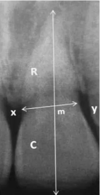
Lind19 developed a radiographic method for
mea-suring the relative length of the root, defined as the ratio between the length of the root and the length of the crown (R/C) (Fig 1). In a sample of 1,038 Swed-ish children, aged eleven or over, the normative value of R/C was 1.63 for males and 1.55 for females,
with-out any statistical difference.15 The root is considered
short when R/C < 1.1:1, in other words, when the
root is of the same size or smaller than the crown.5,15
There are several diagnostic possibilities for oc-clusal teeth with short roots. Several reasons explain the undertaking of a differential diagnosis for teeth with short roots, especially due to the frequency of
publications reporting wrong diagnosis10 (Fig 2):
• Incomplete root formation: a physiological
con-dition, tooth erupting and incomplete apex.13
• SRA: congenitally short root, complete root
formation, pointed or rounded apexes. Evi-dent bilateral involvement found only in per-manent dentition. It mainly affects the central incisors and second premolars, but it may also
be generalized.2,3,12,19
• External apical root resorption: decrease in
apical root length ater full root development. The apical format is typically rounded and ir-regular. It is triggered by various factors, in-cluding: orthodontic treatment, alveolodental trauma, periapical lesions, pericoronal follicle of
teeth with deviant eruption, among others.10
• Dentin dysplasia type 1: short root due to
atypi-cal dentinogenesis. It is associated with pulp obliteration and root surrounded by bone radio-lucence in cavity-free teeth. There is a possibility of loss and early exfoliation and the involvement
of both deciduous and permanent dentitions.28
• Post-trauma root hypoplasia in the deciduous
predecessor: short root due to interruption of normal dentinogenesis as a result of alveolodental
trauma. Generally unilateral involvement.30
Thus, SRA difers from other conditions character-ized by short roots. The diagnosis is randomly made through image, since the condition is asymptomatic. Clinical and radiographic indings are normal, except for shorter roots (Fig. 3). The low collagenolytic ac-tivity in periodontal tissues is diferent from cases of
periodontal disease or active root resorption.4 In cases
of severe root foreshortening, there may be an increase in tooth mobility, indicating loss of periodontal attach-ment and secondary occlusal trauma. Undoubtedly, the concomitant presence of SRA and apical dental resorp-tion makes the diagnosis more complex.
ETIOLOGY OF SRA
The etiology of SRA has not been fully deter-mined. Isolated cases of unknown origin have been
reported, and have been categorized as idiopathic.11
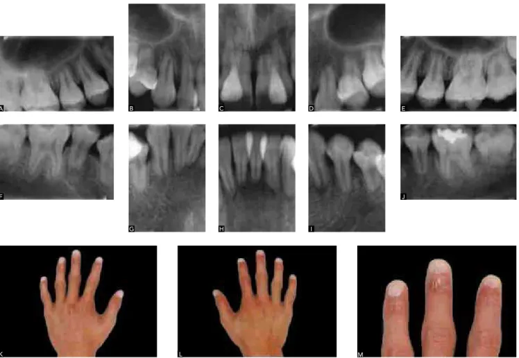
Figure 2 - Short roots due to: A) incomplete root formation, B) external apical root resorption, C) alveolodental trauma, and D) short root anomaly.
Figure 3 - Three-dimensional computed microtomography of proximal sur-faces (distal and mesial) of lower premolar with short root anomaly. In red, the dental pulp, and in white, the enamel (microtomographic X-ray Sky Scan 1076, NRecon and CTan software).
taurodontism, pulp obliteration, supernumerary
teeth, root dilaceration and ectopic eruption.2,11,28,35
In spite of that, the occurrence of SRA was not high-er in people with palate fissures, among whom thhigh-ere
is a high prevalence of dental abnormalities.1
A possible biological basis can be understood from the
regulatory role of the Nic gene.24
Immunohistochem-istry and in situ hybridization in rats has attributed the
formation of short abnormal roots to the absence of the Nic gene, responsible for the diferentiation of
odonto-blasts.24 Genetic sustainability is also enhanced by
bilat-eral involvement in the same individual, with a possibility
of generalized or multiple involvement.12,27 There have
been reports associating SRA with systemic changes or
syndromes, as in certain types of growth deiciency,26,35
scleroderma,14 hypoparathyroidism,16 tumoral
calcino-sis,8 congenital dyskeratosis34 and in syndromes, such as
Down,25 Stevens-Johnson,31 Turner,21 Klinefelter,33 and
Laurence-Moon-Bardet-Biedl17 (Fig. 4).
Others have been described as having a congenital origin, compatible with the term anomaly, because the roots are short as a result of a disturbance during
dental development.2,3,12,19
In this context, environmental and genetic influ-ences have been suggested. The influence of
alveolo-dental trauma,30 masticating stress5 and
chemother-apy/radiotherapy22 have all been reported as
inhibi-tors of root formation. However, evidence suggests that SRA has strong genetic origins, shown by
strik-ing family history.2,12,19 Nevertheless, because of the
apparent heterogeneity of the lineage, no definitive conclusions regarding the type of inheritance could be reached, so nonsyndromic SRA could be included
in the dominant and recessive autosomal models.2,12,19
In addition, the genetic theory is reinforced by the intense correlation of SRA with other tooth devel-opment anomalies, particularly hypodontia,
micro-dontia (cone-shaped lateral incisor), dens invaginatus,
clinical examination to assess the degree of tooth mobility, because if it is excessive, it will restrict the onset or continuity of orthodontic movement. Orth-odontic treatment planning should allow an objective
and coherent correction of the malocclusion.27,29 Light
intermittent forces with longer intervals between ac-tivation are recommended for the control of dental
resorption.18 Root resorption repair seems to depend
on time, with longer intervals between activations
producing more extensive repair.9 In addition, light
forces generate an adequate momentum/force ratio (M/F) resulting from the cervical dislocation of the center of resistance. Periodic periapical radiographs should be taken to monitor orthodontic movement in teeth with critical root length (Figs 5 and 6).
Implications of orthodontic movement in teeth with SRA
Orthodontic movement in teeth with short roots tends to generate a higher risk of root resorption,
probably inluenced by a lower R/C ratio.17 A test
car-ried out with the inite element method showed that short roots concentrate greater mechanical stress in
the middle of the root,23 which would supposedly be
harmful to the cementoblast layer because of extensive
hyalinization coming from the periodontal ligament.10
At first, orthodontic movement is only
contra-indicated for teeth with SRA in extreme cases.12
In most cases, clinical and radiographic
monitor-ing can be used to control dental resorption.20,27,29
The procedure should initially start with a careful
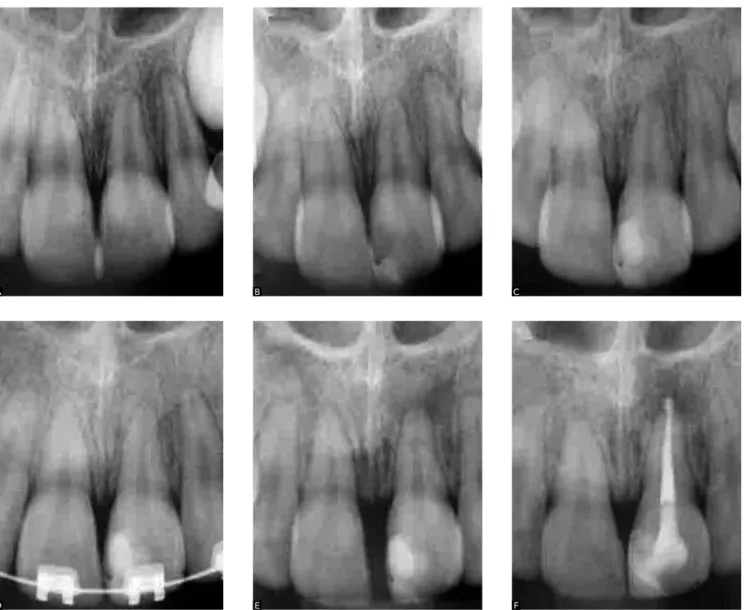
Figure 5 - Radiographic sequence of maxillary central incisors with short root anomaly (SRA). A) Mixed dentition indicating early stages of SRA; B) before, C) dur-ing and D, E, F) after orthodontic movement, showdur-ing relative control of external apical root resorption .
A B
E
C
Figure 6 - Radiographic sequence of upper sec-ond premolars with short root anomaly (SRA). A) Mixed dentition indicating the early stages of SRA; B) before, C) during and D,E,F) after orth-odontic movement, showing relative control of external apical root resorption.
Orthodontic movement of teeth with little inser-tion seems to be faster not because of more intense bone remodeling , but because of excessive inclina-tion . Despite poor evidence, post-treatment relapse seems to be immediate. Case reports involving orth-odontic movement in teeth with decreased insertion,
as in cases of advanced periodontal disease6 or after
an apicectomy,32 have recommended the use of a
permanent fixed splint. The same procedure should be recommended in cases of movement in incisors with SRA, in order to contain excessive tooth
mo-bility and ensure long-term stamo-bility.20
CASE REPORT
A brown-skinned female, aged 12 years and 11 months, complaining about dental esthetics, sought the clinic of Orthodontics specialization course in the Foundation for Scientific and Technological
De-velopment in Dentistry, at the College of Dentistry, University of São Paulo (USP) requesting orthodon-tic evaluation. The patient and her parents were most concerned about the presence of infralabial inclined canines. The anamnesis showed good general health, no history of dental or facial trauma and no previ-ous orthodontic treatment. At the time, family dental history was not an object of investigation.
The facial examination showed a brachyfacial pat-tern without any obvious skeletal asymmetry, convex facial proile, small nasal projection, and an acceptable nasolabial angle. The smile analysis showed adequate exposure of the upper incisors, taking her age into consideration, but indicated a slight dental midline de-viation to the right and crowding of the upper canines in a marked infralabial inclined position of the right canine (#13) (Fig 7). The intraoral evaluation showed good dental health and healthy periodontal tissues.
A B
E C
As for the dental pattern, it presented Class I relation-ship of right molars. The dental examination presented let subdivision Class II (1/2) malocclusion. The upper
right canine erupted in an infralabial inclined position,
while the upper let canine was partially erupted due to space deiciency. Overjet and overbite were moderate and both the upper and lower dental midlines were devi-ated to the right side (Figs 8 and 9). In addition to these aspects, the upper arch was triangular-shaped due to the absence of canines in the dental arch. The shape of the lower jaw was adequate, but with a slight asymmetry in the position of canines and molars, probably attributed to their displacement as a result of the clinical absence of the right second lower premolar (#45). The second decidu-ous let lower molar (#75) was still in place (Fig. 10).
The cephalometric analysis showed a mild Class II
skeletal pattern (ANB = 4.5o), with the maxilla well
positioned in relation to the cranial base (SNA = 83o)
and slight retrusion of the mandible in relation to
the cranial base (SNB = 78.5o), with balanced facial
growth (SN-GoGn = 32o), slightly protruded and
la-bially inclined upper incisors (1-NA = 6.5 mm and
1.NA = 25o) and obvious compensation of the
low-er incisors (1-NB = 7.0 mm and 1.NB = 33o). The
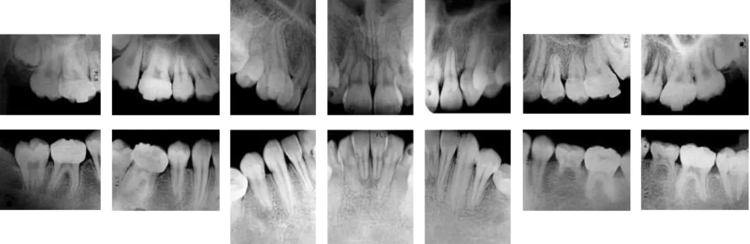
examination of the extraoral radiographs confirmed the absence of significant skeletal asymmetry and the agenesis of the second lower left and right premolars (#35 and 45), as well as the lower right third molar (#48) (Fig 11). The examination of the periapical ra-diographs showed the presence of short root anomaly for the central incisors (#11, 21, 31 and 41) and upper premolars (#15, 14, 24 and 25), and apical root dilac-eration for the upper right incisors (#12) (Fig 12).
Considering that the problem of crowded canines was one of the main reasons why the patient sought orth-odontic treatment, straightening and leveling of teeth
Figure 7 - Initial extraoral photographs.
Figure 9 - Intraoral photograph of the infralabial inclination of the upper canine.
Figure 10 - Initial intraoral occlusal photographs.
Figure 12 - Initial periapical radiographs.
Figure 13 - Intermediate intraoral photographs.
and correction of the dental midlines were the aims of the treatment. Molar and canine normal relation were achieved , and overbite and overjet were reduced. In addition, it was decided to close the space correspond-ing to the misscorrespond-ing let lower premolar.
Treatment plan and progress were performed with ixed orthodontic appliances on the upper and lower arch-es, preceded by the extraction of the irst upper premolars (#14 and 24), and the installation of Klöehn headgear (350 g/14 hours). Correction of midline deviations was aided by open coil springs, anterior retraction and inferior recipro-cal mechanics to close the agenesis space of the let second lower premolar (#35) (Figs 13 and 14).
Extraction space was then closed and orthodon-tic detailing performed with the inclusion of second
molars and individual bends and torques in the 0.019 x 0.025-in stainless steel arch (Fig 15). Radiographic monitoring was carried out in resorption critical areas, especially ater agenesis space closure (Figs 16 and 17). Retention was planned by means of a wraparound re-movable plate and a ixed intercanine bar with 0.028-in orthodontic wire for the lower teeth.
The esthetic and functional goals proposed at the onset of treatment were achieved, due to the meticu-lous use of orthodontic mechanics, radiographic mon-itoring at various stages of the treatment and longer intervals between clinical sessions. As final outcomes, good occlusal relationship between the arches, Class I relationship between molars and canines, and normal levels of overbite and overjet were achieved (Fig 18).
Figure 15 - Intermediate intraoral photographs of finishing phase.
Figure 18 - Final intraoral photographs.
Figure 19 - Final intraoral (occlusal) photographs. Figure 17 - Intermediate extraoral teleradiograph and panoramic radiographs.
These results provided occlusal balance and excur-sive mandibular movements without interference, and periodontal tissues in good conditions. Dental arch morphology and facial balance were also considered sat-isfactory at the end of the treatment (Figs 19 and 20). The inal extraoral radiographs and cephalometric su-perimposition are shown in Figures 21 and 22.
Figure 20 - Final extraoral photographs.
Figure 22 - Cephalometric superimposition before (black) and after (red) orthodontic treatment. A) total; B) partial; C) facial profile and line S.
Figure 23 - Final intraoral (periapical) radiographs.
CONCLUSION
Orthodontic movement of teeth with SRA tends to increase the risk of root resorption, however, treatment is contraindicated in extreme cases, only. Differential diagnosis and an understanding of the etiological reasons for teeth with short roots enable orthodontic movement which requires biomechani-cal adaptations, periodic radiographic monitoring, clinical monitoring of tooth mobility and permanent retention, especially for the incisors.
Pre-treatment Post-treatment
A B C
Acknowledgements
1. Akcam MO, Evirgen S, Uslu O, Memikoglu UT. Dental anomalies in individuals with cleft lip and/or palate. Eur J Orthod. 2010;32(2):207-10. 2. Apajalahti S, Arte S, Pirinen S. Short root anomaly in families and its
association with other dental anomalies. Eur J Oral Sci. 1999;107(2):97-101. 3. Apajalahti S, Holtta P, Turtola L, Pirinen S. Prevalence of short root anomaly
in healthy young adults. Acta Odontol Scand. 2002;60(1):56-9.
4. Apajalahti S, Sorsa T, Ingman T. Matrix metalloproteinase -2, -8, -9, and -13 in gingival crevicular luid of short root anomaly patients. Eur J Orthod. 2003;25(4):365-9.
5. Ando S, Kiyokawa K, Nakashima T, Shinbo K, Sanka Y, Oshima S, et al. Studies on the consecutive surgery of succedaneous and permanent dentition in the Japanese children. 4. Behavior of short-rooted teeth in the upper bilateral central incisors. J Nihon Univ Sch Dent. 1967;9(2):67-82. 6. Arun T, Sayinsu K, Nalbantgil D. Orthodontic approach for patients with
severe periodontal disease. World J Orthod. 2005;6(3):275-80.
7. Borgström MK, Riise R, Törnqvist K, Granath L. Anomalies in the permanent dentition and other indings in 29 individuals with Laurence-Moon-Bardet-Biedl syndrome. J Oral Pathol Med. 1996;25(2):86-9.
8. Burkes Jr EJ, Lyles KW, Dolan EA, Giammara B, Hanker J. Dental lesions in tumoral calcinosis. J Oral Pathol Med. 1991;20(5):222-7.
9. Cheng LL, Türk T, Elekdağ-Türk S, Jones AS, Yu Y, Darendeliler MA. Repair of root resorption 4 and 8 weeks after application of continuous light and heavy forces on premolars for 4 weeks: a histology study. Am J Orthod Dentofacial Orthop. 2010;138(6):727-34.
10. Consolaro A, Martins-Ortiz MF, Consolaro RB. O primeiro estudo sobre hereditariedade relacionada com as reabsorções dentárias em Ortodontia: uma análise crítica do trabalho de Newman. Rev Dental Press Ortod Ortop Facial. 2004;9(2):110-22.
11. Desai RS, Vanaki SS, Puranik RS, Rashmi GS, Nidawani P. An unusual combination of idiopathic generalized short-root anomaly associated with microdontia, taurodontia, multiple dens invaginatus, obliterated pulp chambers and infected cyst: a case report. J Oral Pathol Med. 2006;35(7):407-9.
12. Edwards DM, Roberts GJ. Short root anomaly. Br Dent J. 1990;169(9):292-3. 13. Estrela C, Valladares-Neto J, Bueno MR, Guedes AO, Porto OCL, Pécora JD. Medidas lineares dos estágios de desenvolvimento da dentição permanente humana usando Tomograia Computadorizada de Feixe Cônico: um estudo preliminar. Dental Press J Orthod. 2010;15(5):44-78.
14. Foster TD, Fairburn EA. Dental involvement in scleroderma. Br Dent J. 1968;124(8):353-6.
15. Jakobsson R, Lind V. Variation in root length of the permanent maxillary central incisor. Scand J Dent Res. 1973;81(4):335-8.
16. Jensen SB, Illum F, Dupont E. Nature and frequency of dental changes in idiopathic hypoparathyroidism and pseudohypoparathyroidism. Scand J Dent Res. 1981;89(1):26-37.
17. Kjaer I. Morphological characteristics of dentitions developing excessive root resorption during orthodontic treatment. Eur J Orthod. 1995;17(1):25-34.
REFERENCES
18. Levander E, Malmgren O, Eliasson S. Evaluation of root resorption in relation to two orthodontic treatment regimes. A clinical experimental study. Eur J Orthod. 1994;16(3):223-8.
19. Lind V. Short root anomaly. Scand J Dent Res. 1972;80(2):85-93.
20. Marques LS, Generoso R, Armond MC, Pazzini CA. Short-root anomaly in an orthodontic patient. Am J Orthod Dentofacial Orthop. 2010;138(3):346-8. 21. Midtbo M, Halse A. Root length, crown height, and root morphology in
Turner syndrome. Acta Odontol Scand. 1994;52(5):303-14.
22. Nasman M, Björk O, Söderhäll S, Ringden O, Dahllöf G. Disturbances in the oral cavity in pediatric long-term survivors after diferent forms of antineoplastic therapy. Pediatr Dent. 1994;16(3):217-23.
23. Oyama K, Motoyoshi M, Hirabayashi M, Hosoi K, Shimizu N. Efects of root morphology on stress distribution at the root apex. Eur J Orthod. 2007;29(2):113-7.
24. Park J-C, Herr Y, Kim H-J, Gronostajski RM, Cho M-I. Nic gene disruption inhibits diferentiation of odontoblasts responsible for root formation and result in formation of short and abnormal roots in mice. J Periodontol. 2007;78(9):1795-802.
25. Prahl-Andersen B, Oerlemans J. Characteristics of permanent teeth in persons with trisomy G. J Dent Res. 1976;55(4):633-8.
26. Shaw L. Short root anomaly in a patient with severe short-limbed dwarism. Int J Paediatr Dent. 1995;5(4):249-52.
27. Silva Filho OG, Mateus SRM, Silva VB, Oliveira GAG. Nanismo radicular. Ortodontia 2007;40(4):305-11.
28. Steidler NE, Radden BG, Reade PC. Dentinal dysplasia: a clinicopathological study of eight cases and review of the literature. Br J Oral Maxillofac Surg. 1994;22(4):274-86.
29. Tanaka OM, Knop LH, Shintcovsk RL, Hirata TM. Treatment of a patient with severely shortened maxillary central incisor roots. J Clin Orthod. 2008;42(12):729-31.
30. Tewari N, Pandey RK. Root hypoplasia: an unusual sequel to primary tooth trauma. Dent Traumatol. 2010;26(1):115-7.
31. Thornton JB, Worley SL. Short root anomaly in a patient with a history of Stevens-Johnson syndrome: report of case. ASDC J Dent Child. 1991;58(3):256-9.
32. Valladares Neto J, Costa SP, Estrela C. Orthodontic-surgical-endodontic management of unerupted maxillary central incisor with distoangular root dilaceration. J Endod. 2010;36(4):755-9.
33. Varrela J, Alvesalo L. Taurodontism in 47 XXY males: an efect of the extra X chromosome on root development. J Dent Res. 1988;67(2):501-2. 34. Yavuzyilmaz E, Yamalik N, Yetgin S, Kansu O. Oral-dental indings in
dyskeratosis congenital. J Oral Pathol Med. 1992;21(6):280-4.