State University of Campinas – Campinas/SP – Brasil. Email: [email protected]
Received for publication on September 22, 2006. Accepted for publication on October 16, 2006.
CLINICAL SCIENCES
ENDOSCOPIC STUDY OF THE INTRANASAL OSTIUM
IN EXTERNAL DACRYOCYSTORHINOSTOMY
POSTOPERATIVE. INFLUENCE OF SALINE
SOLUTION AND 5-FLUOROURACIL
Marilisa Nano Costa, Ana Maria Marcondes, Eulalia Sakano, Newton Kara-José
Costa MN, Marcondes AM, Sakano E, Kara-José N. Endoscopic study of the intranasal ostium in external dacryocystorhinostomy postoperative. Influence of saline solution and 5-fluorouracil. Clinics. 2007;62(1):41-6.
PURPOSE: To study, through endoscopy, the postoperative structural changes of the intranasal ostium following external
dacryocystorhinostomy and to evaluate the influence of saline solution and 5-fluorouracil.
METHODS: Fifty patients were distributed into the following groups: Group SS—dacryocystorhinostomy and an injection of
saline solution during surgery (13 patients); Group 5-FU1—dacryocystorhinostomy and an injection of 5 fluorouracil during surgery (17 patients); Group C—dacryocystorhinostomy only (11 patients); Group 5-FU3—dacryocystorhinostomy and 3 injections, 1 during surgery and 1 on the third and fifth postsurgical days (9 patients).
RESULTS: Pair-wise group comparisons using the nonparametric Mann-Whitney test revealed that there was a significant reduction of the ostium area only in Group 5-FU1 vs. Group SS on the 60th postoperative day (
P<.01); however, a comparative study among the 4 groups using the Kruskal-Wallis test showed no significant changes in the ostium area on the 60th postoperative day. The ostium area within groups at the 30th vs 60th postoperative day was significantly reduced for Group C (
P < .05; Mann-Whitney test); no significant changes were found for the other groups.
DISCUSSION:These results suggest that the use of 5-fluorouracil in external dacryocystorhinostomy does not significantly
influence the final size of the surgical fistula as determined 2 months postsurgery.
KEYWORDS: Fluorouracil. Dacryocystorhinostomy. Endoscopy. Dacryocystitis.
INTRODUCTION
Dacryocystorhinostomy is a surgical technique in which an anastomosis is made between the lacrimal sac and the nasal cavity. It is indicated for most of chronic dacryocistitis. The percentage of success with dacryo-cystorhinostomy presented by several authors is usually greater than 90%, regardless of the surgical technique used.1,2
In spite of technical advances, recurrences may occur within 4 to 6 weeks after surgery.3 Anatomic conditions and
affections that may lead to lack of success include a small lacrimal sac and ethmoid cells in the bone trepanation area,4
Paget’s disease,5 sarcoidosis,6 nasal fracture, medium
tur-binate hypertrophy, septal deviation, and nasal cavity syn-echia.7
The most frequent postoperative conditions associated with recurrences are obstruction of the intranasal ostium by healing or granulation tissue, nasal cavity synechia,8
in-adequate size and position of the osteotomy,9 mucosa flap
fall,7 and incomplete lacrimal sac opening.10
In dacryocystorhinostomy, several materials, such as a polyethylene tubing,11 silicone sponge,12 silicone tubing,2
and absorbable gelatin sponge,13 have been used in the
At the time of writing, antimetabolites are frequently used when treating a number of ocular pathologies char-acterized by the proliferation of non-neoplastic cells; 5-fluorouracil (5-FU) is one of such drugs, because it has a powerful inhibitory effect on mitosis. Responses are gen-erally good when compared to other antimetabolites and 5-FU has been shown to have a powerful inhibitory effect on growing fibroblasts.14
The Fluorouracil Filtering Surgery Study Group (FFSS), formed in the United States in 1989, evaluated the results of trabeculectomy with the use of subconjunctival 5-FU. Sub-conjunctival 5-FU injections were recommended after trabeculectomy in patients with uncontrolled glaucoma with poor prognosis, especially with aphakic glaucoma, and in patients with previous surgical failure.15 The FFSS concluded
after 5 years of follow-up that 5-FU greatly improves the surgical prognosis of trabeculectomy.16 Use of 5-FU is a safe
and effective method by which a significant number of fail-ing filtration blebs can be rescued from failure.17
This study aimed at studying, through endoscopy the postoperative structural changes of the intranasal ostium that take place following external dacryocystorhinostomy, as well as to evaluate the influence of the use of saline so-lution versus 5-FU on the evoso-lution of the ostium, seeking a complementary alternative for the treatment of dacryo-cystitis with a poor prognosis.
MATERIALS AND METHODS
Patients with dacryocystitis were selected from the Oph-thalmology Clinic of the State University of Campinas Clinic Hospital (Brazil). The hospital’s Ethics Commission approved the research protocol.
Ophthalmologic, clinical, laboratory, and nasal endos-copy exams were performed for all the patients before the surgery. Patients with nasal affections, such as severe sep-tal deviation, turbinate hypertrophy, nasal fractures, and other lacrimal system problems, were excluded from the study.
Fifty patients underwent external dacryocystorhinostomy associated with the use or not of 5-FU. We adopted the sur-gical technique that uses only 2 anterior flaps from the lac-rimal sac mucosa and the nasal mucosa, respectively. We standardized the size of the osteotomy at 10 mm for all the patients. The suture of the mucosa flaps was done with vycryl 5-0, and the stitches were placed at an adjacent subcutane-ous tissue level to prevent the flaps from falling.
Patients were randomly distributed into 4 groups as fol-lows: Group SS— dacryocystorhinostomy and an injection of saline solution during the surgery (13 patients); Group 5-FU1—dacryocystorhinostomy and an injection of 5-FU
during the surgery (17 patients); Group C—dacryocystorhi-nostomy only (11 patients); Group 5-FU3—dacryocystorhi-nostomy and 3 injections of 5-FU, 1 injection during sur-gery and 1 on the third and fifth postoperative days (9 pa-tients).
Preparation of the solution for Group 5-FU1 prior to the surgery was as follows: 1 mL of 5-FU (250 mg/10 mL) was added to 4 mL of 0.9% saline solution; 0.50 mL (2.5 mg) of this solution was injected into the nasal mucosa at the end of the surgery. The saline solution used in this prepa-ration was also used for Group SS. At the end of the surgi-cal procedure, 0.50 mL of the appropriate solution was in-jected into the nasal mucosa beside the inferior edge of the ostium with an insulin syringe and needle.
The solution for Group 5-FU3 was prepared as follows: 4 mL of 5-FU (250 mg/10 mL) was added to 6 mL of 0.9% saline solution, and 0.50 mL (5 mg) was injected as previ-ously described into the nasal mucosa at the end of sur-gery. The same dose was repeated in the third and fifth post-operative days. For those postpost-operative treatments, 10% Xylocaine spray was administered as the topical anesthetic, and subsequently, the 5-FU solution was injected into the nasal mucosa using a 30-gauge needle and an insulin sy-ringe into the same area where it had been injected during the surgical procedure. The total dose used over the 3 in-jections was 15 mg.
The patients were examined on the 7th, 30th, and 60th
postoperative days, when they underwent ophthalmologic, probe, and irrigation exams of the lacrimal system, as well as endoscopic evaluation of the surgical area of the nasal cavity. We performed vertical and horizontal measurement of the ostium with an L measurer divided into millimeters on one side (Figure 1). A rigid Hopkins telescope of 4 mm diameter and with a 30° optical axis (Storz model) was used, which allowed clear viewing of the nasal cavity. En-doscopy was performed on the patients in the dorsal decu-bitus position after topical anesthesia with nasal packing
soaked in 2.5% neotutocaine solution with 1/1000 adrena-line. In order to evaluate the dimensions of the surgical os-tium, an approximate calculation of the area was performed based on the predominant shape found at endoscopy. The determination of the area was based on the formula for the calculation of an elliptic area: A = r/4 x V x H, using the measurement of 2 opposed diameters of the ostium (Fig-ure 1). By convention, the horizontal diameter (H) was the one parallel to the lacrimal sac, and the vertical diameter (V) was the one perpendicular to the first one.
The ostium area on the 60th postoperative day was
com-pared among the 4 groups using the nonparametric Kruskal-Wallis test. Pair-wise comparisons between groups regard-ing the area of the ostium on the 60th postoperative day
were using the nonparametric Mann-Whitney test. Within-group changes at the 30th vs 60th postoperative day were
tested with the nonparametric Mann-Whitney test. A P value less than .05 was taken to indicate a significant dif-ference.
RESULTS
Endoscopic evaluation
The incidence of nasal mucosa flap fall was as follows: 1 case (7.7%) in Group SS, 10 cases (59%) in Group 5-FU1, 3 cases (27%) in Group C, and 5 cases (56%) in Group 5-FU3. In none of the cases was there a fall of the entire flap with occlusion of the surgical ostium.
An oval ostium shape predominated in all the groups as follows: 10 cases (77%) in Group SS, 8 cases (47%) in Group 5-FU1, 10 cases (91%) in Group C, and 5 cases (56%) in Group 5-FU3. The remaining cases presented a round ostium shape.
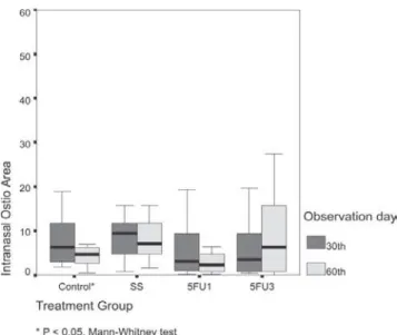
The comparative study between the 4 groups using the Kruskal-Wallis test did not show significant changes on the 60th postoperative day; however, the pair-wise comparisons
revealed that the ostium area of Group SS was significantly larger than that of Group 5-FU1 (P < .01, Mann-Whitney test) (Figure 2).
Regarding changes in the ostium area between the 30th
and the 60th days, an increase of the ostium area occurred
in 3 cases (23%) in Group SS (SS), 5 cases (29%) in Group 5-FU1,1 case (9.1%) in Group C, and 3 cases (33%) in Group 5-FU3. A decrease in the ostium area was observed in 4 cases (31%) in Group SS, 9 cases (53%) Group 5-FU1, 1 case (9.1%) in Group C, and 3 cases (33%) in Group 5-FU3; There was no change in the ostium area for 12 cases as follows: 6 cases (46%) in Group SS, 3 cases (18%) in Group 5-FU1, 1 case (9.1%) in Group C, and 2 cases (22%) in Group 5-FU3. The change in area from the 30th to the
60th postoperative day was significantly reduced only in
Group C (P < .05, Mann-Whitney test) (Figure 3). No other significant differences were found regarding this parameter. Granulation tissue was present at the intranasal ostium level in all the groups as follows: 6 cases (46%), 10 cases (59%), 3 cases (27%), and 3 cases (33%) in Groups SS, 5-FU1, C, and 5-FU3, respectively. Extraction of the granu-lation tissue was performed only in symptomatic patients with an ostium blockage that impeded lacrimal drainage as follows: 3 cases in Group SS, 4 cases in Group 5-FU1, and 1 case in Group 5-FU3. In all of theses cases there was epiphora and disappearance of secretion.
Persistent epiphora was observed in 4 patients. The en-doscopic evaluation in the first week showed the presence of a bone fragment in the surgical fistula in 1 of these cases (Group C), which was removed on the same occasion. In another symptomatic patient, a patent lacrimal system was
Figure 2 - The graph shows the postoperative intranasal ostium areas by treatment group on the 60th postoperative day
observed. We suspected that the lacrimation was due to a maxillary sinusitis, since the endoscopy showed the pres-ence of secretion from the main ostium of the maxillary sinus. In the other 2 cases, we observed a concentric in-crease of healing tissue that greatly dein-creased the size of the ostium in one of the patients (Group 5-FU1), and com-pletely occluded it in the other (Group 5-FU3).
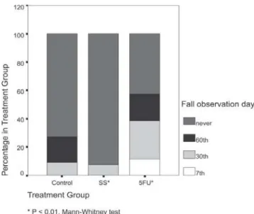
Comparing the number of flap falls among the several groups (Figure 4), there were no significant differences dur-ing the observation period. When the data was reorganized into 3 categories (saline solution, control, and 5-FU; Fig-ure 5), there were significantly more flap falls in the 5-FU group compared to saline solution group (Group SS) (Mann-Whitney test, P < .01).
There was no significant difference concerning the oc-currence of granulation tissue among the groups (P = .361). Among the 50 patients who underwent dacryocystorhi-nostomy, only 1 experienced a total ostium occlusion by the healing tissue, and there was persistent epiphora in 2 others. The presence of ostium with a much-reduced area was observed for these last cases.
No nasal, ocular, or systemic complications from the use of 5-FU were observed in this study.
DISCUSSION
In this study, the size of the final surgical ostium was always smaller than the initial surgical orifice. This result is similar to that of Linberg et al,18 who showed that an
ample osteotomy did not necessarily result in a larger in-tranasal ostium. Those authors attributed the reduction of the ostium area to the contracture of the wound during the healing process. Although in the present study the size of the osteotomy was the same in all the patients, there was great variation of the ultimate intranasal ostium among the subjects of the same group, suggesting the presence of sev-eral factors that could influence the final result.
The distinct decrease in the healed intranasal ostium area when compared to the surgical opening is not surpris-ing, considering normal healing processes. Ross19 described
the stages of the healing process as follows: hemostasis, inflammation, and remodeling. The healing contracture takes place during the last stage of the process.
If the size of the final ostium is related to the healing process, it is valid to assume that the use of a drug that inhibits fibroblast proliferation would result in a larger os-tium area. The option to use 5-FU was based on the fact that these antimetabolites have produced positive results in ocular surgeries, when the main objective is to interfere with the healing process.
A single dose of 5-FU has been shown to be effective in the treatment of massive periretinal proliferation, as is shown in the experimental work of Blumenkrans et al.20
This antimetabolite has also been used to improve surgi-cal prognosis in the eyes of human patients with glau-coma.15 Until 1989, most researchers used a 105-mg dose
in patients with high-risk glaucoma; later, reduced dosages were used, producing similar results with much fewer side effects.21;22 The occurrence of a flap fall in the 5-FU groups
occurred regardless of the dosage and the frequency of dos-ing. Flap falls have been cited as the cause of recurrence, as they block the surgical ostium.7 Endoscopy showed that
the flap could partially return to the nasal mucosa plane. A complete flap fall was not present in any of the cases.
Kao et al23 observed that the ostium area was
signifi-Figure 4 - Percentage of flap falls on 7th, 30th, and 60th days after surgery, by 4 groups (C, SS, 5-FU1, and 5-FU3)
cantly reduced in patients in whom only endonasal dacryo-cystorhinostomy was performed, compared to those that also received an intrasurgical injection of mitomycin. The authors concluded that this was due to the use of the an-timetabolite. The evaluation via helical computed tomo-graphic dacryocystography of the effect of intrasurgical mitomycin in patients undergoing external dacryocystorhi-nostomy revealed that the use of this antimetabolite resulted in larger ostium areas.24 In contrast, in the present study,
the results obtained at the 60-day follow-up evaluation re-vealed that the ostium area was distinctly larger in patients receiving a saline solution injection than in patients receiv-ing an 5-FU injection. Additionally, the number of flap falls of the nasal mucosa was less frequent in patients receiv-ing a saline injection compared to those receivreceiv-ing 5-FU in-jections.
Group C (control group) presented the natural evolu-tion of the ostium using the technique of 2 anterior flaps, characterized by the consistent occurrence of a distinct de-crease in area of the surgical ostium as well as the occur-rence of flap falls. Since all possible determinant causes of the flap fall, such as the use of quickly absorbing thread and inadequate fixation of sutures, were avoided, the most probable causes of this occurrence were the growing and thickening of the healing tissue or the partial return of the nasal mucosa to its original position.
The number of nasal mucosa flap falls was significantly greater in the groups receiving 5-FU injections compared to the group receiving a saline solution injection. The pro-portion of flap falls in the Group C was smaller than that in the 5-FU groups and larger than that in Group SS; how-ever, this variation was not statistically significant.
The within-group evaluation showed that the decrease
in the area between the 30th and 60th postoperative days was
statistically significant only in Group C. This finding indi-cates that the smaller decrease in area could be the influ-ence of the mechanical factor of the injection, rather than the effect of 5-FU.
In the present study, the endoscopic evaluation revealed that the occurrence of epiphora was not related to the os-tium size.
We can assume that in the 2 symptomatic cases in which the lacrimal pathway was patent, a much reduced ostium area could have hindered the proper lacrimal drain-age in these patients. The persistence of a bone fragment in the surgical area and the presence of ethmoid cells in the osteotomy area that were observed in these cases could have caused the healing exacerbation, significantly decreas-ing the ostium. In the only case where there was complete surgical ostium occlusion, the presence of ethmoid cells during the surgery was verified. According to Picó,4
eth-moid cells constitute anatomic variations that predispose surgical failure.
Endoscopy was crucial for clearing up persistent epi-phora, providing information for correct diagnosis and treat-ment. Good results were obtained with the removal of granulation tissue or hematic crust that was blocking the central ostium area.
According to Piaton et al25 and Bakri et al,26 the use of
5-FU in patients undergoing laser dacryocystorhinostomy did not improve the surgical prognosis compared to the re-sults obtained with patients who underwent only the sur-gical treatment. Accordingly, the results of this study indi-cate that the use of 5-FU in external dacryocystorhinostomy is also without a significant influence in the final size of the surgical fistula at the 60 days postsurgery.
RESUMO
Costa MN, Marcondes AM, Sakano E, Kara-José N. Estu-do enEstu-doscópico Estu-do óstio intranasal no pós-operatório de dacriocistorrinostomia externa e influência do uso de so-lução salina e de 5 fluorouracil. Clinics. 2007;62(1):41-6.
OBJETIVO: Estudar, através da endoscopia, as alterações
estruturais pós-operatórias do óstio intranasal na dacriocis-torrinostomia externa, e avaliar a influência da solução sa-lina e do 5 fluorouracil.
MÉTODOS: Cinquenta pacientes foram distribuídos nos
seguintes grupos: Grupo SS– 13 pacientes submeteram-se à dacriocistorrinostomia com uso de solução salina; Gru-po 5-FU1- 17 pacientes submeteram-se à
dacriocistor-rinostomia e injeção de 5 fluorouracil; Group C—11 paci-entes submeteram-se apenas à dacriocistorrinostomia; Gru-po 5-FU3- 9 pacientes submeteram-se à dacriocistor-rinostomia associada a três injeções de 5 fluorouracil.
RESULTADOS: A comparação pareada pelo teste não
paramétrico de Mann-Whitney revelou redução significa-tiva da área do óstio apenas na comparação Grupo 5-FU1 vs. Grupo SS, no 60º dia após a cirurgia (P<.01). No
en-tanto, um estudo comparativo entre os 4 grupos usando o teste de Kruskal-Wallis não revelou diferenças significati-vas na área do óstio no 60º dia após a cirurgia. Na
compa-ração intra-grupos, a área do óstio no 30 º vs 60 º dia após
(P < .05; teeste de Mann-Whitney); nenhuma outra com-paração apresentou diferenças significativas.
DISCUSSÃO: O resultado deste estudo sugere que o uso
de 5 fluorouracil na dacriocistorrinostomia externa não in-fluencia significantemente no tamanho final da fístula
ci-rúrgica durante os dois meses de avaliação.
UNITERMOS: Fluorouracil. Dacriocistorrinostomia.
Endoscopia. Dacriocistite.
REFERENCES
1. Dupuy-Dutemps L, Bourguet J. Procédés plastiques des dacryocystorhinostomies. Ann Oculist. 1921;159:241-61.
2. Older J. Routine use of a silicone stent in a dacryocystorhinostomy. Ophthalmic Surg. 1982;13:911-15.
3. Dayal Y. External dacryocystorhinostomy. Am J Ophthalmol. 1961;51:514-17.
4. Picó G. A modified technique of external dacryocystorhinostomy. Am J Ophthalmol. 1971;72:679-90.
5. Hurwitz J. Failed dacryocystorhinostomy in Paget’s disease. Can J Ophthalmol. 1979;291-3.
6. Weingarten R , Goodman E. Late failure of a dacryocystorhinostomy from sarcoidosis. Ophthalmic Surg. 1981;12:343-6.
7. Costa M, Sakano E. O valor da endoscopia nasal na semiologia lacrimal. Arq Brás Oftalmol. 1989;52:140.
8. Bosshard C. Endoskopie der nase als hilfe furdie Tranenwegschirurgie. Klin Monatsbl Augenheilkd. 1982;180:303-7.
9. Vergez A, Herbert F. A propos de la dacryocystorhinostomie. Bull Soc Ophtalmol. 1978;273-6.
10. Welham R, Henderson P. Results of dacryocystorhinostomy analysis of causes for failure. Trans Ophthalmol. 1973;93:601-9.
11. Huggert A, Sundmark E. Treatment of lacrimal obstruction. Am J Ophthalmol. 1965;60:603-10.
12. Mirabile T, Tucker C. Dacryocystorhinostomy with silicone sponge. Arch Ophthalmol. 1979;74:235.
13. Leone C. Gelfoam-thrombin dacryocystorhinostomy stent. Am J Ophthalmol. 1982;94:412.
14. Blumenkranz M, Clafin A. Selection of therapeutic agents for intraocular proliferative disease: cell culture evaluation. Arch Ophthalmol. 1984;102:598-604.
15. The Fluorouracil Filtering Surgery Study Group. Fluorouracil filtering surgery study one-year follow-up. Am J Ophthalmol. 1989;108:625-35.
16. Fluorouracil Filtering Surgery Study Group. Five year follow-up of the Fluorouracil Filtering Surgery Study Group. 1996;121:349-66. 17. Broadway C, Jones N, Bunce C, Thiagarajan M, Khan T. Needle revision
of failing and failed trabeculectomy blebs with adjunctive 5-fluorouracil: survival analysis. 2004;111:664-73.
18. Linberg J, Anderson R, Bumsted R. Study of intranasal ostium external dacryocystorhinostomy. 1982;100:1758-62.
19. Ross R. Wound healing. Sci Am. 1969;220:40-50.
20. Blumenkranz M, Ophir A, Clafin A, Hajek A. Fluorouracil for the treatment of massive periretinal proliferation. Am J Ophthalmol. 1982;94:458-67.
21. Jampel H. Trabeculectomy with 5-fluorouracil for adult inflammatory glaucoma. Am J Ophthalmol. 1990;109:168-73.
22. Krug J, Melamed S. Adjunctive use of delayed and adjustable low dose 5-fluorouracil in refractory glaucoma. Am J Ophthalmol. 1990;109:412-18.
23. Kao C, Liao L, Tseng H, Chen S, Houk K. Dacryocystorhinostomy with intra-operative mitomycin. 1997;104:86-91.
24. Gonzalvo J, Fuertes I, Fernandez J, Hernandez G, Rabinal F, Honrubia M. External DCR with mitomycin: clinical and anatomical evaluation with helical computed tomography. Arch Soc Esp Oftalmol. 2000;75:611-7.
25. Piaton J, Keller P, Limon S, Quenot S. Revision of failed dacryocystorhinostomy using the transcanalicular approach. Results of 118 procedures. J Fr Ophtalmol. 2000;24:265-73.