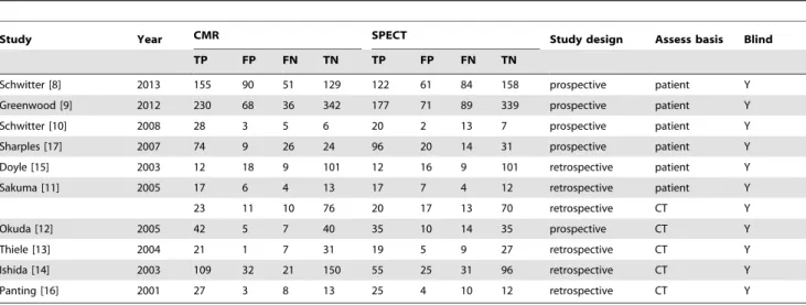
Direct comparison of cardiovascular magnetic resonance and single-photon emission computed tomography for detection of coronary artery disease: a meta-analysis.
Texto
Imagem


Documentos relacionados
En la tabla 8, se presenta la relación entre los porcentajes de la composición corporal (masa grasa, masa muscular, androide y ginoide) con los niveles de actividad
raphy and single-photon emission computed tomography have revealed asymmetrical ac- tivation of parahippocampal and hippocam- pal regions (6,7), while qualitative morpho-
Radiological propedeutics of femoroacetabular impingement in times of computed tomography and magnetic resonance imaging: what a radiologist needs to know..
Cardiac magnetic resonance imaging (CMRI) and cardiac computed tomography (CCT) are noninvasive imaging methods that serve as useful tools in the diagnosis of coronary artery
For this purpose, a dedicated tomographic imaging system based on SPECT (Single Photon Emission Computed Tomography, a diagnostic medical imaging modality used in nuclear medicine)
Key words: Juvenile idiopathic arthritis; temporomandibular joint; computed tomography; magnetic resonance
A new autopsy technique consists of the internal examination of death bodies using computed tomography (CT) and magnetic resonance imaging (MRI), without opening
To compare the presence of post-operative residual disease by magnetic resonance imag- ing (MRI) and [18F]fluorothymidine (FLT)-positron emission tomography (PET)-computer
