Brazilian Journal of Physics, vol. 35, no. 3B, September, 2005 785
Development of a Tomographic System for Online Dose Measurements in BNCT
(Boron Neutron Capture Therapy)
A. Valda, D. M. Minsky, A. J. Kreiner,∗ A. A. Burlon, and H. Somacal
Escuela de Ciencia y Tecnolog´ıa (UNSAM), San Mart´ın, Buenos Aires, Argentina and
Dpto. de F´ısica, Centro At´omico Constituyentes, Comisi´on Nacional de Energ´ıa At´omica, Buenos Aires, Argentina
Received 19 July, 2005
Within our activities on accelerator-based boron neutron capture therapy (BNCT) carried out at the Tandar Laboratory (Comisi´on Nacional de Energ´ıa At´omica, Argentina) we present here the study and design of a tomographic imaging system for the measurement of the spatial distribution of the absorbed dose during a BNCT session. The10B(n,α)
7Li boron neutron capture reaction produces a 478 keV gamma ray in 94% of the cases.
In BNCT a large fraction of this radiation escapes from the patient body. Its detection is thus very attractive for a noninvasive boron concentration measurement and an online absorbed dose evaluation since the dose absorbed by the tumor and healthy tissue strongly depends on the boron uptake and the neutron flux. For this purpose, a dedicated tomographic imaging system based on SPECT (Single Photon Emission Computed Tomography, a diagnostic medical imaging modality used in nuclear medicine) is proposed since standard SPECT cameras cannot be used due to the photon energy (478 keV) and the particular background gamma field in BNCT. A detection system (collimator and detectors) is proposed. Monte Carlo numerical simulations are used for an implementation of a statistical algorithm used for the tomographic image reconstruction.
I. INTRODUCTION
Boron neutron capture therapy (BNCT) [1] is a radiation therapy under development for the treatment of some cancers like melanoma and glioblastoma multiforme. It is performed in two steps: first, a stable isotope of boron (10B) is admi-nistered to the patient via a carrier drug and then the patient is irradiated with an epithermal neutron beam. 10B will then undergo the capture reaction10B(n,α)
7Li (10B capture cross section for thermal neutrons: 3840b). The emitted charged particles (∼1 MeV) have a high linear energy transfer (LET)
and are lethal only to the cells in close proximity to the re-action point because their range is ∼10µm. Reactor-based
epithermal neutron beams have been developed to treat pati-ents (e. g. [2] - [4]). As an alternative to reactors, the appli-cation of accelerator-based neutron sources for BNCT (AB-BNCT) is being proposed at a number of laboratories (e. g. [5] - [8]). Among other advantages over reactor sources, ac-celerators can produce more appropriate neutron spectra with low fast neutron and gamma ray contamination of the therapy beam.
The dosimetric aspects of BNCT are very complex due to the several interactions of neutrons with the different nuclei appearing in healthy and tumor tissue. Nevertheless the absor-bed dose in the tumor is dominated by the10B capture reac-tion. Usually the evolution of the10B concentration in tumor and in healthy tissue is obtained indirectly from blood samples taken from the patient before, after and during the BNCT tre-atment since tumor-to-blood and healthy tissue-to-blood10B concentrations are inferred from previous biodistribution stu-dies of the 10B carrier. Hence a better measurement of10B concentration during a BNCT treatment will result in a better
∗Also CONICET, Argentina
knowledge of the dose received by the patient and its rela-tion to the treatment response. The external detecrela-tion of the 478 keV prompt-gamma radiation (linear attenuation coeffici-ent for soft tissue is∼0.1 cm−
1) with spatial discrimination should serve as the basis for an online dose measurement in a noninvasive way. In this sense, a device called gamma-ray telescope has been developed and is in use at the BNCT faci-lity in Petten, the Netherlands [8]. In Japan, Kobayashi et al. [9] have presented a feasibility study of a tomographic sys-tem based on single photon emission tomography (SPECT). In this work we propose a tomographic system, also using the SPECT principle, based on pixelated scintillator detectors. In the first section we evaluate the prompt-gamma production is-sued from the capture in10B based on expected results of AB-BNCT applied to brain tumors. The next section is devoted to the description of the collimator and radiation detector of the proposed system. Finally we assess the imaging performance of the system by Monte Carlo numerical simulations using a simple phantom as radiation source. An iterative statistical al-gorithm (Expectation- Maximization Maximum-Likelihood) is used for the single slice reconstruction.
II. ASSESSMENT OF478keV PROMPT-GAMMA
PRODUCTION IN BNCT
In BNCT the dose delivered to the tumor is usually limi-ted by the maximum dose that the healthy tissue can receive which is dependent on its boron concentration (nonspecific drug uptake) and on the neutron beam quality. From our pre-vious work [10] on accelerator-based BNCT applied to brain tumors and usingp-boronphenylalanine (BPA) as the boro-nated compound, it is possible to estimate a tumor dose of 40 RBE-Gy in a BNCT treatment. The10B capture reaction dose component accounts for about 80% of this value. Consi-dering a mean energy per capture reaction of 2.31 MeV, one
786 A. Valda et al.
per cubic centimeter of tumor. This is the maximum number of photons available for the online dose measurement; owing to the attenuation in soft tissue, this number should be cor-rected by a factor of about 0.37. Concentration measurements
require the use of tomographic approaches, the most appropri-ate for this application is that based on the nuclear medicine image modality known as SPECT, for Single Photon Emission Tomography [11].
III. SYSTEM DESIGN
In SPECT, a bidimensional position sensitive detector (the gamma camera) detects the gamma rays emitted from the pa-tient. The gamma camera is composed of the collimator, the radiation detector itself and a coding electronics. The collima-tor selects the direction of incidence of the gamma rays into the detector. It consists of a large number of holes in an absor-bing material like lead. The direction of the holes determines the direction of the accepted radiation (usually perpendicu-lar to the detection plane). The geometrical characteristics of the collimator mostly determine the imaging properties of the system (spatial resolution and sensitivity). In clinical SPECT the detector is a continuous NaI(Tl) crystal optically coupled to a set of photomultiplier tubes (PMT). The relative signal amplitudes from the PMTs is electronically analyzed (Anger logic) in order to give the spatial position of the impact of the gamma ray on the detector.
A. Collimator
The round-hole parallel collimator in lead was designed on the basis of geometrical calculations and estimates of the pho-ton attenuation in collimator material. The expression used for the geometrical spatial resolution (Rg) was
Rg=
φ(e+d)
e (1)
whereφis the collimator diameter,ethe collimator thickness (length of holes) anddthe distance from the point whereRgis calculated to the collimators entrance face. The septum thick-ness or thickthick-ness of material between adjacent holes (t) was calculated by numerically solving the equation:
t l =
2φ+t
e2+ (2φ+t)2
(2)
wherelis the length of lead that attenuates a given amount of 478 keV photons (linear attenuation coefficient 1.97 cm−
1). The geometrical point source sensitivity (εg) was obtained by evaluating the solid angle subtended by all hole apertures, which depends onφ,e,d andt (sinceφ+t is the center-to-center hole separation). The collimator geometry is then ob-tained by maximizingεgfor a given spatial resolutionRg, at a given distancedand allowing a given septal attenuation.
6x10⁻³
5
4
3
2
1
geometrc efficiency (%)
14 13 12 11 10 9 8 7
spatial resolution (mm)
Collimator thickness (mm):
40 80 100 140 160
(a)
3.4x10⁻³
3.2
3.0
2.8
2.6
2.4
2.2
2.0
geometric efficiency (%)
160 140 120 100 80 60 40
collimator thickness (mm)
(b)
FIG. 1: (a) Collimator geometric efficiency as a function of spa-tial resolution for different collimator thicknesses. Distance source-collimator: 20 cm, septal attenuation: 94%. The vertical dotted line intersects the curves at 1 cm of spatial resolution. (b) Geometric ef-ficiency as a function of collimator thickness for 1 cm of spatial re-solution (other parameters as in (a))
For this application we chosel=1.54 cm (which provides
an attenuation of 94%). Figure 1.a shows a family of effici-ency vs. resolution curves for several collimator thicknesses; the resolution is taken atd=20 cm. ConsideringRg=1 cm at 20 cm from the collimator, it is possible to see that the ef-ficiency is maximum (εg=3.5·10
−3%) fore=9
.4 cm.
Fi-gure 1.b presents a complementary visualization of this re-sult. However, with the aim of diminishing the weight of the collimator structure we have chosen to work in a suboptimal situation withe=8 cm that givesεg=3.4·10−
3% (2% less than the optimum). In order to largely cover a transaxial field-of-view of 20 cm, a collimator of 23.7 cm in lateral length was
chosen; such a collimator has 37 parallel holes.
B. Detectors
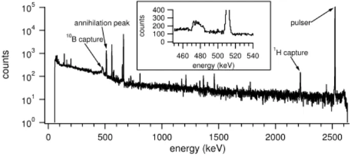
In previous work [12] on the characterization of a neutron beam obtained from an accelerator, we have measured with a high-purity germanium (HPGe) detector typical gamma ray spectra during the irradiation of a cylindrical head phantom containing a small (1.6 cm in diameter, 2 cm in height)
cy-linder with boron (Fig. 2). The choice of the radiation de-tector is related to the presence of the 511 keV annihilation peak near the 478 keV prompt gamma peak. The annihilation peak is originated as a consequence of electron-positron pair creation after interaction of high energy gamma rays, mostly 2.23 MeV issued from neutron capture in hydrogen. In
re-Brazilian Journal of Physics, vol. 35, no. 3B, September, 2005 787
100
101
102
103
104
105
counts
2500 2000
1500 1000
500 0
energy (keV)
10B capture
annihilation peak
1
H capture pulser 400
300 200 100 0
counts
540 520 500 480 460
energy (keV)
FIG. 2: HPGe spectrum obtained during the irradiation of a cylindri-cal head phantom containing a small cylinder with boron. Relevant peaks are indicated. The inset is a closer view of the region around the 478 and 511 keV peaks. The broadening of the10B capture peak is caused by the Doppler effect since the prompt gamma is emitted “in flight” by the7Li ion.
solution of 1.8% at 662 keV (FWHM=12 keV) but the
pre-sent technology limits the fabrication of these crystals to about 5 mm in thickness, which provides an intrinsic efficiency of only 4%. On the other hand, the recently developed LaCl3(Ce) crystal, a high light yield scintillator (49 photons/keV), can be constructed with a more appropriate thickness. For exam-ple 2.5 cm of LaCl3(Ce) provide an intrinsic efficiency of
about 16% . Even if LaCl3(Ce) has worse energy resolution than CdZnTe, it is however good enough for our application (FWHM=28 keV for 662 keV) [13].
In order to have appropriate statistics for the tomographic reconstruction during the treatment time it is necessary to sur-round the patient with a sufficient number of detector arrays, which will provide the projection data. Considering the to-tal number of prompt gamma events produced in 1 cm3tumor (cf. II) and 5 arrays of LaCl3(Ce) detectors coupled to the col-limator described above (each having a detection efficiency of 2·10−6), a total of 2·104photons/cm3of 478 keV could be detected. This array should be successively positioned at 4 different angles around the patient, as we will see in the next section, giving a total of 1000 photons per position and per array issued from a tumor of 1 cm3 during the whole treat-ment time. Therefore, if one wants to monitor the treattreat-ment at its mid-time point, it should be possible to have a statistical fluctuation less than 10% in the determination of the prompt gamma production in a tumor of 1 cm3.
IV. NUMERICAL SIMULATIONS AND TOMOGRAPHIC
RECONSTRUCTION
A. Acquisition data
The simulations were performed using the Monte Carlo si-mulation code MCNP-4c [14]. Two phantom sources were used to simulate the acquired (projection) data: (i) a set of four parallel “hot” cylinders with diameters 0.5 , 1, 1.5 and
2 cm and (ii) the same set immersed in a 20 cm diameter cy-linder for simulating a background source with a size compa-rable to a human head. In the last case the10B concentration
b)
Detector iPixel j
a)
9°
FIG. 3: Geometric configurations considered in the Monte Carlo si-mulations. (a) Simulation of the projection data of the phantoms: there are 37 detectors in each of the 20 angular positions of the array. (b) Calculation of the elementi jof the system matrix.
in the four small cylinders was the same and the cylinders-to-background concentration ratio was 4 : 1, like in a typical BNCT case. One-dimensional projections were taken around the cylinders in order to reconstruct bi-dimensional transaxial planes (the three-dimensional volume can be reconstructed by stacking independent bi-dimensional planes). Each angular position of a detector plane is composed of 37 line projections according to the collimator-detector described in the previous section. A total of 20 angular positions (evenly spaced at 9 degrees) were considered (Figure 3.a) giving a total of 740 projection data. The number of simulated events is such that the statistical fluctuations of the counting process can be ne-glected.
B. Image reconstruction
788 A. Valda et al.
-10 -8 -6 -4 -202468 10 -10
-8 -6 -4 -2 0 2 4 6 8 10
[c
m
]
[cm]
-10 -8 -6 -4 -202468 10 -10
-8 -6 -4 -2 0 2 4 6 8 10
[c
m
]
[cm]
-10 -5 0 5 10 0
20 40 60 80 100 120 140 160
Distance from center [cm]
P
rojec
tion
Ang
le [D
egr
e
es
]
-10 -5 0 5 10 0
20 40 60 80 100 120 140 160
Distance from center [cm]
P
rojec
tion
Ang
le [D
egr
e
e
s
]
-10 -8 -6 -4 -202468 10 -10
-8 -6 -4 -2 0 2 4 6 8 10
[cm]
[c
m
]
-10 -8 -6 -4 -202468 10 -10
-8 -6 -4 -2 0 2 4 6 8 10
[c
m
]
[cm] [cm]
[cm]
FIG. 4: Simulated reconstructed images of the hot-rod phantoms: without background (upper row) and with background (4:1) (lower row). From left to right: original phantom, sinogram and reconstruc-ted image.
V. CONCLUSIONS
A tomographic device for spatially resolved online dosime-try in BNCT has been proposed. Monte Carlo numerical
si-mulations are used to evaluate its imaging performance. First results indicate that it would be possible to recover boron con-centrations in tumors with 2 cm in diameter. As the accuracy of this determination depends on the counting statistical fluc-tuations, future numerical work points in this direction. We are also working in the correction for in-phantom photon atte-nuation. Anthropomorphic phantoms will be used to explore the system performance better.
Acknowledgments
The authors wish to thank the TANDAR personnel for their collaboration. Special thanks are addressed to the END group (CNEA Physics Department) members, M. E. Caraballo, M. Debray, M. Davidson, J. Davidson, J. M. Kesque, M. Ozafr´an, and M. E. V´azquez for their contribution to the experimental aspects of this work. We gratefully acknowledge the support provided by the National Agency for the Promotion of Science and Technology, ANPCyT.
[1] J. A. Coderre and G. M. Morris, Radiat. Res.151, 1 (1999) and references therein.
[2] R. Rogus, O. Harling, and J. Yanch, Med. Phys.21, 1611 (1994). [3] C. Vroegindeweij, P. Watkins, and R. L. Moss; Frontiers in neu-tron capture therapy; M. F. Hawthorne, K. Shelly and R. J. Wi-ersema, eds. (New York: Kluwer Academic/Plenum Publishers), 375 (2001).
[4] H. R. Blaumann, S. J. Gonz´alez, J. Longhino et al., Med. Phys.
31, 70 (2004).
[5] R. Klinkowstein, R. Schefer, J.C. Yanch, et al., Proceedings of the 7th International Symposium on Neutron Capture Therapy for Cancer, Vol. 1; B. Larsson, J. Crawford and R. Weinreich, eds.; (Amsterdam, Elsevier Science B. V.), 522 (1997).
[6] A. A. Burlon, A. J. Kreiner and A.Valda; Proceedings of the Sixth Mexican Symposium; L. M. Montano Zetina and G. Her-rera Corral, eds. (AIP Conference Proceedings), 54 (2002). [7] C. N. Culbertson, S. Green, A. J. Mason et al., Applied Radiation
and Isotopes61, 733 (2004).
[8] W. F. A. R. Verbakel, W. Sauerwein, K. Hideghety, and F.
Stecher-Rasmussen, Int. J. Radiation Oncology Biol. Phys.55, 743 (2003).
[9] T. Kobayashi, Y. Sakurai, and M. Ishikawa, Med. Phys.27, 2124 (2000).
[10] A. A. Burlon, A. J. Kreiner, A. A. Valda, and D. M. Minsky, Applied Radiation and Isotopes61, 811 (2004).
[11] See for example P. Suetens, “Fundamentals of medical ima-ging”; Cambridge University Press, 2002.
[12] A. A. Burlon, A. J. Kreiner, A. A. Valda et al., Nucl. Instr. and Meth. B229, 144 (2005).
[13] E. V. D. van Loef, W. Mengesha, J. D. Valentine et al., IEEE Trans. Nucl. Sci.50, 155 (2003).
[14] J. F. Briesmeister, “A general Monte Carlo n- particle trans-port code”, Los Alamos, NM, Los Alamos National Laboratory, (2000).