Rev Odontol UNESP. 2013 Nov-Dec; 42(6): 432-438 © 2013 - ISSN 1807-2577
ORIGINAL ARTICLE
Inluence of whitening gel on pulp chamber temperature
rise by in-oice bleaching technique
Inluência do gel clareador no aumento da temperatura intra-câmara pulpar pela
técnica do clareamento em consultório
Sandro Cordeiro LORETTO
a, Mylena Ranieri LIBDY
a, Fabiola do Socorro da Rocha RIBEIRO
a,
Esther Marina França BRAGA
a, Karina Gama Kato CARNEIRO
a,
Mário Honorato SILVA E SOUZA JÚNIOR
aaFaculdade de Odontologia, UFPA – Universidade Federal do Pará, Belém, PA, Brasil
Resumo
Introdução: O clareamento dental representa uma manobra conservadora na recuperação estética de dentes com alterações cromáticas. Contudo, os tratamentos clareadores são passíveis de causar efeitos adversos, quando não bem planejados e executados. Objetivo: Este estudo avaliou a influência do gel clareador no aumento da temperatura intra-câmara pulpar através da técnica de clareamento dental fotoativado realizado em consultório.
Material e método: Um incisivo central superior humano foi seccionado na porção radicular 3 mm abaixo da junção cemento-esmalte. O canal radicular foi alargado para permitir a introdução do sensor do termômetro na câmara pulpar, a qual foi preenchida com pasta térmica,favorecendo a transferência de calor das paredes dentárias para o sensor do termômetro digital com termopar tipo K (MT- 401A) durante o clareamento. Três agentes clareadores fotossensíveis (peróxido de hidrogênio a 35%) foram utilizados, sendo: Whiteness HP (FGM), Whiteness HP Maxx (FGM) e Lase Peroxide Sensy (DMC). Um aparelho fotopolimerizador de led (Flash Lite – Discus Dental) foi empregado para a ativação dos géis clareadores. Seis ciclos de clareamento foram realizados para cada grupo testado. Os resultados foram submetidos à ANOVA de um critério e ao teste t (LSD) (α≤0,05). Resultado: A menor média de variação de temperatura (°C) foi observada com o Lase Peroxide Sensy (0,20), enquanto que a maior com o Whiteness HP (1,50). Conclusão: Os géis clareadores Whiteness HP e Whiteness HP Maxx interferiram significativamente no aumento da temperatura intra-câmara pulpar durante o clareamento, sendo esta variação dependente do tipo de gel clareador empregado.
Descritores: Clareamento dental; temperatura alta; cavidade pulpar.
Abstract
Introduction: Dental bleaching is a conservative method for the aesthetic restoration of stained teeth. However, whitening treatments are likely to cause adverse effects when not well planned and executed. Objective: This study evaluated the influence of whitening gel on temperature rise in the pulp chamber, using the in-office photoactivated dental bleaching technique. Material and method: The root portion of an upper central human incisor was sectioned 3mm below the cemento-enamel junction. The root canal was enlarged to permit the insertion of the K-type thermocouple sensor (MT-401) into the pulp chamber, which was filled with thermal paste to facilitate the transfer of heat during bleaching. Three photosensitive whitening agents (35% hydrogen peroxide) were used: Whiteness HP (FGM), Whiteness HP Maxx (FGM) and Lase Peroxide Sensy (DMC). An LED photocuring light (Flash Lite – Discus Dental) was used to activate the whitening gels. Six bleaching cycles were performed on each group tested. The results were submitted to one-way ANOVA and LSD t-test (α≤0.05). Result: The lowest mean temperature variation (°C) was detected for Lase Peroxide Sensy (0.20), while the highest was recorded for Whiteness HP (1.50). Conclusion: The Whiteness HP and Whiteness HP Maxx whitening gels significantly affected the temperature rise in the pulp chamber during bleaching, and this variation was dependent on the type of whitening gel used.
INTRODUCTION
Currently, several clinical forms may be available in the attempt to reverse coloring changes in the teeth, and the choice of one or more methods depending on the etiology of the color change. hus, the following possibilities may be mentioned: in-oice bleaching, in-home bleaching, combination of home and oice techniques, use of polyethylene whitening strips, bleaching associated with the use of diferent light sources, and toothpastes with whitening potential1,2.
Among these, in-oice bleaching presents an appropriate alternative to in-home bleaching, especially in cases of severe discoloration, discoloration of a single tooth, lack of patient cooperation, or also if faster treatment is desired3-5.
hus, when acceleration of the bleaching process is desired, the whitening agent may be activated by heat, potentiating its action. he idea of potentiated bleaching dates from 1918, when Abbot reported the use of a high-intensity light source to increase the temperature of the hydrogen peroxide. In most cases, light or laser heat sources are used to increase the temperature of the whitening product on the tooth surface. It is common, currently, to ind highly detailed reports of so-called photoactivated (photopotentiated) whitening treatments where additional light is used together with the whitening agent5.
he process by which the release of hydroxyl radicals from the peroxide is accelerated by the increase in temperature is known as thermocatalysis. An increase in the speed of decomposition of the peroxide occurs during this process and, consequently, there is an improvement in the eicacy of the whitening treatment. Also, this potentiation may be due to the direct excitation of the hydrogen peroxide by the high-frequency light, which allows its breakdown (decomposition) into two hydroxyl radicals through a mechanism known as photolysis5.
Diferent light sources with a wide variety of wavelengths may be used to activate whitening products, especially: conventional halogen lights, LEDs (light emitting diodes), xenon plasma arcs, and the lasers5. To this end, LEDs have been widely used to emit
energy in a narrow range of the visible spectrum, guaranteeing greater safety in heat generation6-10.
However, although traditionally considered as light sources that do not generate heat11, LEDs have been questioned recently
as to their capacity to raise the temperature of the tooth5,12,13.
Asmussen, Peutzfeldt14 suggest that this variation of results is
related to the technological development of dental LEDs, since the irst generation had power density values notably less than the devices used nowadays (high-power LEDs).
In addition, it should be remembered that every increase in the temperature of the whitening gel consequently brings about an increase in the temperature of the tooth surface and the pulp chamber15. Such an increase may be responsible for pathological
alterations in the pulp organ. Zach, Cohen16 observed that an
increase on the order of 5.5°C would be capable of generating irreversible pulp damage (pulp necrosis).
hus, diferent whitening gels have been marketed with advertised photosensitive potential, mainly due to the addition of dyes capable of absorbing the heat coming from the light source: for example, carotene17. However, this possibility still is
not well documented and the beneit of using additional light in the bleaching process is questioned in recent publications5,8,18-21.
herefore, regard the frequent introduction of new techniques, materials and equipment designed for dental bleaching, and the wide popularity of photoactivated whitening therapies, the objective of the present study is to evaluate the inluence of whitening gels, activated with a light source (high-power LED), on the change in pulp chamber temperature.
MATERIAL AND METHOD
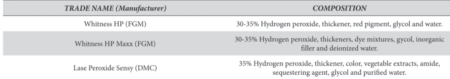
hree (3) photosensitive whitening agents were used in this study: Whiteness HP (FGM Produtos Odontológicos, Joinville, SC, Brasil), Whiteness HP Maxx (FGM Produtos Odontológicos, Joinville, SC, Brasil) and Lase Peroxide Sensy (DMC, São Carlos, SP, Brasil) (Table 1). A high-intensity LED source, Flash Lite 1401 (Discus Dental Inc., Culver City, CA, USA), was used to activate the whitening gels.
One (1) upper central human incisor, free of caries and restorations and extracted for periodontal reasons, was selected for this purpose. he tooth donor was fully informed about the nature of the study and signed the Free Informed Consent Form, thereby authorizing the use of the tooth. he present study was submitted to the Committee for Ethics in Research on Human Beings at the Health Sciences Institute (ICS) of the Federal University of Pará (UFPA), and was approved under number 110/10 – CEP-ICS/UFPA.
he selected tooth was disinfected with a 0.1% thymol solution for one week, submitted to prophylaxis with pumice and water and then was sectioned in the root portion approximately 3 mm below the cemento-enamel junction using a double-faced diamond disc at slow rotation (KG Sorensen, Cotia, SP, Brazil). Pulp remnants located in the chamber and root canal were removed by irrigation with Milton solution (0.5% sodium
Table 1. Whitening gels used in the present study
TRADE NAME (Manufacturer) COMPOSITION
Whitness HP (FGM) 30-35% Hydrogen peroxide, thickener, red pigment, glycol and water.
Whitness HP Maxx (FGM) 30-35% Hydrogen peroxide, thickeners, dye mixtures, gycol, inorganic iller and deionized water.
Lase Peroxide Sensy (DMC) 35% Hydrogen peroxide, thickener, color, vegetable extracts, amide,
hypochlorite) and saline. he root canal was enlarged using a # 2135 diamond tipped bur (KG Sorensen) to allow the introduction of the sensor of the thermometer into the pulp chamber.
he measurement of temperature variation in the central incisor pulp chamber was done using a digital thermometer attached to a K-type thermocouple (MT-401A). he pulp chamber was illed with thermal paste (Implastec Eletroquímica, Votorantim, SP, Brazil) in order to allow the heat transfer from the tooth walls to the thermocouple sensor during whitening procedures. he thermal paste was replaced at the end of each experimental group.
he sensor was inserted into the pulp chamber and positioned in the uppermost part thereof. he location of the sensor was controled using a periapical radiograph. A heavy silicon base was made to support the tooth during the whitening procedure, allowing the buccal surface to remain perpendicular to the tip of the LED.
A metallic marker, 5mm in height, was itted to this base, to standardize the distance from the tip of the LED to the buccal surface of the tooth.
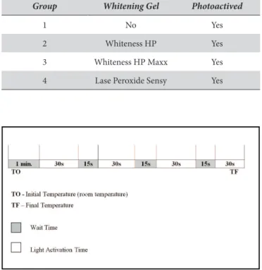
he groups were assigned according to the whitening gel used, as shown in Table 2.
he bleaching cycle followed the sequence described below (Figure 1), except for Group 1 (Control), where the initial minute of delay (necessary for the gel to penetrate into the teeth) was not observed. A digital timer was used for more precise control of the bleaching cycles. Six bleaching cycles were performed for each experimental group.
Before starting each bleaching cycle, the light intensity was measured using a radiometer, and the light output was constant at 600±10 mW/cm2.
In groups 2, 3 and 4, the manipulation of the whitening gels followed strictly the manufacturers’ instructions. A 1mm thick uniform layer was applied to the surface of the enamel, measured by a periodontal probe. One-minute application interval was observed to allow the whitening agent to penetrate into the dental substrates.
Next, the bleaching cycle was performed as shown in Figure 1. At the end of each cycle the whitening gel was exchanged, allowing the whole system return to the initial temperature (25°C). he inal temperatures of each whitening cycle were recorded, the values were subtracted from the initial temperature, and the data for the changes in temperature (∆T) for each experimental group were thus obtained.
Ater, the data were submitted to One-Way ANOVA and t-test (Least Signiicant Diference) at the 5% level of signiicance (p≤0.05).
RESULT
Descriptive statistics (mean and standard deviation) were obtained for the temperature variation (°C) in the pulp chamber during the photoactivated whitening treatment (Table 3). It can be pointed out that the lowest mean variation was obtained with Group 4 (Lase Peroxide Sensy – DMC), whereas the greatest mean was found with Group 2 (Whiteness HP – FGM). Also, the groups in which the whitening gel was applied to the tooth surface showed greater means of temperature variation when compared to the control group (Group1), with the exception of Group 4.
Table 4 (one-way ANOVA) shows that, at the level of signiicance adopted (α≤0.05) and considering the p-value found (p ≤ α), signiicant diference among the groups was demonstrated. Subsequent analysis was performed for this reason.
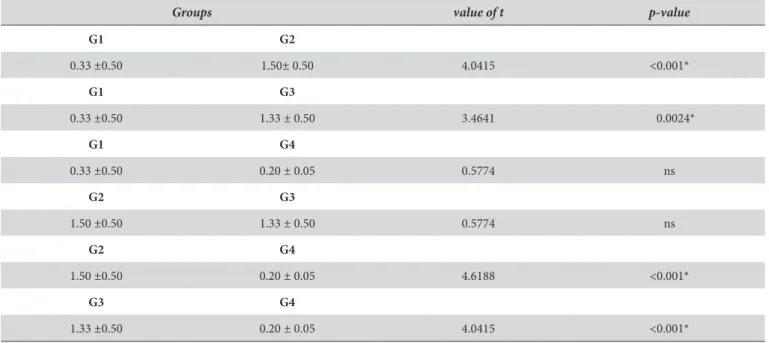
Table 5 shows the comparisons, in pairs, of the mean values of temperature variation (°C) of the groups tested, making it possible to show signiicant diferences among G1 and G2, G1 and G3, G2 and G4, and G3 and G4 (p<0.05).
DISCUSSION
In-oice dental bleaching is a highly popular therapy in dentistry. he procedure, using this technique, frequently involves the application of materials based on high concentrations of hydrogen peroxide, ranging from 30 to 35%5,22. Also, in an attempt
to reduce the total treatment time, activation of the whitening gels with diferent light sources through a technique known as power bleaching was proposed. his is based on heating the whitening product and, consequently, accelerating the bleaching reaction which allows the faster generation of free oxygen radicals that potentiate the action on the pigmented molecules5,22,23.
However, it must be pointed out that light activated whitening treatments, in general, are accompanied by an increase in temperature both on the tooth surface as well as inside the pulp chamber, which may contribute to changes in the health of the pulp, causing a simple increase in sensitivity up to extreme situations such as necrosis5. hus, the whitening
gel play an important role, since the temperature increase
Table 2. Division of groups in the present study
Group Whitening Gel Photoactived
1 No Yes
2 Whiteness HP Yes
3 Whiteness HP Maxx Yes
4 Lase Peroxide Sensy Yes
depends on the amount and the type of pigments (coloring) added to the products9, which was the object of the present
study. his study proposes to investigate the inluce of the whitening material on the behavior of the pulp chamber in relation to the temperature.
he change in pulp chamber temperature was measured using a digital thermometer with a K-type thermocouple (MT-401A), which has a nickel-chrome negative arm and a nickel-aluminum positive arm. he temperature change between the two alloys produces a voltage that is proportional to the temperature increase15,22,24-27.
Also, under the controlled conditions of an in vitro study, only one tooth (upper central incisor) was used during the tests to standardize the procedures and avoid uncontroleled variations due to the diferences existing between the thicknesses of the dentin and the enamel24,26-27.
Furthermore, the thermal paste was used to permit the transfer of heat from the tooth walls to the thermocouple sensor during the whitening procedures, acting as the pulp tissue. his paste was developed to permit a perfect coupling and to eliminate the air between the assemblies, thus illing the micro-gaps that might exist and increasing the thermal performance of the heat generating system28.
Moreover, it is worth noting the distance of the light tip, since its inluence on the intensity of the light that reaches the tooth surface and, therefore, the speed of the bleaching reaction and the heat that it may generate. he present study adopted 5mm distance between the tip of the light and the surface of the tooth covered by the whitening gel, which allowed a greater increase in temperature6,28.
In the present study, the thickness of the whitening gel applied to the tooth surface was standardized at 1mm, as recommended
Table 4. Analysis of variance for the mean values of temperature variation (°C)
Source of Variation GL SQ QM F
Whitening Gel 3 8.333 2.778 11.1111
Error 20 5 0.25
(p) = 0.000314* ---
---One-way ANOVA. *Signiicant at 5%.
Table 3. Descriptive statistics (mean and standard deviation) of temperature variation (°C)
Group Whitening Gel Mean Standard Deviation
1 No 0.33 ±0.50
2 Whiteness HP 1.50 ±0.50
3 Whiteness HP Maxx 1.33 ±0.50
4 Lase Peroxide Sensy 0.20 ±0.05
Table 5. Means of temperature variation (°C) in relation to the whitening gel used
Groups value of t p-value
G1 G2
0.33 ±0.50 1.50± 0.50 4.0415 <0.001*
G1 G3
0.33 ±0.50 1.33 ± 0.50 3.4641 0.0024*
G1 G4
0.33 ±0.50 0.20 ± 0.05 0.5774 ns
G2 G3
1.50 ±0.50 1.33 ± 0.50 0.5774 ns
G2 G4
1.50 ±0.50 0.20 ± 0.05 4.6188 <0.001*
G3 G4
1.33 ±0.50 0.20 ± 0.05 4.0415 <0.001*
by the manufacturers (FGM and DMC). Also, this is the value used in a prior evaluation27 since, clinically, the thickness of the
gel used on the vestibular surface of the tooth depends on its consistency. hat is, the more luid the gel, the less the thickness.
Six bleaching cycles were performed on each group in the study since the standard deviation for evaluations of this nature is considered low, as seen in the present study and other similar investigations15,25. Also, each bleaching cycle began with
a one-minute delay with the gel on the tooth surface to permit penetration into the teeth.
Another characteristic observed was the 15-second interval between cycles, which is done in clinical practice to avoid overheating the dental pulp, since the dissipation of energy is rapid and occurs, on average, ater 10 seconds29. he amount of
time adopted for light exposure (30 seconds) is recommended and used for activations with LED equipment15,22.
In fact, equipment with high energy output, as in the case of high power photocuring units, are able to transfer heat to the dental tissues. his process is common in daily dental practice30-33.
he heat generated deep within the dental substrates may cause damage to the hard tissues (enamel and dentin) and sot tissue (pulp), in addition to inducing sensitivity (pain) caused by the temperature increase34. he pulp, which is responsible
for maintaining tooth vitality, is vulnerable to heat. When the increase in intrapulp temperature exceeds 5.5°C, irreversible damage is induced in the pulp tissue16.
Even considering cool bulbs, LEDs are capable of heating the gel and tooth surface and, consequently, increasing the temperature of the pulp chamber. Several authors working with LEDs with power density between 500 and 950mW/cm2, using
whitening gel and following the recommended protocol, have observed the increase in temperature6,35. his output may be
applied to the data obtained in the present study in which the light from the LED, with a mean intensity of 600mW/cm2, raised
the temperature for all groups studied, although not surpassing the critical value of 5.5°C necessary to cause changes in the pulp16.
herefore, exposure to the light raised the temperature in the diferent groups studied. However, higher values were noticed when whitening gels were applied, except for the group in which Lase Peroxide Sensy (DMC) was used. In addition, the data observed for control group showed lower temperatures than the other groups, which is consistent with the literature9,22,24.
he results, above, may be explained by the interaction between the light or heat with the chemical composition of the whitening product. Briely, when the light is projected onto the bleaching gel only a small fraction is absorved and its energy converted into heat, since the hydrogen peroxide shows no absorption in the visible spectrum but only in the ultraviolet spectrum. herefore, in an attempt to increase light absorption and, consequently, its conversion to heat, the whitening products are mixed with speciic dyes such as carotene5. his is probably
the action mechanism in photoactivated (photopotentiated) bleaching techniques. he red-orange coloring of the carotene
increases the absorption of blue light, such as emitted by the LED source used in the present study.
Such a claim, however, is contrary to the results obtained with the Lase Peroxide Sensy (DMC) whitening gel in the present study. his gel with red-orange coloring is, according to the manufacturer, composed of 35% hydrogen peroxide, a photosensitive agent, a thickener, coloring, vegetable extracts (annatto and juá), amide, a chelating agent, glycol and water. At the beginning of the bleaching reaction the gel had a reddish color that, during the activation period, becomes more orangish.
he amount of coloring in the whitening gel may support the above results. hus, an increase in the concentration of the coloring results in a signiicant increase in the temperature of the gel and, therefore, in the tooth and the pulp chamber24.
It is possible that the amount of coloring added to the Lase Peroxide Sensy (DMC) gel is less when compared to the other whitening products used. Torres, Celaschi*also airmed that the attraction of the diferent pigments present in the several brands of whitening gels also varies, resulting in diferent heating levels of the gels. hese variations may be due to lower light absorption by some colors, or due to the presence of a lower concentration in some products, which indicates that the heating results produced both in the gel and the pulp chamber will depend on the type of gel used.
he results obtained with the Whiteness HP (FGM) and Whiteness HP Maxx (FGM) gels may be explaind by the capacity of the whitening gel to absorb the radiant energy (light) and convert it into thermal energy (heat) through a process known as the photothermal efect. he highest values of temperature variation associated with these two products may be connected, again, to the type of coloring present as well as to the appropriate concentration of the coloring.
Ferreira**, evaluating the behavior of the same gels irradiated with diferent wavelengths in the visible spectrum, concluded that regardless of the various wavelengths used, there was a signiicant increase in light absorption. he green wavelength promoted greater temperature increase when compared to the wavelengths of the orange, red and control groups. his probably occurred due to the presence of the purple coloring, with peak absorption of 521nm, in the whitening gels evaluated. In the present study, the Whiteness HP (FGM) and Whiteness HP Maxx (FGM) groups also showed signiicant temperature increase when irradiated with the blue light, as was found in the literature6,35.
Still, it should be emphasized that the lowest temperature variation was observed with the Whiteness HP Maxx (FGM) whitening gel, when compared to the Whiteness HP (FGM), even though this diference was not shown to be signiicant. Although the gels show similar formulations, according to information from the manufacturer the Whiteness HP Maxx
* Torres CRG, Celaschi S. Uso de fontes de energia no clareamento dental. In: Torres CRG, Borges AB, Kubo CH, Gonçalves SEP, Araújo RM, Celaschi S, et al. Clareamento dental com fontes híbridas LED/LASER. São Paulo: Santos; 2007. cap. 4, p. 35-51.
contains inorganic iller that acts as a barrier and collector of heat waves. his barrier causes the heat waves to be used by the gel to accelerate the bleaching, preventing them from reaching the pulp directly and increasing its temperature.
In summary, it is valid to repeat the importance of knowledge of the technique and the action mechanism of the whitening materials, as well as to comply with the instructions of the manufacturers. Furthermore, it is incumbent upon the professional to have a critical view of products available in the marketplace, to identify their active ingredients, and to use them correctly in the diferent situation encountered in routine, clinical dental practice.
CONCLUSION
Within the limitations of the present study, it was possible to conclude that:
• he light emitted by the high-power LED device led to an
increase in the pulp chamber temperature;
• he whitening gel applied to the tooth surface potentiated the
absorption of the heat emitted by the light source, which was veriied by the increase in the pulp chamber temperature;
• Possible diferences in the composition of the whitening
gels, as well as the concentration of photosensitive colorings, inluenced the absorption of heat emitted by the light source, which was veriied by distinct increases in pulp chamber temperature for the diferent groups studied.
REFERENCES
1. Sarrett DC. Tooth whitening today. J Am Dent Assoc. 2002;133(11):1535-8. PMid:12462698.
2. Kugel G, Ferreira S. he art end science of tooth whitening. J Mass Dent Soc. 2005; 53(4):34-7. PMid:15828604.
3. Barghi N. Making a clinical decision for vital tooth bleaching: at-home or in-oice? Compend Contin Educ Dent. 1998;19(8):831-8. PMid:9918107.
4. Perdigão J, Baratieri LN, Arcari GM. Contemporary trends and techniques in tooth whitening: a review. Pract Proceed Aesthet Dent. 2004;16(3):185-92. PMid:15199693.
5. Buchalla W, Attin T. External bleaching therapy with activation by heat, light or laser - a systematic review. Dent Mater. 2007;23(5):586-96. http://dx.doi.org/10.1016/j.dental.2006.03.018
6. Carrasco TG, Carrasco-Guerisoli LD, Fröner IC. In vitro study of the pulp chamber temperature rise during light-activated bleaching. J Appl Oral Sci. 2008;16(5):355-9. http://dx.doi.org/10.1590/S1678-77572008000500010
7. Ontiveros JC. In-oice bleaching with adjunct light. Dent Clin North Am. 2011; 55(2):241-53. http://dx.doi.org/10.1016/j.cden.2011.01.002 8. Kivanç BH, Arisu HD, Ulusoy OIA, Saglan BC, Görgül G. Efect of light-activated bleaching on pulp chamber temperature rise: an in
vitro study. Aust Endod J. 2012;38(2):76–9. http://dx.doi.org/10.1111/j.1747-4477.2010.00271.x
9. Eldeniz AU, Usumez A, Usumez S, Ozturk N. Pulpal temperature rise during light-activate bleaching. J Biomed Mater Res B Appl Biomater. 2005;72(2):254-9. http://dx.doi.org/10.1002/jbm.b.30144
10. Dogan A, Hubbezoglu I, Dogan OM, Bolayir G, Demir H. Temperature rise induced by various light curing units through human dentin. Dent Mater J. 2009;28(3):253-60. http://dx.doi.org/10.4012/dmj.28.253
11. Harrington L, Wilson HJ. Determination of radiation energy emitted by light activation units. J Oral Rehabil. 1995;22(5):377-85. http:// dx.doi.org/10.1111/j.1365-2842.1995.tb00788.x
12. Farah WJ, Peter MJ. LED light-curing units. he Dental Advisor. 2005;71(10):710-11.
13. Oberholzer TG, Makofane ME, Preez IC du, George R. Modern high powered led curing lights and their efect on pulp chamber temperature of bulk and incrementally cured composite resin. Eur J Prosthodont Rest Dent. 2012;20(2):50-5.
14. Asmussen E, Peutzfeldt A. Temperature rise induced by some light emitting diodes and quartz tungsten halogen curing units. Eur J Oral Sci. 2005;113(1):96-8. http://dx.doi.org/10.1111/j.1600-0722.2004.00181.x
15. Sulieman M, Addy M, Rees JS. Surface and intrapulpal temperature rises during tooth bleaching: an in vitro study. Br Dent J. 2005;199(1):37-40. http://dx.doi.org/10.1038/sj.bdj.4812558
16. Zach L, Cohen G. Pulp response to externally applied heat. Oral Surg Oral Med Oral Pathol. 1965;19:515–30. http://dx.doi. org/10.1016/0030-4220(65)90015-0
17. Hein DK, Ploeger BJ, Hartup JK, Wagstaf RS, Palmer TM, Hansen LD. In-Oice vital tooth bleaching – what do lights add?. Compend Contin Educ Dent. 2003; 24(4):340-52.
18. Michida SMA, Passos SP, Marimoto ARK, Garakis MCV, Araújo MAM. Intrapulpal temperature variation during bleaching with various activation mechanisms. J Appl Oral Sci. 2009;17(5):436-9. http://dx.doi.org/10.1590/S1678-77572009000500016
19. Mollica FB, Rocha DM, Travassos AC, Valera MC, Araújo MAM. Temperature variation in pulp chamber during dental bleaching in presence or absence of light activation. Rev Odonto Ciênc. 2010;25(4):382-5. http://dx.doi.org/10.1590/S1980-65232010000400011 20. Batista GR, Barcellos DC, Borges AB, Torres CRG. Analysis of the pulp chamber temperature of teeth submitted to light activation with
and without bleaching gel. World J Dent. 2011;2(1):23-7. http://dx.doi.org/10.5005/jp-journals-10015-1048
23. Rosentiel S, Gegauf A, Johnston W. Duration of tooth color change ater bleaching. J Am Dent Assoc. 1991;122(4):54-9.
24. Baik JW, Rueggeberg FA, Liewehr FR. Efect of light-enhanced bleaching on in-vitro surface and intrapulpal temperature rise. J Esthet Restor Dent. 2001;13(6):370-8. http://dx.doi.org/10.1111/j.1708-8240.2001.tb01022.x
25. Sulieman M, Addy M, Rees JS. Surface and intrapulpal temperature rises during tooth bleaching: an in vitro study. Br Dent J. 2006;200(11):631-4. http://dx.doi.org/10.1038/sj.bdj.4813644
26. Zhang C, Wang X, Kinoshita J, Zhao B, Toko T, Kimura Y, et al. Efects of KTP laser Irradiation, Diode Laser and LED on tooth bleaching: a comparative study. Photomed Surg. 2007;25(2): 91-5. http://dx.doi.org/10.1089/pho.2006.2025
27. Yazic AR, Khanbodaghi A, Kugel G. Efects of an in-oice bleaching system (ZOOM ™) on pulp chamber temperature in vitro. J Contemp Dent Pract. 2007;8(4):19-26.
28. Torres CR, Canappele TM, Arcas FC, Borges AB. In vitro assessment of pulp chamber temperature of diferent teeth submitted to dental bleaching associated with LED/laser and halogen lamp aplliance. Gen Dent. 2008;56(5):481-6.
29. Reingewirtz Y, Szumukler-Moncler S, Senger B. Inluence of diferent parameters on the bone heating and drilling time in implantology. Clin Oral Implants Res.1997;8(3):189-97. http://dx.doi.org/10.1034/j.1600-0501.1997.080305.x
30. Hannig M, Bott B. In-vitro pulp chamber temperature rise during composite resin polymerization with various light-curing sources. Dent Mater. 1999;15(4):275-81.
31. Uhl A, Mills RW, Jandt KD. Polymerization and light-induced heat of dental composites cured with LED and halogen technology. Biomaterials. 2003;24(10):1809-20. http://dx.doi.org/10.1016/S0109-5641(99)00047-0
32. Martins GR, Cavalcanti BN, Rode SM. Increases in intrapulpal temperature during polymerization of composite resin. J Prosthet Dent. 2006;96(5):328-31. http://dx.doi.org/10.1016/j.prosdent.2006.09.008
33. Uhl A, Volpel A, Sigusch BW. Inluence of heat from light curing units and dental composite polymerization on cells in vitro. J Dent. 2006;34(4):298-306. http://dx.doi.org/10.1016/j.jdent.2005.07.004
34. Lin M, Xu F, Lu TJ, Bai BF. A review of heat transfer in human tooth - experimental characterization and mathematical modeling. Dent Mater. 2010;26(6):501-13. http://dx.doi.org/10.1016/j.dental.2010.02.009
35. Coutinho DS, Silveira Jr L, Nicolau RA, Zanin F, Brugnera Jr A. Comparison of temperature increase in in vitro human tooth pulp by diferent light sources in dental whitening process. Lasers Med Sci. 2009;24(2):179-85. http://dx.doi.org/10.1007/s10103-008-0546-2
CONFLICTS OF INTEREST
he authors declare no conlicts of interest.
CORRESPONDING AUTHOR
Sandro Cordeiro Loretto
Av. Conselheiro Furtado, 2312, Torre Oásis, apto. 401, Cremação, 66040-100 Belém - PA, e-mail: [email protected]