a v a i l a b l e a t w w w . s c i e n c e d i r e c t . c o m
j o u r n a l h o m e p a g e : w w w . e l s e v i e r . c o m / l o c a t e / e j p s
PLA-PEG nanocapsules radiolabeled with
99m
Technetium-HMPAO: Release properties and
physicochemical characterization by atomic force
microscopy and photon correlation spectroscopy
Maira Alves Pereira
a, Vanessa Carla Furtado Mosqueira
b,∗,
Jos´e M ´ario Carneiro Vilela
c, Margareth Spangler Andrade
c,
Gilson Andrade Ramaldes
d, Valbert Nascimento Cardoso
aaDepartamento de An ´alises Cl´ınicas, Faculdade de Farm ´acia, Universidade Federal de Minas Gerais, Av. Ant ˆonio Carlos 6627,
Belo Horizonte, MG 31270-901, Brazil
bDepartamento de Farm ´acia, Escola de Farm ´acia, Universidade Federal de Ouro Preto, Rua Costa Sena, 171 Centro, Ouro Preto,
MG 35400-000, Brazil
cFundac¸ ˜ao Centro Tecnol ´ogico de Minas Gerais (CETEC), Avenida Jos´e C ˆandido da Silveira 2000, Belo Horizonte, MG 31170-000, Brazil
dDepartamento de Produtos Farmacˆeuticos, Faculdade de Farm ´acia, Universidade Federal de Minas Gerais,
Av. Ant ˆonio Carlos 6627, Belo Horizonte, MG 31270-901, Brazil
a r t i c l e
i n f o
Article history:
Received 4 November 2006 Received in revised form 15 August 2007
Accepted 18 September 2007 Published on line 29 September 2007
Keywords:
Nanocapsules
Atomic force microscopy
99mTechnetium-HMPAO
Physicochemical characterization Photon correlation spectroscopy Radiotracer
a b s t r a c t
The present work describes the preparation, characterization and labelling of conventional and surface-modified nanocapsules (NC) with99mTc-HMPAO.The size, size distribution and
homogeneity were determined by photon correlation spectroscopy (PCS) and zeta potential by laser doppler anemometry. The morphology and the structural organization were eval-uated by atomic force microscopy (AFM). The stability and release profile of the NC were determined in vitro in plasma. The results showed that the use of methylene blue induces significant increase in the encapsulation efficiency of99mTc-HMPAO, from 24.4 to 49.8% in
PLA NC and 22.37 to 52.93% in the case of PLA-PEG NC (P< 0.05) by improving the complex sta-bilization. The average diameter of NC calculated by PCS varied from 216 to 323 nm, while the average diameter determined by AFM varied from 238 to 426 nm. The AFM analysis of diameter/height ratios suggested a greater homogeneity of the surface-modified PLA-PEG nanocapsules compared to PLA NC concerning their flattening properties. The in vitro release of the99mTc-HMPAO in plasma medium was faster for the conventional PLA NC
than for the surface-modified NC. For the latter, 60% of the radioactivity remained associ-ated with NC, even after 12 h of incubation. The results suggest that the surface-modified
99mTc-HMPAO-PLA-PEG NC was more stable against label leakage in the presence of proteins
and could present better performance as radiotracer in vivo.
© 2007 Elsevier B.V. All rights reserved.
∗Corresponding author. Tel.: +55 31 35591638; fax: +55 31 35591628. E-mail address:[email protected](V.C.F. Mosqueira).
1.
Introduction
Over the last few decades, several radiopharmaceuticals have been developed for the detection of infection and inflam-mation (Oyen et al., 1996a; Chianelli et al., 1997; Rennen et al., 2001). There is an ongoing search for new and better radiopharmaceuticals having high sensitivity and specificity (Laverman et al., 1999). Accurate and rapid detection of infec-tion and inflammatory lesions facilitate both the elucidainfec-tion of the course of the disease and the installation of a tailored therapeutic regimen. One approach involves the use of radi-olabeled nanocarriers to target infection and inflammation improving their scintigraphic acquisitions, because they could be able to permeate to the interstitial space passing through the leaky capillaries of the inflammatory regions, by passive targeting. Long-circulating (StealthTM) nanocarriers can
theo-retically allow radiotracer delivery to sites outside MPS (Oyen et al., 1996b).
Nanocapsules (NC) are submicronic nanoparticulate carri-ers composed of an oily core surrounded by a polymeric wall with lipophilic and/or hydrophilic surfactants at the interface (Legrand et al., 1999). The ability of NC to improve the bio-pharmaceutical properties of lipophilic substances has been demonstrated (Barratt, 2000; Mosqueira et al., 2006; Sasatsu et al., 2006) and has generated great interest for use as diagnos-tic agents. However, conventional nanopardiagnos-ticles are rapidly cleared from the circulation by phagocytes of the mononuclear phagocyte system (MPS) (Gref et al., 1994). Surface modifi-cation of nanocapsules with hydrophilic polymers, such as polyethyleneglycol (PEG), resulted in decreased recognition by the MPS, thereby increasing the half-life of their circulation in the blood stream (Mosqueira et al., 1999, 2001b).
Technetium-99m d,l-hexamethylpropyleneamine oxime (99mTc-HMPAO) is a radiopharmaceutical widely used to
obtain scintigraphic images, especially for cerebral
diagno-sis.99mTc-HMPAO is especially used for blood cells labelling
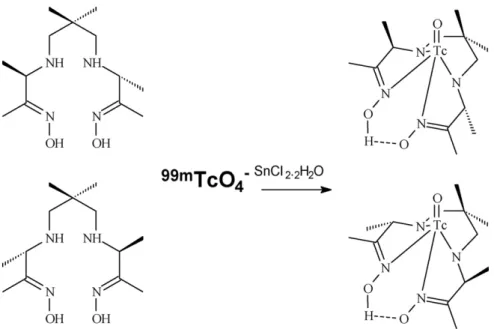
to obtain images of infections and inflammations because of its lipophilic properties (Martin-Comin and Prats, 1999). As shown inFig. 1, radiolabeling ofd,l-HMPAO may give rise to two99mTc-labelled enantiomers with almost identical
prop-erties (Vanderghinste et al., 2003). The employment of the
99mTc-HMPAO complex in the preparation of new types of
radiopharmaceuticals such as the radiolabeled liposomes has been studied by several authors (Goins et al., 1993; Phillips, 1998; Laverman et al., 1999; Boerman et al., 2000). However, studies with liposomes have been limited because of inherent problems such as low encapsulation efficiency, poor storage stability (Soppimath et al., 2001) and a high incidence of side effects after i.v. administration (Brouwers et al., 2000; Dams et al., 2000; Szebeni et al., 2000, 2002).
The NC presents high entrapment efficiency for lipophilic drugs, a low polymer content and a low inherent toxicity when compared with liposomes and nanospheres (Mosqueira et al., 2006). Nevertheless, before utilizing a NC formulation for diag-nosis, it must be very well characterized. Optimal preparations consist of a small (200–300 nm), monodispersed nanocapsule population, stable in biological media, principally in blood.
Ruozi et al. (2005) obtained information about the dimen-sions and homogeneity of liposomes using classical photon correlation spectroscopy (PCS) and atomic force microscopy (AFM). Recently, the AFM method was also used to charac-terize nanocapsules prepared with biodegradable polymers (Mosqueira et al., 2005; Leite et al., 2005; Assis et al., 2007).
The aim of the present work was the preparation and char-acterization of the conventional and the sterically stabilized NC using poly-d,l-lactide (PLA) polymer and the copolymer of monomethoxypolyethyleneglycol-co-poly-d,l-lactide (PLA-PEG), respectively, for the encapsulation of 99mTc-HMPAO
complex. Furthermore, it was also investigated the labelling efficiency of NC with99mTc-HMPAO complex and its release
profiles in physiological media.
2.
Materials and methods
2.1. Materials
Soy lecithin (Epikuron®170) was purchased from Lucas Meyer
(France). Poly(d,l-lactic acid), PLA50,with an average
molecu-lar weight (Mw) of 75,000(g/mol) and the surfactant poloxamer 188 were provided by Sigma–Aldrich (USA). Miglyol 810N (caprylic/capric triglycerides) was received as a gift from Hulls (Germany). PLA-PEG (PLA of 49,000 g/mol containing approxi-mately 10% PEG covalently grafted with a Mw of 5000 g/mol) was provided by Alkermes (USA) and used without further purification. The HMPAO (d,l-hexamethylpropyleneamine oxime) was obtained by reconstitution of a Ceretec kit (Amer-sham Inc., UK) and99mTc (pertechnetate form) was obtained
as a sodium solution from a99Mo generator (IPEN/Brazil). All
the solvents used were analytical grade, and other chemicals were commercially available reagent grade and used without further purification. MilliQ®
water (Simplicity 185, Millipore, Bedford, USA) was used throughout.
2.2. Methods
2.2.1. Preparation of nanocapsules
Conventional nanocapsules (PLA NC) were prepared accord-ing to the method described byFessi et al. (1989)and those sterically stabilized with PEG were prepared by the method described byMosqueira et al. (2001a). In the conventional NC preparation poly(d,l-lactic acid) (Mw 75,000) was used together with Poloxamer 188, a surfactant containing polyethyleneg-lycol chains on its chemical structure. Surface-modified NC was prepared using the diblock polymer of PLA-PEG without poloxamer surfactant. In the PLA-PEG NC the PEG chains are covalently linked to NC surface. Briefly, the polymer (0.6%, w/v) was dissolved in acetone (2 mL) containing 0.75% (w/v) of soy lecithin (Epikuron®170) and 2.5% (v/v) of Miglyol®810N. In
the case of conventional NC, this organic solution was poured into the external aqueous phase (4 mL) containing 0.75% (w/v) of poloxamer 188 with stirring. The solvents were evaporated under reduced pressure up to 2 mL volume (Fisatom Rotary Evaporator, Brazil).
2.2.2. 99mTc-HMPAO-labelled nanocapsules preparation
An HMPAO kit (Ceretec®
, Amersham Inc., UK) containing 0.5 mg HMPAO and 4.0g SnCl2was reconstituted with 2 mL of
0.9% NaCl solution. The mixture was divided into four aliquots of 0.4 mL each and stored at−70◦C under a nitrogen
atmo-sphere. Aliquots (0.4 mL) of HMPAO were incubated with 296 MBq of Na99mTcO
4 for 5 min and 250L of methylene blue solution was added. Methylene blue was used to stabilize the lipophilic 99mTc-HMPAO complex (Barthel et al., 1999). The
radiolabeled complex was then dissolved in the organic phase (acetone) during the preparation of the nanocapsules. The fol-lowing steps of the procedure were carried out as described above to obtain the99mTc-HMPAO nanostructures.
2.2.3. Purification of99mTc-HMPAO-NC
99mTc-HMPAO that was not associated with NC was separated
from the99mTc-HMPAO loaded in NC by gel filtration through a
Sephadex G-25 (Pharmacia®
, Sweden) column. A 1 mL aliquot of a colloidal suspension of 99mTc-HMPAO-NC was layered
onto a 1.5 cm×15 cm Sephadex G-25 column, and the sample was eluted with saline. One-millilitre fractions were collected and counted in a scintillation counter (Capintec, INC-CRC.15R, USA). An aliquot (1 mL) containing unloaded NC and free
99mTc-HMPAO was passed through the column to determine
the difference in the separation profiles of the preparations. The small amounts of99mTc-HMPAO complex used in this
work were soluble in water (manufacturer, Ceretec-Amersham Health). The purified99mTc-HMPAO-NC was used in further
characterization experiments.
2.2.4. 99mTc-HMPAO encapsulation efficiency and release
The encapsulation efficiency of99mTc-HMPAO in the
nanocap-sules was calculated by the difference between the total activity of the lipophilic complex in the colloidal suspension and the free 99mTc-HMPAO found in the external aqueous
phase (Eq. (1)). The ultrafiltrate (external aqueous phase) was obtained by ultrafiltration/centrifugation of 400L of NC suspension at 1800×gfor 15 min in an AMICON device (Microcon, Millipore®, molecular weight cut-off: 10,000). The
99mTc-HMPAO released from NC formulations was determined
in 70% rat plasma under sink conditions at different times. Aliquots containing 3.7 MBq of99mTc-HMPAO-PLA-PEG NC and 99mTc-HMPAO-PLA NC (200L) were incubated with 500 mL of
plasma (70%) at 37◦C, under moderate stirring, in separated
tubes for each time point. After 30, 120, 480 and 720 min sam-ples (200L) were collected to radioactivity determination in a gamma counter, after external phase separation from the total NC suspension, using the ultrafiltration/centrifugation technique with Ultrafree®units, 0.1
m pore size (Millipore®).
The separation membrane used in the release study allowed the elution of the free 99mTc-HMPAO complex or that was
associated with plasmatic protein, while the99mTc-HMPAO
entrapped in NC remained in the membrane upper compart-ment. An additional experiment was performed in plasma medium with99mTc-HMPAO encapsulated in PLA-PEG NC
dur-ing NC preparation or99mTc-HMPAO incubated with unloaded
PLA-PEG NC. This analysis was evaluated in time periods of up to 720 min. These experiments were also performed at 37◦C
with moderate stirring. The non-encapsulated99mTc-HMPAO
was encountered in the ultrafiltrate, and the encapsulated
99mTc-HMPAO was retained by the ultrafiltration membrane.
The encapsulation efficiency of radioactivity into the NC was determined from following Eq.(1), whereA= activity:
(%)=A
total−Aultrafiltrate
Atotal ×100 (1)
2.2.5. Nanocapsules characterization
expressed as mean±standard deviation for at least three dif-ferent batches of each nanocapsule formulation.
2.2.5.2. Atomic force microscopy imaging. AFM observation was performed in air at room temperature, on a Dimen-sion 3000 apparatus, as well as on Multimode Equipment, both monitored by a Nanoscope IIIa controller from Digi-tal Instruments (Santa Barbara, CA, USA). A droplet (5L) of sample was deposited on a freshly cleaved mica surface, spread and partially dried with a stream of argon. The images were obtained in tapping mode, using commercial silicon probes, from NanosensorsTM, with cantilevers having a length
of 228m, resonance frequencies of 75–98 kHz, spring con-stants of 29–61 N/m and a nominal tip curvature radius of 5–10 nm. The scan rate used was 1 Hz. Dimensional analyses were performed using the “section of analyses” program of the system. A minimum of ten images from each sample was analyzed to assure reproducible results. The values represent an average±standard deviation of approximately 40 particle measurements.
2.2.5.3. Zeta potential analyses. The zeta potential was deter-mined by laser doppler anemometry (LDA) in a Zetasizer HS3000 (Malvern Instruments, Malvern, UK). The samples were analyzed following dilution by 1:1000 in 1 mM NaCl at a conductivity of approximately 120±20S/cm2. Values
reported are the mean±standard deviation of at least three different batches of each NC formulation.
2.2.6. Statistics
All experiments were performed in triplicate and were expressed as mean values±standard deviations, except as otherwise stated. Mean sizes, zeta potential and drug release data at each time point were compared by an ANOVA test using the Prim 4.0 program while considering a probability of 5% to be significant.
3.
Results and discussion
NC containing 99mTc-HMPAO were prepared by the
inter-facial deposition of the preformed polymer followed by solvent evaporation (Fessi et al., 1989), a simple and rapid method of obtaining nanocapsules containing a radioactive tracer suitable for intravenous administration. All formula-tions described here produced particles smaller than 500 nm and greater than 200 nm. Normal tissues contain capillaries with tight junctions that are less permeable to nanosized particles (Litzinger et al., 1994). The different types of NC pro-duced in this work are intended to be used as radiotracers of inflammation sites where the capillaries are very leaky. In this way, the sizes found were considered suitable for the further purpose. Two types of nanocapsules containing99m
Tc-HMPAO were developed in this work: conventional PLA NC and surface-modified PLA-PEG NC. Surface-modified nanocap-sules (PLA-PEG NC) were included in this study because their uptake by the mononuclear phagocyte system is substantially delayed, thereby increasing the time during which they circu-late in the blood stream (Mosqueira et al., 2001b).
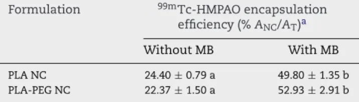
Table 1 – Effect of methylene blue on the encapsulation efficiency of99mTc-HMPAO complex in polymeric NC
Formulation 99mTc-HMPAO encapsulation
efficiency (%ANC/AT)a
Without MB With MB
PLA NC 24.40±0.79 a 49.80±1.35 b PLA-PEG NC 22.37±1.50 a 52.93±2.91 b
Identical letters indicate that there is no significant difference between the data. The data represent the mean±S.D. (n= 3) for each determination.
a A
NCis activity associated to the NC andATis the total activity in NC suspension (Bq).
3.1. Nanocapsule characterization
A better encapsulation efficiency of the99mTc-HMPAO
com-plex in the nanocapsules was obtained using methylene blue as a stabilizer. According to the results shown inTable 1, the encapsulation efficiency was around 50% for both prepa-rations (PLA NC and PLA-PEG NC). On the other hand, the encapsulation efficiency of99mTc-HMPAO in NC preparations
without methylene blue was greatly reduced. Thus, the sta-bility of the lipophilic99mTc-HMPAO complex was enhanced
by methylene blue, as has been reported in a previous work (Barthel et al., 1999; Sobal and Sinzinger, 2001).
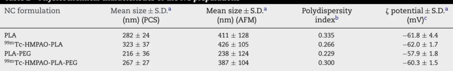
The particle size, polydispersity index and zeta potential of prepared conventional and surface-modified NC contain-ing99mTc-HMPAO are presented inTable 2. The polydispersity
indices showed that the different formulations prepared by the nanoprecipitation method were homogeneous with respect to the polydispersity index and can be considered monodisperse (≤0.3). The PLA-PEG NC showed reduced sizes compared to the PLA NC by the both methods used to deter-mine NC mean sizes (Table 2). These results are in agreement with the data obtained byMosqueira et al. (2001a) for PLA and PLA-PEG NC, however, the NC sizes were slightly dif-ferent from those previously reported, probably because the average molecular weights of the polymers were different and obtained from other suppliers. The mean diameters of unloaded and 99mTc-HMPAO loaded NC were very similar
and no significant difference were found among the prepa-rations.99mTc-HMPAO encapsulation in PLA and PLA-PEG NC
did not induce any significant increase in the mean diame-ter and in the polydispersity of the colloidal NC suspensions. Some authors have demonstrated that the mean diameter of nanoparticles has an influence on the biodistribution studies because particles larger than 300 nm and lower than 70 nm are rapidly taken up by MPS accumulating in the spleen and liver. On the other hand, particles within the 150–200 nm range were founded to be longest circulating (Litzinger et al., 1994; Owens and Peppas, 2006). Based on this observation, the long-circulating PLA-PEG NC, with sizes around 200 nm, could be particularly interesting to reach the loose junctions of the endothelium of inflammatory or infectious foci, considering the mean sizes measured by PCS. In fact, biodistribution stud-ies performed in our laboratory (data not published) showed that99mTc-HMPAO-PLA-PEG NC were able to identify
Table 2 – Physicochemical characteristics of the NC preparations NC formulation Mean size±S.D.a
(nm) (PCS)
Mean size±S.D.a (nm) (AFM)
Polydispersity
indexb potential
±S.D.a (mV)c
PLA 282±24 411±128 0.335 −61.8±4.4
99mTc-HMPAO-PLA 323±37 426±105 0.266 −62.0±1.7
PLA-PEG 216±36 238±124 0.229 −57.9±1.8
99mTc-HMPAO-PLA-PEG 267±27 387±104 0.300 −60.3±1.5
The entrapment of99mTc-HMPAO did not significantly affect either the mean diameter or the zeta potential of the nanocapsules (P> 0.05). a Standard deviation (n= 3) of the population that was reported by the instrument.
b Monodispersed samples (≤0.3).
c Measurement after 1:1000 dilution in 1 mM NaCl (conductivity, 120±20S/cm).
intravenously in rats. Both types of PEG chains associated with a NC surface, adsorbed (Poloxamer 188) or covalently grafted (PLA-PEG copolymer), seemed to stabilize the association of
99mTc-HMPAO with the nanostructures, thereby preventing
their aggregation, because no signs of physical instability were observed during the experiments even when PLA or PLA-PEG NC were incubated in 70% rat plasma release media.
Zeta potential results (Table 2) showed that the unloaded PLA NC and 99mTc-HMPAO loaded NC formulations
exhib-ited a negative charge with values ranging from −57.9 to
−62.0 mV. The entrapment of 99mTc-HMPAO did not
signifi-cantly affect either the mean diameter or the zeta potential of the nanocapsules (P> 0.05) indicating that probably the com-plex was mainly associated with the oily core of NC. These values were typically observed for colloidal carriers containing PLA polymer, where values of−50.9 to−51 mV were found for PLA-POLOX NC and PLA-PEG NC 45–5 kDa (10% PEG), respec-tively, prepared from polymers with slightly different Mw and using the same method (Mosqueira et al., 2001a, 2006). The negative zeta potential imparted by the PLA polymer and lecithin was not masked by the presence of PEG chains with its hiding properties, probably because the high contents of lecithin in the NC formulation contributed to the mainte-nance of the negative surface charge, as has been previously discussed byMosqueira et al. (2001a, 2006). However, even when these negatively charged PLA-PEG NC were used in vivo (Mosqueira et al., 2001b) they showed long-circulating properties, probably because lecithin layers surrounding NC masks the real zeta potential at NC interfaces. In general, nanospheres, without lecithin and oil, suffer a much higher effect of PEG on the zeta potential than NC (Govender et al., 2000).
AFM is a powerful technique, with dimensional resolu-tions from 1 up to 100 ˚A, that provides a unique possibility for visualizing nanoparticles in a natural environment, without sample manipulation (Neves et al., 1998). While PCS measures hydrodynamic radius, the AFM technique allows direct mea-surement of size in samples in a partially dried state, deposited on freshly cleaved mica plates, which permits simultaneous characterization of particle shape and structure. It is advan-tageous because the size, structure and shape are important parameters in the design of nanostructures used in diag-nosis by an intravenous route. All NC preparations showed a spherical form on the mica surface in the AFM images (Figs. 2 and 3). The NC maintained their structures unaltered for up to 1 week after drying. The NC presented a
homoge-neous distribution in height and in three-dimensional images (Fig. 3). The average sizes of unloaded PLA NC obtained by AFM were 411±128 nm, while those containing99mTc-HMPAO
were 426±105 nm (n= 40). Similarly, when the NC size was determined by PCS, no significant differences were observed between unloaded NC and those containing the99mTc-HMPAO
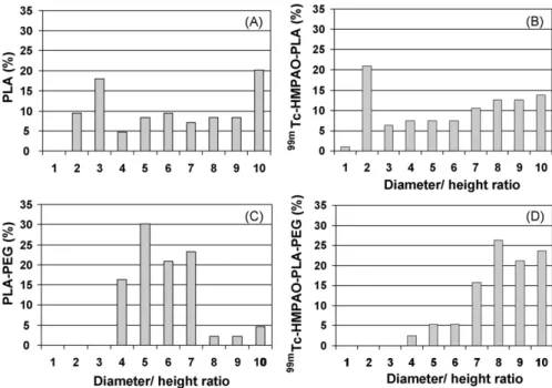
complex. However, the diameters calculated by AFM were observed to be larger than those measured by PCS for both NC types. This observation can be explained by a possible NC flat-tening on mica that takes place after drying process with argon flow. The PLA NC images (Fig. 4) show nanostructures and layers deposited on the mica with different heights, resulting in complex images when compared with PLA-PEG. These few nanometer layers (2–10 nm) were attributed to poloxamer and phospholipids organized in lamellar structures, as could be observed in the analysis images obtained from these controls (poloxamer and lecithin). Similar observations were previ-ously reported byLeite et al. (2005). NC in all samples appears deposited on these layers or immersed in them (Fig. 4). On the other hand, PLA-PEG NC (Fig. 2C and D) presents more homogeneous distribution of particles with the absence of the low height layered structures. It could be explained by the absence of poloxamer in the PLA-PEG formulation that reduces the complexity of forms in the system. The average diame-ter calculated for PLA-PEG NC was 238±124 nm (n= 40) and for99mTc-HMPAO-PLA-PEG NC was 387±104 nm (n= 40). The
mean size differences were not significant (P> 0.05). In fact, the flattening process was probably related to variations in poly-meric wall thickness and homogeneity of polymer deposition in the process of NC preparation, as previously speculated by
Fig. 2 – AFM topographic (height) images A (PLA NC) and C (PLA-PEG NC) and phase images B (PLA NC) and D (PLA-PEG NC) showing spherical nanocapsules obtained in tapping mode. Scan sizes are shown in the figure.
as nanospheres, or even nanoemulsions (without polymeric wall) could occur. This result agrees with those observed by
Mosqueira et al. (2001a), which demonstrated the complex-ity of the nanostructures formed by the nanoprecipitation method and the increase in homogeneity of PLA-PEG NC pre-pared without poloxamer using ultracentrifugation in Percoll®
gradient method. The PLA-PEG NC presented a more homo-geneous wall with diameter/height ratios of 4–7 (Fig. 5C and D).99mTc-HMPAO associated with PLA-PEG NC increased the
NC dispersion heterogeneity and diameter/height difference (7–10), probably because it produced alterations in the struc-ture of NC wall. Poloxamer seemed also to play a role in the formation of different types of nanostructures in NC formula-tions and in the appearance of complex layers in AFM images that probably increase the heterogeneity of PLA NC (Fig. 4). The evidence of NC wall formation and the determination of its thickness was elegantly performed byR ¨ube et al. (2005)using the small angle neutron scattering technique applied to NC prepared by the same nanoprecipitation method used in this paper.
In PLA-PEG NC images obtained by AFM, a halo was observed around the NC (Fig. 2D). The phase images suggest that this phenomenon could be generated by a hydration layer around sterically stabilized PLA-PEG NC, probably because a small amount of water are firmly fixed to the hydrophilic groups by hydrogen bonds at NC surface and was not com-pletely removed by the fast argon flow that spread the sample on mica. It is noteworthy to point out that this halo was not observed by AFM in PLA NC stabilized with poloxamer and appears particularly in the phase images of PLA-PEG NC (Fig. 2D). This structure in halo form suggests the presence of covalently grafted PEG in this preparation.
Fig. 3 – AFM image: three-dimensional view of unloaded PLA nanocapsules spread on mica and its homogenous dispersion of size.
3.2. 99mTc-HMPAO release from nanocapsules
The in vitro release profiles of99mTc-HMPAO from PLA and
PLA-PEG nanocapsules are shown inFig. 6. The experiments were preformed under sink conditions to avoid the interfer-ence of the complex solubility in the release rate. In this study a fast release of radioactivity for all NC types was observed in the first 30 min. Approximately 33% of 99m
Tc-HMPAO was released in plasma medium for PLA-PEG NC. This result can be explained by the release of an adsorbed part of the99mTc-HMPAO weakly bound to the NC surface, or by a fast
equilibrium changes in the partition between internal oily core and release medium. Plasma probably acts as a driving force that promotes99mTc-HMPAO release from the NC in the burst
phase. In the case of NC, a fast release of drug in the first few minutes after dilution in release media was interpreted by several authors as drug adsorbed at the NC surface (Lopes et al., 2000; Mosqueira et al., 2001b, 2006). However, the release profile of99mTc-HMPAO was biphasic, because after the initial
burst phase (30 min) only 7% of the radioactivity was released from NC up to 12 h (Fig. 6). Phillips (1998) has shown that liposomes are able to retain99mTc-HMPAO in plasma at 37◦C
for as much as 90 h. However, comparative and quantitative analysis of the ability of the both nanosystems to entrap this radiotracer have not been performed and should be evaluated in further experiments. On the other hand, when the99m
Tc-HMPAO was only incubated with the already formed PLA-PEG
Fig. 4 – AFM image of99mTc-HMPAO-PLA nanocapsules in the “section analysis” software showing nanocapsule height in black arrows, and different nanometric layers deposed on mica indicated by red and green arrows (height). The measured sizes are indicated in tables with the same colours. Scan size: 7.5m×7.5m. (For interpretation of the references to color
Fig. 5 – Percentage of diameter/height ratios of nanocapsules preparations calculated from 40 particles measurements using “section analysis software” of the Nanoscope IIIa AFM microscope. In A: unloaded PLA NC; B:99mTc-HMPAO-PLA NC; C: unloaded PLA-PEG NC; D:99mTc-HMPAO-PLA-PEG NC. The higher ratios indicate higher degrees of nanocapsule flattening on mica surface.
NC, 70% of the radioactivity was released in the first 30 min and 90% in 120 min, suggesting that in this case99mTc-HMPAO
was not truly encapsulated into the NC structure, being rapidly desorbed when in contact with the release media.Lopes et al. (2000), in a similar experiment, observed a total release of the ethionamide after incubation with preparations of nanocap-sules and nanospheres. Based on these results, they proposed that the drug might be mainly bound to the particle’s sur-face. After the initial burst,99mTc-HMPAO that seems to be
Fig. 6 – Release profiles of99mTc-HMPAO encapsulated in PLA() and in PLA-PEG NC () and release profile
99mTc-HMPAO just incubated with preformed PLA-PEG NC
() in the 70% rat plasma/saline medium at 37◦C (3.4g
HMPAO/mL of release medium) using 0.1m ultrafiltration
membrane pore (Millipore®) to separate the nanocapsules from the external medium.
firmly associated to PLA-PEG NC structure, release less than 10% of the total radioactivity in the first 12 h in medium with 70% of plasma. This result indicates that PLA-PEG NC is more able to retain 99mTc-HMPAO compared to PLA NC. Cruz et al. (2006)also evidenced a biphasic release profile from NC, attributing the burst phase to the presence of the polymer, whereas the presence of the oil increased the half-life of the sustained phase. The release profiles of99mTc-HMPAO from
the NC, despite of the type of the polymer, are in agreement with those cited above, however the rates could not be com-pared because the release media was different. In this way, a relationship between PLA-PEG polymer and the radioactive complex could explain the reduced initial burst, although this is not completely understood. Furthermore, the halo visual-ized in AFM images surrounding PLA-PEG NC could represent a steric barrier that impairs99mTc-HMPAO diffusion from NC
structure to protein binding sites (Fig. 2D). It is another hypoth-esis to explain the differences in the release profiles found between the two types of NC studied. The result obtained with conventional PLA nanocapsules showed that approximately 80% of the radioactivity was released in 120 min of incubation. These data are in agreement with the studies ofMosqueira et al. (2006)where a better ability to retain lipophilic antimalarial drug was observed with PLA-PEG NC when compared with PLA NC stabilized with poloxamer in media containing plasma. Poloxamer seems to play an important role in release profiles of lipophilic drugs from NC, because it was able to increase the percentage and the rate of halofantrine release, when it was added to the PLA-PEG NC formulation (Mosqueira et al., 2006). Therefore, the developed PLA-PEG NC emerged as very suit-able carrier for transporting99mTc-HMPAO radioactive tracer
further pre-clinical experiments for diagnosis of inflammatory processes.
4.
Conclusion
Both types of nanocapsules preparations studied in this work were able to encapsulate the 99mTc-HMPAO with the same
yield. However, the physical and structural properties of PLA-PEG nanocapsules to which PLA-PEG chains are covalently linked were more homogeneous, as shown by AFM. Indeed, the release profiles of these nanocapsules indicate that a part of the radioactivity was probably adsorbed at the NC sur-face and presents a burst effect after dilution. However, the greater part of the radioactivity was truly associated with the NC inner structure, and a reduced release rate was observed even in the presence of plasma in the external medium. The better release profile of99mTc-HMPAO from PLA-PEG NC, the
homogeneity of the polymeric wall and the adequate size indi-cate that this approach could provide an imaging agent for diagnosis.
Acknowledgments
The authors thank CAPES-Brazil for personal financial sup-port for the first author. This work was supsup-ported by the FAPEMIG/NANOBIOMG network. The authors are grateful to Dr. Elenara L. Sena (UFSC-Brazil) for the kind gift of the PLA-PEG polymers purchased from Alkermes. We acknowledge also Dr. Monica C. Oliveira and Danielle N. Assis for their help-ful discussions and Dr. David L. Nelson for English revision of the article.
r e f e r e n c e s
Assis, D.N., Mosqueira, V.C.F., Vilela, J.M.C., Andrade, M.S., Cardoso, V.N., 2007. Release profiles and morphological characterization by atomic force microscopy and photon correlation spectroscopy of99mTechnetium-fluconazole
nanocapsules. Int. J. Pharm.,
doi:10.1016/j.ijpharm.2007.08.002.
Barratt, G., 2000. Therapeutic applications of colloidal drug carriers. Pharm. Sci. Technol. Today 3, 163–171. Barthel, H., Kampfer, I., Seese, A., Dannenber, C., Kluge, R.,
Burchert, W., Knapp, W.H., 1999. Improvement of brain SPECT by stabilization of Tc-99m-HMPAO with methylene blue or cobalt chloride. Nuklearmedizin 38, 80–84.
Boerman, O.C., Laverman, P., Oyen, W.J.G., Corstens, F.H.M., Storm, G., 2000. Radiolabelled liposomes for scintigrafic imaging. Prog. Lipid Res. 39, 461–475.
Brouwers, A.H., De Jong, D.J., Dams, E.T.M., Oyen, W.J.G., Boerman, O.C., Laverman, P., Naber, T.H.J., Storm, G., Corstens, F.H.M., 2000. Tc-99m-PEG-Liposomes for the evaluation of colitis in Crohn’s disease. J. Drug Target 8, 225–233.
Chianelli, M., Mather, S.J., Martin-Comin, J., Signore, A., 1997. Radiopharmaceuticals for the study of inflammatory processes: a review. Nucl. Med. Commun. 18, 437–455. Cruz, L., Soares, L.U., Costa, T.D., Mezzalira, G., Silveira, N.P.,
Guterres, S.S., Pohlmann, A.R., 2006. Diffusion and
mathematical modeling of release profiles from nanocarriers. Int. J. Pharm. 313, 198–205.
Dams, E.T.M., Oyen, W.J.G., Boerman, O.C., Storm, G., Laverman, P., Kok, P.J.M., Buijs, W.C.A.M., Bakker, H., Meer, J.W.M., van der Corstens, F.H.M., 2000.99mTc–PEG liposomes for the
scintigraphic detection of infection and inflammation: clinical evaluation. J. Nucl. Med. 41, 622–630.
Fessi, H., Puisieux, F., Devissaguet, J.P., Ammoury, N., Benita, S., 1989. Nanocapsule formation by interfacial polymer deposition following solvent displacement. Int. J. Pharm. 55, R1–R4.
Goins, B., Klipper, R., Rudolph, A.S., Cliff, R.O., Blumhardt, R., Phillips, W.T., 1993. Biodistribution and imaging studies of Tc-99m-labelled liposomes in rats. J. Nucl. Med. 34, 2160–2168. Govender, T., Riley, T., Ehtezazi, T., Garnett, M.C., Stolnik, S., Illum, L., Davis, S., 2000. Defining the drug incorporation properties of PLA-PEG nanoparticles. Int. J. Pharm. 199, 95–110.
Gref, R., Minmitake, Y., Percchia, M.T., Trubetskoy, V., Torchilin, V., Langer, R., 1994. Biodegradable long-circulating polymeric nanospheres. Science 263, 1600–1603.
Laverman, P., Dams, E.T.M., Oyen, W.J.G., Storm, G., Koenders, E.B., Prevost, R., van der Meer, J.W.M., Corstens, F.H.M., Boerman, O.C., 1999. A novel method to label liposomes with99mTc by
the hydrazino nicotinyl derivate. J. Nucl. Med. 40, 192–197. Legrand, P., Barratt, G., Mosqueira, V., Fessi, H., Devissaguet, J.P.,
1999. Polymeric nanocapsules as drug delivery systems. A review. STP Pharma. Sci. 9, 411–418.
Leite, E.A., Vilela, J.M.C., Mosqueira, V.C.F., Andrade, M.S., 2005. Poly-caprolactone nanocapsules morphological features by atomic force microscopy. Microsc. Microanal. 11, 48–51. Litzinger, D.C., Buiting, A.M.J., van Rooijen, N., Huang, L., 1994.
Effect of liposome size on the circulation time and intraorgan distribution of amphipathic poly(ethylene glycol)-containing liposomes. Biochem. Biophys. Acta Biomembr. 1190, 99–107.
Lopes, E., Pohlmann, A.R., Bassani, V., Guterres, S.S., 2000. Polymeric colloidal systems containing ethionamide: preparation and physico-chemical characterization. Pharmazie 55, 527–530.
Martin-Comin, J., Prats, E., 1999. Clinical applications of radiolabelled blood elements in inflammatory bowel disease. Q. J. Nucl. Med. 43, 74–82.
Mosqueira, V.C.F., Legrand, P., Gref, R., Heurtault, B., Appel, M., Barrat, G., 1999. Interactions between a macrophage cell line (J774A1) and surface-modified poly(d l-lactide) nanocapsules bearing poly(ethylene glycol). J. Drug Target 7, 65–78. Mosqueira, V.C.F., Legrand, P., Gulik, A., Bourdon, O., Gref, R.,
Labarre, D., Barratt, G., 2001a. Relationship between
complement activation, cellular uptake and physicochemical aspects of novel PEG-modified nanocapsules. Biomaterials 22, 2967–2979.
Mosqueira, V.C.F., Legrand, P., Morgat, J.-P., Vert, M., Mysiakine, E., Gref, R., Devissaguet, J., Barratt, G., 2001b. Biodistribution of long-circulating PEG-grafted nanocapsules in mice: effects of PEG chain length and density. Pharm. Res. 18 (10), 1411–1419. Mosqueira, V.C.F., Leite, E.A., Barros, C.M., Vilela, J.M.C., Andrade,
M.S., 2005. Polymeric nanostructures for drug delivery: characterization by atomic force microscopy. Microsc. Microanal. 11, 36–39.
Mosqueira, V.C.F., Legrand, P., Barratt, G., 2006. Surface-modified and conventional nanocapsules as novel formulation for parenteral delivery of halofantrine. J. Nanosci. Nanotechnol. 9/10, 3193–3202.
Neves, B.R.A., Vilela, J.M.C., Andrade, M.S., 1998. Microscopia de varredura por sonda mec ˆanica: uma introduc¸ ˜ao. Cer ˆamica 44, 212–219.
Oyen, W.J.G., Boerman, O.C., Storm, G., van Bloois, L., Koenders, E.B., Claessens, R.A.M.J., Perenboom, R.M., Crommelin, D.J.A., van der Meer, J.W.M., Corstens, F.H.M., 1996a. Detecting infection and inflammation with Tc99m-labeled Stealth®
liposomes. J. Nucl. Med. 37, 1392–1397.
Oyen, W.J.G., Boerman, O.C., van der Laken, C.J., Claessens, R.A.M.J., van der Meer, J.W.M., Corstens, F.H.M., 1996b. The uptake mechanisms of inflammation and infection-localizing agents. Eur. J. Nucl. Med. 23, 459–465.
Phillips, W.T., 1998. Targeting of drugs 6: strategies for stealth therapeutic systems. In: Gregoriadis, McCormack (Eds.), Use of Radiolabelled Liposomes for PEG-Liposome-based Drug Targeting and Diagnostic Imaging Applications. Plenum Press, New York, pp. 109–120.
Rennen, H.J.J.M., Boerman, O.C., Oyen, W.J.G., Corstens, F.H.M., 2001. Imaging infection/inflammation in the new miliennium. Eur. J. Nucl. Med. 28,
241–252.
R ¨ube, A., Hause, G., M ¨ader, K., Kohlbrecher, J., 2005. Core-shell structure of miglyol/poly(d l-lactide)/poloxamer
nanocapsules studied by small-angle neutron scattering. J. Control. Release 107, 244–252.
Ruozi, B., Tosi, G., Forni, F., Fresta, M., Vandelli, M.A., 2005. Atomic force microscopy and photon correlation spectroscopy: two techniques for rapid characterization of liposomes. Eur. J. Pharm. Sci. 25, 81–89.
Sasatsu, M., Onishi, H., Machida, Y., 2006. In vitro and in vivo characterization of nanoparticles made of MeO–PEG amine/PLA block copolymer and PLA. Int. J. Pharm. 317, 167–174.
Sobal, G., Sinzinger, H., 2001. Methylene blue-enhanced stability of (99mTc)HMPAO and simplified quality control—a
comparative investigation. Appl. Radiat. Isot. 54, 633–636. Soppimath, K.S., Aminabhavi, T.M., Kulkarni, A.R., Rudzinski,
W.E., 2001. Biodegradable polymeric nanoparticles as drug delivery devices. J. Control Release 70, 1–20.
Szebeni, J., Baranyi, L., Savay, S., Bodo, M., Morse, D.S., Basta, M., Stahl, G.L., Bunger, R., Alving, C.R., 2000. Liposome-induced pulmonary hipertension: properties and mechanism of a complement-mediated pseudoallergic reaction. Am. J. Physiol. Heart Circ. Physiol. 279, H1319–H1328.
Szebeni, J., Baranyi, L., Savay, S., Milosevits, J., Bunger, R., Laverman, P., Metselaar, J.M., Storm, G., Chanan-Khan, A., Liebes, L., Muggia, F.M., Cohen, R., Barenholz, Y., Alving, C.R., 2002. Role of complement activation in hipersensitivity reactions to Doxil and Hynic PEG liposomes: experimental and clinical studies. J. Liposome Res. 12, 165–172.
Vanderghinste, D., Van Eeckhoudt, M., Terwinghe, C.,