0021-7557/$ - see front matter © 2013 Sociedade Brasileira de Pediatria. Published by Elsevier Editora Ltda. All rights reserved. http://dx.doi.org/10.1016/j.jped.2013.03.001
www.jped.com.br
☆Please, cite this article as: Rodrigues M, D’Amico MF, Patiño FR, Barbieri D, Damião AO, Sipahy AM. Clinical manifestations, treatment, and outcomes of children and adolescents with eosinophilic esophagitis. J Pediatr (Rio J). 2013;89:197−203.
* Corresponding author.
E-mail: [email protected] (M. Rodrigues).
ORIGINAL ARTICLE
Clinical manifestations, treatment, and outcomes of children
and adolescents with eosinophilic esophagitis
☆Maraci Rodrigues
a,*, Maria Fernanda M. D’Amico
b, Fatima Regina Almeida Patiño
c,
Dorina Barbieri
d, Aderson Omar Mourão Cintra Damião
a, Aytan M. Sipahi
eaPhD in Medicine. Assistant Physician, Departamento de Gastroenterologia e Hepatologia, Hospital das Clínicas,
Universidade de São Paulo (USP), São Paulo, SP, Brazil
bPediatric Gastroenterologist. Coordinator of the Pediatric Residency, Complexo Hospitalar do Mandaqui, São Paulo,
SP, Brazil
cPediatric Gastroenterologist, Complexo Hospitalar do Mandaqui, São Paulo, SP, Brazil
dProfessor, Departamento de Pediatria, Instituto da Criança, Hospital das Clínicas, USP, São Paulo, SP, Brazil
eChief, Grupo de Intestino, Departamento de Gastroenterologia e Hepatologia, Hospital das Clínicas, USP, São Paulo,
SP, Brazil
Received 1 February 2012; accepted 28 September 2012
KEYWORDS
Eosinophilic esophagitis; Gastroesophageal relux
Abstract
Objective: This study aimed to describe the clinical, endoscopic, and histologic
characteristics, as well as the response to conventional treatment of pediatric patients with the classical form of eosinophilic esophagitis (EoE).
Methods:Study of clinical, laboratory, endoscopic, and histologic data and response to
conventional treatment of 43 previously followed pediatric patients with the classical form of EoE.
Results: A total of 43 patients diagnosed with EoE were included in the study, of which
PALAVRAS-CHAVE Esofagite eosinofílica;
Reluxo
gastroesofágico
Manifestações clínicas, terapêutica e evolução de crianças e adolescentes com eso-fagite eosinofílica
Resumo
Objetivo:O objetivo deste estudo foi descrever as características clínicas, endoscópicas e
histológicas, assim comoa resposta ao tratamento convencional de pacientes pediátricos com a forma clássica de esofagite eosinofílica (EEo).
Métodos: Levantamento de dados clínicos, laboratoriais, endoscópicos, histológicos e
da resposta ao tratamento convencional de 43 pacientes pediátricos acompanhados previamente com a forma clássica de EEo.
Resultados: Foram incluídos 43 pacientes com diagnóstico de EEo, sendo 37 do
sexo masculino (86%), com idade média de 8,4 anos. Os sintomas mais encontrados foram: náusea, vômito e dor abdominal (100%) em crianças menores de sete anos; e inapetência (60%), queimação retroesternal (52%) e impactação alimentar (48%) em crianças maiores de sete anos e adolescentes. Em relação aos achados endoscópicos, 12 (28%) pacientes apresentavam placas esbranquiçadas na mucosa do esôfago, oito (18,5%) sulcos longitudinais, dois (4,5%) anéis concêntricos, três (7%) sulcos longitudinais e placas esbranquiçadas, e os outros 18 (42%) apresentavam aparência normal da mucosa esofágica. Apesar da resposta favorável inicial, 76,7% dos pacientes necessitaram realizar mais de um ciclo terapêutico com corticoterapia (aerossol ou sistêmica) e dieta (de exclusão ou eliminação dos alérgenos alimentares ou elementares). Persistência do infiltrado eosinofílico foi encontrada em uma parcela dos pacientes, a despeito da resposta clínica favorável.
Conclusões: A forma clássica da EEo apresenta sintomas diferentes segundo a faixa
etária. Parcela expressiva dos pacientes necessitou de mais de um ciclo terapêutico para apresentar remissão clínica. Observou-se melhora endoscópica e histológica; no entanto, a infiltração eosinofílica persistiu em parcela dos pacientes.
© 2013 Sociedade Brasileira de Pediatria. Publicado por Elsevier Editora Ltda. Todos os direitos reservados.
Introduction
Eosinophilic esophagitis (EoE) was described in the late 1970s by Landers et al.,1 and has been identified as a
clinical pathological entity since 1993 by Attwood et al.2
The Brazilian scientific literature in pediatrics is limited to two case series described by Cury et al.3 and by Ferreira
et al.4
Considering the recent and rapid evolution in the understanding of this condition, a group of researchers has updated the latest consensus on EoE,5 characterizing it as
an immunological esophageal disease with chronic evolution and frequent relapses, whose clinical manifestations are related to esophageal dysfunction and, histologically, to the presence of eosinophil accumulation in the esophageal mucosa more than 15 eosinophils per high-power field (HPF),
in the absence of eosinophil infiltration in the gastric and duodenal mucosa. The symptoms and pathological findings should improve with medical treatment, which involves the exclusion of allergens (food or aeroallergens), use of topical corticosteroids, or both therapeutic measures. A subset of patients responsive to proton pump inhibitors (PPIs) has also been described, termed PPI responders, differentiating them from the group of patients with classic EoE, known as non-PPI-responders. All other recognized causes of EoE should be excluded, as there are no symptoms identified at physical examination, serum markers or pathognomonic endoscopic data of the disease.5
The aim of this retrospective study was to describe the clinical, endoscopic, and histological characteristics and response to conventional treatment of pediatric patients with the classical form of EoE.
allergens). Persistence of eosinophil infiltration was found in some patients despite favorable clinical response.
Conclusions:The classic form of EoE typically shows different symptoms according age
range. A significant number of patients required more than one treatment cycle to show clinical remission. Endoscopic and histologic improvement was observed; however, eosinophilic infiltration persisted in some patients.
Methods
This was a retrospective study using data collection in a series of cases addressing the clinical, laboratory, endoscopic, and histological data from the records of 43 patients treated in the period between February, 2004 and September, 2010 with a diagnosis of EoE in the Gastroenterology Outpatient Clinic of the Hospital das Clínicas da Faculdade de Medicina da Universidade de São Paulo (HCFMUSP).
Inclusion criteria
All patients were referred to the specialized service with a history of previous upper endoscopy in their healthcare services of origin, with an initial diagnosis of reflux esophagitis resistant to treatment with PPI for at least three months.
All patients were symptomatic. After obtaining the clinical history, physical examination was performed and personal or family history of first-degree atopy was assessed; subsequently, patients underwent a new upper endoscopy. Two fragments of the esophagus were obtained (one proximal and one distal), as well as two fragments of the stomach and duodenum through upper endoscopy and biopsy forceps for histological evaluation. The number of eosinophils per high-magnification field (x400) was
quantified in at least 10 fields; EoE was considered when ≥
15 eosinophils/HPF5 were found in the esophageal mucosa
in at least 2 high-power fields and normal gastric and duodenal mucosa.
The presence of specific serum immunoglobulin E (IgE) was analyzed (ImmunoCAP®)6 for the suspected food
according to clinical history, to assist in the identification of food allergens involved in the EoE process, according to
the standard method, considered positive when ≥ class II.
Exclusion criteria
All other known causes of EoE were excluded, such as gastroesophageal reflux disease (GERD), celiac disease, Crohn’s disease, infection, hypereosinophilic syndrome, achalasia, drug hypersensitivity, vasculitis, pemphigus vegetans, connective tissue disease, and graft-versus-host disease.
As recommended in the consensus of 2007,7 the possibility
of GERD was ruled out by the lack of response to previous treatment with PPI.
The therapeutic modality adopted by the patients of the present study was in accordance with recent data from the literature on EoE.5,7 PPI is not considered the primary
treatment for EoE, but can be used as adjunctive therapy to relieve some of the symptoms. Systemic corticosteroid therapy was used for children younger than 2 years and topical therapy for the other patients. In the first case, prednisolone 1 mg/kg/day was used, divided in two daily doses for four weeks, with scheduled withdrawal up to the eighth week. In the second case, fluticasone propionate was used by oral route (children 2 to 4 years: 176 mcg daily; children 5 to 10 years: 444 mcg daily; adolescents > 11 years: 880 mcg daily). A suitable topical dose was administered in the form of an oral spray, divided in two
doses per day. Patients were instructed not to use the spacer and that the content of the spray must be swallowed and not aspirated, and patients were not allowed to eat or drink for at least 30 minutes after medication. Treatment with fluticasone was carried out for three months.
Dietary therapy (removal of specific antigens or elementary diet) was patient-specific and considered as an effective part of the treatment, with adequate calories, vitamins, and micronutrients of the received diet at baseline and at follow-up.7
Patients were followed through regular visits to the outpatient clinic every two months, where parents and patients were asked about symptoms, treatment adherence, and side effects of drug therapy. The monitoring of upper endoscopy was recommended after 9 to 12 months of treatment initiation.
This study was approved by the Ethics Committee of the Department of Gastroenterology of HCFMUSP.
Results
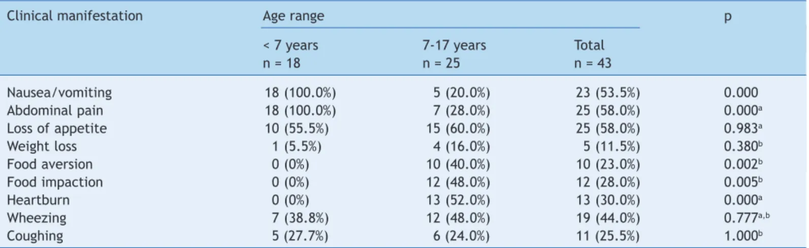
Of the 43 patients, 37 were males (86%), and 41 were defined by their parents as white and two as black. Patient’s age ranged from 1 month to 17 years, with a mean of 8.4 years. Approximately 44% of patients were identified by clinical history as having respiratory allergies, and 23% of first-degree relatives had respiratory, gastrointestinal, or skin atopy. The clinical manifestations of the 43 patients are shown in Table 1. Nausea, vomiting, and abdominal pain were more frequent in patients aged less than 7 years, while food aversion, food impaction, and heartburn were more prevalent in patients aged 7 to 17 years.
The search results for specific serum IgE (ImmunoCAP®) are summarized in Table 2. Of the 43 patients studied, 18 (41.9%) had sensitization to more than one food protein or aeroallergens, 2 (4.6%) were sensitive to one studied protein, and the remaining 23 (53.5%) patients presented no sensitization.
Regarding the endoscopic findings of the 43 patients on admission, whitish plaques on the esophagus mucosa predominated in 12 (27.9%), longitudinal grooves in 8 (18.6%) patients, whereas in 18 (41.2%) the macroscopic esophageal aspect was normal. At the microscopy, the quantification of eosinophils/HPF varied from 15-100 eosinophils per HPF, with the highest concentration of eosinophils in the lesions characterized as whitish plaques, as shown in Table 3. It was also observed that the proportion of patients with the number of eosinophils between 15 and 30 per field in normal endoscopies (44.4%, 8/18) was higher than in those with endoscopic findings suggestive of EoE with higher eosinophil count (8.0%, 2/23, p = 0.009, Fisher’s exact test).
In four patients younger than 2 years, hypoallergenic formulas were used: protein hydrolyzate (2 cases) and amino acid formula (2 cases). In one child older than 2 years of age, a free amino acid formula was used, due to nutritional problems.
Of the 43 cases studied, 41 received topical glucocorticoids, and only two patients received oral corticosteroids, due to the young age. All 43 patients received acid suppression with PPIs as adjuvant treatment.
33 (76.7%) of 43 patients received at least two courses of treatment, because when diet and medication were discontinued, the symptoms returned within four to eight weeks; however, after the second course of treatment, all patients were asymptomatic.
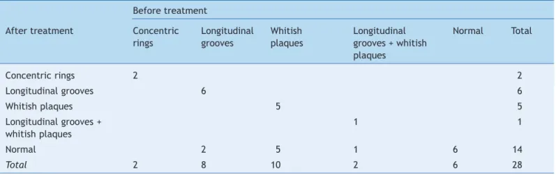
Of the 43 patients, 28 agreed to undergo a control endoscopy. Table 4 shows that the six patients who had a normal endoscopy at baseline remained with a normal endoscopy, while of the 22 who initially had endoscopic
Table 1 Symptom distribution in 43 patients according to age range.
Clinical manifestation Age range p
< 7 years 7-17 years Total
n = 18 n = 25 n = 43
Nausea/vomiting 18 (100.0%) 5 (20.0%) 23 (53.5%) 0.000
Abdominal pain 18 (100.0%) 7 (28.0%) 25 (58.0%) 0.000a
Loss of appetite 10 (55.5%) 15 (60.0%) 25 (58.0%) 0.983a
Weight loss 1 (5.5%) 4 (16.0%) 5 (11.5%) 0.380b
Food aversion 0 (0%) 10 (40.0%) 10 (23.0%) 0.002b
Food impaction 0 (0%) 12 (48.0%) 12 (28.0%) 0.005b
Heartburn 0 (0%) 13 (52.0%) 13 (30.0%) 0.000a
Wheezing 7 (38.8%) 12 (48.0%) 19 (44.0%) 0.777a,b
Coughing 5 (27.7%) 6 (24.0%) 11 (25.5%) 1.000b
aChi-squared test with Yates’ correction. bFisher’s exact test.
Table 2 Number of patients with eosinophilic esophagitis with positive IgE antibody (RAST) to aeroallergens and food allergens according to age range.
Speciic IgE Age range p
< 7 years 7-17 years Total
n = 18 n = 25 n = 43
Cow’s milk 8 (44.0%) 7 (28.0%) 15 (34.9%) 0.428a,b
Egg white 1 (5.5%) 2 (8.0%) 3 (7.0%) 1.000b
Fish 1 (5.5%) 1 (4.0%) 2 (4.5%) 1.000b
Peanuts 2 (11.0%) 1 (4.0%) 3 (7.0%) 0.562b
Soybean 2 (11.0%) 2 (8.0%) 4 (9.0%) 1.000b
Wheat 0 (0%) 0 (0%) 0 (0%) Non-analyzable
House dust 4 (22.0%) 12 (48.0%) 16 (37%) 0.159a
aChi-squared test with Yates’ correction. bFisher’s exact test.
Table 3 Association between endoscopic lesion severity and intensity of eosinophilic iniltrate in 43 patients with eosinophilic esophagitis.
Eosinophil counts per high-power ield
Total 15-30 31-50 > 50
Concentric rings 2 1 1 0
Longitudinal grooves 8 0 4 4
Whitish plaques 12 0 0 12
Longitudinal grooves + whitish plaques 3 1 2 0
Normal 18 8 8 2
abnormalities, eight had normal esophageal mucosa in the second endoscopy (McNemar’s test, p = 0.013). Regarding eosinophil count, initially all patients had increased eosinophil count (10 patients with 15 to 30 eosinophils; 10 with 31-50 eosinophils; and 8 with eosinophil count > 50). All had fewer than 15 eosinophils in the second biopsy after treatment.
Discussion
The Brazilian scientific literature on EoE in the pediatric population is limited to two case series described by Cury et al.3 and by Ferreira et al.,4 highlighting symptom
refractoriness to standard treatment of GERD in both articles. Another report8 emphasized EoE as differential
diagnosis of achalasia. In the present study, the clinical and endoscopic manifestations and response to therapy of 43 patients treated in the period between February 2004 and September 2010 and whose final diagnosis was the classic form of EoE are described. The subgroup of patients with EoE responsive to PPIs, in relation to symptoms and histopathological findings were not included, as all patients were combined before the inclusion of this subgroup in the updated EoE criteria.5
Additionally, the updated consensus included a small number of patients with fewer than 15 eosinophils/HPF, who are treated with PPIs, provided they show other signs of eosinophilic inflammation, including eosinophilic microabscesses, accumulation of eosinophils in the superficial layer of the esophageal mucosa, or extracellular eosinophilic granules. The reasons pointed out by researchers were the possibility of inadequate biopsies, sample errors, and coping with chronic disease or partial response to treatment. The present study did not evaluate these histological findings, considering only the infiltration of at least 15 eosinophils/HPF.5
The mean age of the patients at diagnosis was 8.4 years (1 month to 17 years); patients were predominantly males (86%), and the majority were white. Although it appears to be an absolute majority, racial classification by skin color is controversial, especially in Brazil, where miscegenation
is high. The mean age of disease prevalence was similar to that in the literature.9 There is also agreement in the
literature on male predominance.10-15
It is interesting to stress that this disease has been described in all continents, except for Africa, probably due to the absence of the gene(s) responsible for the disease or absence of environmental factors.16
Another aspect to highlight is that these patients were treated during a six-year period, and most patients were referred from the Pediatric Gastroenterology outpatient clinic of the Complexo Hospitalar do Mandaqui, corresponding to the frequency of 7 patients/year; however, there are no data on the total of patients presenting with suspected GERD/year and responders to PPIs during this period, among which the subset of EoE responders to PPIs, who did not participate in this study, was found.
Some authors have called attention to the increasing prevalence of EoE in children and adolescents.11,12,17
When comparing the prevalence of symptoms in the present study with those of other authors, an agreement in the variability in clinical presentation according to age was observed; most symptoms of nausea, vomiting, and abdominal pain were observed in children younger than 7 years, while heartburn, food aversion, and food impaction were mostly observed in children older than 7 years and adolescents.7,9,10,13,14
Regarding the presence of atopy, respiratory symptoms were observed, such as recurrent wheezing (44%) and cough (25.5%), which reinforced the suspicion of EoE; these findings are similar to those found by other authors.12,14
Endoscopic findings suggestive of EoE at the diagnosis were observed in 58% of case, and apparently normal esophageal mucosa were found in 42% of cases. Other studies showed endoscopic alterations suggestive of EoE in greater proportions, around 73% of cases.7,10,14 Although
none of these endoscopic findings is pathognomonic of EoE, finding at least one of them is strongly suggestive of the disease. It is noteworthy that some of the present patients had an initial diagnosis of monilial esophagitis, due to the endoscopic finding of whitish plaques on the esophageal mucosa.
Table 4 Evolution analysis of endoscopic indings in 28 patients with eosinophilic esophagitis who underwent control evaluation nine to 12 months after treatment initiation.
Before treatment
After treatment Concentric Longitudinal Whitish Longitudinal Normal Total rings grooves plaques grooves + whitish
plaques
Concentric rings 2 2
Longitudinal grooves 6 6
Whitish plaques 5 5
Longitudinal grooves + 1 1
whitish plaques
Normal 2 5 1 6 14
Regarding the histological aspects, at least two fragments were obtained from the esophageal mucosa for the quantification of eosinophils/HPF. The literature recommends obtaining at least one fragment in each esophageal segment (proximal-medium and distal) as esophageal inflammation may be focal and involve apparently healthy areas.5,7 This procedure increases the
diagnostic sensitivity to 97% of cases.18
Eosinophilic infiltration of 15-30 per HPF was found in 89% of apparently normal mucosa, probably because the eosinophilic inflammatory infiltrate had not yet reached the mucosa and thus had not manifested any lesions suggestive of the disease.
The determination of specific IgE only assists in identifying IgE-mediated food allergy of type I or immediate type.6 The detection of specific IgE has been considered
indicative of sensitization to food, confirmed only after its exclusion, with improvement of all symptoms and return of symptoms after oral provocation test.19 Regarding the
research of aeroallergens through tests involving specific IgE, there have been only case reports in adults showing its probable role in the triggering of EoE.20 Among the patients
evaluated in this study, the presence of sensitization by cow’s milk protein was found in 15 (34.9%) patients, and by aeroallergens in 16 (37.0%) patients identified through ImmunoCAP. The frequency of sensitization (positive ImmunoCAP) to inhalants and foods in this group of patients was 46.5%, which should be compared with previously published Brazilian data in normal and atopic children of 25.8% and 79%, respectively.21
The treatment of these patients involved dietary therapy, with the exclusion of the main allergenic foods (cow’s milk, soy milk, eggs, fish, peanuts, and wheat) when allergens were not identified through clinical history or ImmunoCAP performed as indicated in literature.5,7,22
The use of the elemental formula diet removes all potential food allergens, and is especially indicated for infants, whose oral intake tolerance is increased.23
After histological confirmation of EoE, in addition to dietary therapy, patients received topical steroids with spray inhalers (41/43) or by oral route for children younger than 2 years (2/43). Studies have shown that the use of systemic or topical corticosteroids has been effective in resolving the clinical and pathological manifestations of EoE, but its systemic use must be reserved for severe cases.14,24,25
The use of topical corticosteroids for the maintenance treatment of EoE in adults and children has not been established, as well as dose, frequency, and mode of administration, as these formulations were not designed for use in the esophagus.5,7,24 The doses suggested by
the current literature vary from 440 to 880 mcg/day for children and 880-1760 mcg/day for adolescents and adults, continuously for six to eight weeks.
28 of 43 patients underwent control endoscopy; clinical, endoscopic, and histological improvement was observed between six and nine months after treatment, with a decrease in the number of eosinophils/HPF to approximately 0-12 eosinophils/HPF.
It is noteworthy the fact that 75% of patients remained temporarily asymptomatic after finishing the first
treatment cycle and withdrawing medication; however, after exposure to food allergens, they started to present the same symptoms again, requiring a second treatment cycle. The clinical outcome of the present patients with EoE, although chronic, was satisfactory. There was no loss of patients during outpatient follow-up; however, parents or guardians of the patients did not always accept the performance of a third upper endoscopic assessment.
Treatment duration has not been established, nor has the importance of treating asymptomatic patients who remain with histological signs of EoE after initial treatment and the frequency of endoscopic controls during follow-up.5
Patients gained quality of life, reflected by changes in feeding behavior reported by the parents, demonstrating improved mood and better social behavior, and began to have meals with their families once again. The families felt gratified and many felt embarrassed of having wrongly classified their children as being too selective and as having a social disorder.
In patients with the classic form of EoE, symptoms vary according to age, with nausea, vomiting, and abdominal pain most often found in young children, and lack of appetite, heartburn, and food impaction in older children and adolescents. Patients with suspected EoE should undergo biopsies in the proximal and distal locations of the esophagus, stomach, and duodenum, in order to attain a complete diagnosis. A favorable response to treatment from the clinical and histological standpoint was achieved by all patients, requiring at least two treatment cycles in 76.7% of 43 patients.
Conlicts of interest
The authors have no conflicts of interest to declare.
References
1. Landres RT, Kuster GG, Strum WB. Eosinophilic esophagitis in a patient with vigorous achalasia. Gastroenterology. 1978;74: 1298-301.
2. Attwood SE, Smyrk TC, Demeester TR, Jones JB. Esophageal eosinophilia with dysphagia. A distinct clinicopathologic syndrome. Dig Dis Sci. 1993;38:109-16.
3. Cury EK, Schraibman V, Faintuch S. Eosinophilic iniltration of the esophagus: gastroesophageal relux versus eosinophilic esophagitis in children: discussion on daily practice. J Pediatr Surg. 2004;39:e4-7.
4. Ferreira CT, Vieira MC, Vieira SM, Silva GS, Yamamoto DR, Silveira TR. Eosinophilic esophagitis in 29 pediatric patients. Arq Gastroenterol. 2008;45:141-6.
5. Liacouras CA, Furuta GT, Hirano I, Atkins D, Attwood SE, Bonis PA, et al. Eosinophilic esophagitis: updated consensus recommendations for children and adults. J Allergy Clin Immunol. 2011;128:3-20.
6. Hamilton RG. Clinical laboratory assessment of immediate-type hypersensitivity. J Allergy Clin Immunol. 2010;125:S284-96. 7. Furuta GT, Liacouras CA, Collins MH, Gupta SK, Justinich C,
8. Silva Segundo GR. Acalasia do esôfago e esofagite eosinofílica. J Pediatr (Rio J). 2005;81:185-6.
9. Assa’ad AH, Putnam PE, Collins MH, Akers RM, Jameson SC, Kirby CL, et al. Pediatric patients with eosinophilic esophagitis: an 8-year follow-up. J Allergy Clin Immunol. 2007;119:731-8. 10. Liacouras CA, Spergel JM, Ruchelli E, Verma R, Mascarenhas M,
Semeao E, et al. Eosinophilic esophagitis: a 10-year experience in 381 children. Clin Gastroenterol Hepatol. 2005;3:1198-206. 11. Liacouras CA. Eosinophilic esophagitis in children and adults. J
Pediatr Gastroenterol Nutr. 2003;37:S23-8.
12. Noel RJ, Putnam PE, Rothenberg ME. Eosinophilic esophagitis. N Engl J Med. 2004;351:940-1.
13. Gill R, Durst P, Rewalt M, Elitsur Y. Eosinophilic esophagitis disease in children from West Virginia: a review of the last decade (1995-2004). Am J Gastroenterol. 2007;102:2281-5. 14. Orenstein SR, Shalaby TM, Di Lorenzo C, Putnam PE, Sigurdsson
L, Mousa H, et al. The spectrum of pediatric eosinophilic esophagitis beyond infancy: a clinical series of 30 children. Am J Gastroenterol. 2000;95:1422-30.
15. Fogg MI, Ruchelli E, Spergel JM. Pollen and eosinophilic esophagitis. J Allergy Clin Immunol. 2003;112:796-7.
16. Beals JK. Eosinophilic esophagitis in children associated with TSLP gene. Medscape [accessed 25 Dec 2012]. Available from: http://www.medscape.com/viewarticle/718089
17. Prasad GA, Alexander JA, Schleck CD, Zinsmeister AR, Smyrk TC, Elias RM, et al. Epidemiology of eosinophilic esophagitis over three decades in Olmsted County, Minnesota. Clin Gastroenterol Hepatol. 2009;7:1055-61.
18. Walsh SV, Antonioli DA, Goldman H, Fox VL, Bousvaros A, Leichtner AM, et al. Allergic esophagitis in children: a clinicopathological entity. Am J Surg Pathol. 1999;23:390-6.
19. Solé D, Silva LR, Rosário Filho NA, Sarni RO; Sociedade Brasileira de Pediatria; Associação Brasileira de Alergia e Imunopatologia. Consenso Brasileiro sobre Alergia Alimentar: 2007. Rev Bras Alerg Imunopatol. 2008;31:65-89.
20. Spergel JM, Andrews T, Brown-Whitehorn TF, Beausoleil JL, Liacouras CA. Treatment of eosinophilic esophagitis with speciic food elimination diet directed by a combination of skin prick and patch tests. Ann Allergy Asthma Immunol. 2005;95: 336-43.
21. Naspitz CK, Solé D, Jacob CA, Sarinho E, Soares FJ, Dantas V, et al. Sensibilização a alérgenos inalantes e alimentares em crianças brasileiras atópicas, pela determinação in vitro de IgE total e especíica - Projeto Alergia (PROAL). J Pediatr (Rio J). 2004;80:203-10.
22. Kagalwalla AF, Sentongo TA, Ritz S, Hess T, Nelson SP, Emerick KM, et al. Effect of six-food elimination diet on clinical and histologic outcomes in eosinophilic esophagitis. Clin Gastroenterol Hepatol. 2006;4:1097-102.
23. Kelly KJ, Lazenby AJ, Rowe PC, Yardley JH, Perman JA, Sampson HA. Eosinophilic esophagitis attributed to gastroesophageal relux: improvement with an amino acid-based formula. Gastroenterology. 1995;109:1503-12.
24. Teitelbaum JE, Fox VL, Twarog FJ, Nurko S, Antonioli D, Gleich G, et al. Eosinophilic esophagitis in children: immunopathological analysis and response to luticasone propionate. Gastroenterology. 2002;122:1216-25.
ERRATUM
Erratum
on
‘‘Clinical
manifestations,
treatment,
and
outcomes
of
children
and
adolescents
with
eosinophilic
esophagitis’’
夽
Errata
de
‘‘Manifestac
¸ões
clínicas,
terapêutica
e
evoluc
¸ão
de
crianc
¸as
e
adolescentes
com
esofagite
eosinofílica’’
Maraci
Rodrigues
a,∗,
Maria
Fernanda
M.
D’Amico
b,
Fatima
Regina
Almeida
Pati˜
no
c,
Dorina
Barbieri
d,
Aderson
Omar
Mourão
Cintra
Damião
a,
Aytan
M.
Sipahi
eaPhDinMedicine.AssistantPhysician,DepartamentodeGastroenterologiaeHepatologia,HospitaldasClínicas,Universidadede
SãoPaulo(USP),SãoPaulo,SP,Brazil
bPediatricGastroenterologist.CoordinatorofthePediatricResidency,ComplexoHospitalardoMandaqui,SãoPaulo,SP,Brazil
cPediatricGastroenterologist,ComplexoHospitalardoMandaqui,SãoPaulo,SP,Brazil
dProfessor,DepartamentodePediatria,InstitutodaCrianc¸a,HospitaldasClínicas,USP,SãoPaulo,SP,Brazil
eChief,GrupodeIntestino,DepartamentodeGastroenterologiaeHepatologia,HospitaldasClínicas,USP,SãoPaulo,SP,Brazil