Atrial fibrillation with high ventricular rate in emergency room:
What’s the best strategy for treatment?
ALEXANDREDE MATOS SOEIRO1*, TATIANADE CARVALHO ANDREUCCI TORRES LEAL1, MARIA CAROLINA FERESDE ALMEIDA SOEIRO1,
CARLOS VICENTE SERRANO JR.1, MÚCIO TAVARES OLIVEIRA JR.1
1Unidade Clínica de Emergência, Instituto do Coração (InCor), Hospital das Clínicas, Faculdade de Medicina da Universidade de São Paulo (HC-FMUSP), São Paulo, SP, Brazil
S
UMMARYStudy conducted at Instituto do Coração (InCor), Hospital das Clínicas, Faculdade
de Medicina da Universidade de São Paulo (HC-FMUSP), São Paulo, SP, Brazil
Article received: 7/30/2016
Accepted for publication: 10/19/2016
*Correspondence:
Address:Av. Dr. Enéas de Carvalho Aguiar, 44
São Paulo, SP – Brazil Postal code: 05403-900
http://dx.doi.org/10.1590/1806-9282.62.09.879
Atrial ibrillation (AF) is the most common arrhythmia in clinical practice and can lead to signiicant decline in functional status and quality of life among affected patients. The risk of developing AF increases with age and the presence of structural heart disease. Thus, the attendance of patients with high ventricu-lar response to AF is common, which makes knowledge of its management mandatory. In this context, the choice of heart rate and/or rhythm control therapy is fundamental and complex, with multiple possibilities. Thus, this review aims to assist in the management of these patients, systematizing their care.
Keywords: atrial ibrillation, emergency, arrhythmia.
I
NTRODUCTIONAtrial ibrillation (AF) is the most common arrhythmia in clinical practice and is characterized by the absence of P waves and irregular interval between QRS complexes on the electrocardiogram (ECG).1-3
AF can lead to a signiicant reduction in the func-tional status and quality of life of the affected patients. It increases mortality between 1.5 and 1.9 times on the account of hemodynamic deterioration caused by in-creased heart rate (HR), loss of atrioventricular (AV) syn-chrony and progressive dysfunction of the left atrium and ventricle, in addition to increasing the risk of stroke and other embolic events triggered by atrial thrombi.1-5
The risk of developing AF increases with age and with the presence of structural heart disease. Prevalence in-creases from 0.1% in adults less than 55 years to 8% in those aged 80 years or older. It is higher among men pared with women (1.1% x 0.8%); and among white com-pared with black individuals (2.2% x 1.5%).2,4,5
Arterial hypertension (SAH) and coronary artery dis-ease (CAD) (post acute myocardial infarction or ischemic cardiomyopathy) are the most common comorbidities present in patients with AF in developed countries. Rheu-matic heart disease, although currently uncommon in developed countries, has a much greater association with AF. AF is an infrequent form of manifestation of acute
myocardial infarction or ischemia in the absence of oth-er signs and symptoms of CAD.1-5
AF and heart failure (HF) often occur together, and a cause and effect relationship between the two is com-mon. In addition, AF is associated with pulmonary dis-orders including chronic obstructive pulmonary disease (COPD) and pulmonary embolism. Similarly, both the clinical and subclinical forms of hyperthyroidism are associated with an increased risk of AF. Other risk factors related to AF include chronic renal failure, genetic factors, autonomic dysfunction, hypomagnesemia, alcohol con-sumption, and drugs such as theophylline, adenosine and digitalis.1-5
The classiication proposed by the ACC/AHA/ESC for AF divides it into ive subtypes:1-3
• First detected or diagnosed episode independent of its duration and presence of any symptoms.
• Paroxysmal: recurrent (more than two episodes) that terminate spontaneously lasting up to 7 days (usually ceases within 24 hours).
• Persistent: Episodes lasting longer than 7 days that re-quire pharmacological or electrical cardioversion to return to sinus rhythm.
• Permanent: when it is not possible to maintain sinus rhythm after cardioversion, or when it has been deci-ded not to attempt cardioversion for several factors (e.g. elderly, asymptomatic, AF with low or normal ven-tricular response and large left atrium).
Thus, the presence of a patient with high ventricular re-sponse AF is frequent, and makes the knowledge of its management compulsory. In this context, the choice of heart rate and/or rhythm control therapy is fundamental and complex, with multiple possibilities. This review therefore aims at assisting in the approach of these pa-tients, systematizing their care.
C
LINICAL PICTURENot all patients with AF are symptomatic and among those who are, there is a wide variety of symptoms. Infor-mation such as onset of symptoms, timing of diagnosis, frequency and duration of episodes and severity of symp-toms is very important for the therapeutic decision. Epi-sodes can be precipitated by exercise, emotions or acute intake of large amounts of alcohol. In other cases, they may be precipitated during sleep or after a meal.1-5
The typical symptoms associated with AF are palpita-tions, tachycardia, fatigue, weakness, dizziness, reduced exercise capacity, increased urinary volume, dyspnea.1-3 Follow-up studies of patients with AF have shown that approximately 90% of these individuals have recurrent episodes of AF, although more than 90% of the events are not recognized by them. Nevertheless, asymptomatic episodes lasting more than 48 hours are not uncommon, occurring in 17% of patients.4,5
More severe symptoms include restless dyspnea, an-gina, pre-syncope, syncope (infrequent). Some patients may also have thromboembolic events and/or stroke as their irst presentation.4,5
F
IVE STEPS FOR THE SYSTEMATIC APPROACH OFAF
IN THE EMERGENCY ROOM1) Recognize/ward off clinical instability related to AF It is not always easy to recognize whether AF is the main cause of clinical instability presented by the patient. Anam-nesis and physical examination should be performed in all patients with AF, seeking signs of hemodynamic instability (hypotension, signiicant pulmonary congestion, history of syncope, mental confusion, angina/chest pain), adequate use of medications, signs/symptoms of infection, time of onset of symptoms, presence of structural heart changes (systolic HF and/or left ventricular hypertrophy) and coro-nary artery disease.1-3
Those presenting signs of hemodynamic instability at baseline (hypotension, signiicant pulmonary conges-tion, history of syncope, mental confusion, angina/chest pain – these being attributable to arrhythmia [usually in HR greater than 150 to 160 beats per minute]) should be taken to the emergency room for peripheral venous access, continuous monitoring and oxygen therapy, and promptly submitted to synchronized electrical cardiover-sion with 100 to 200 J, regardless of the time of onset. A bolus dose of 10,000 units of unfractionated heparin
is recommended prior to this emergency cardioversion. In such cases, after the reversal, associated diseases such as CAD, valvopathy and ventricular dysfunction should always be investigated. The same is true for patients with AF and pre-excitation with elevated HR or
hemodynam-ic instability.1-3
2) Remove secondary causes that induce high ventricular response The main cause for high ventricular response AF should always be evaluated, especially in those patients with refrac-tory HR control or who already have permanent/persistent AF and presented recent lack of HR control even while correctly using the medications. When drug adherence is appropriate, drug or alcohol use, hyperthyroidism, anemia, hydroelectrolytic disorders (especially hypo or hyperkalemia and hypomagnesemia), infections and pulmonary throm-boembolism should always be considered. In these cases, all patients should perform a chest x-ray and, in women, even if asymptomatic, the collection of type I urine and uroculture must be performed.1-3
A recently published retrospective study evaluated the management of patients with high ventricular response AF in the ER, either according to the rhythm control or reversal strategy or not, when the patient arrives at the site. In about 30% of patients, the cause of high ventricular response AF was sepsis. The rate of adverse events among patients undergoing control/reversal strategies versus un-controlled strategy was 40.7% x 7.1%, and only about 20% of patients achieved adequate control with therapy when a secondary cause was present. This reinforces the idea of investigating the secondary cause before adopting comple-mentary measures in the initial approach, unless the patient presents with hemodynamic instability.6
3) Obtain proper control of HR
The results of two large multicentric studies, AFFIRM and RACE, demonstrated that both rhythm control and ventricular rate control strategies are associated with sim-ilar rates of mortality and severe comorbidities. Generally, the choice of one or another strategy takes into account factors such as the age of the patient and the presence of symptoms associated with AF with an impact on the qual-ity of life, and reduction of the left ventricular systolic function attributed to the presence of the arrhythmia.7,8
Thus, theoretically, the initial and main objective adopted in the care of patients in the emergency room who do not present clinical instability related to AF is HR control, especially in those with more than 48 hours of symptoms, multiple comorbidities or heart disease (left ventricular dysfunction, left ventricular hypertrophy and left atrium > 50 mm).1-3 When controlling HR, unrestrict-ed control maintenance (HR < 110 bpm) provunrestrict-ed to be similar to the restricted control (HR < 80 bpm), and the most lexible measure was also adopted in the emergency sector approach.7 Exception is made for patients with left ventricular dysfunction, symptomatic mitral stenosis or coronary stenosis, in whom HR control should be more rigorous due to underlying heart disease.1-3
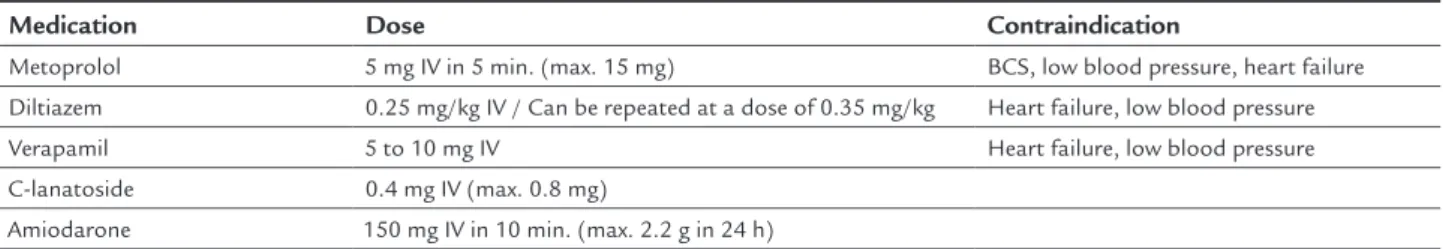
A heart rate control strategy generally requires drugs that reduce AV node conduction, such as B-blockers, non-dihydropyridine calcium channel blockers or digoxin, either alone or in combination (Table 1).1-3
B-blockers and/or calcium channel blockers (diltiazen and verapamil) are generally the drugs of choice for ini-tiation of therapy. It has been proven that drugs such as diltiazen, esmolol, metoprolol and verapamil are supe-rior in the control of HR compared to digitalis and ami-odarone.9-13 In patients without ventricular dysfunction we may use intravenous metoprolol (maximum dose of 15 mg), diltiazen 0.25 mg intravenously (repeat 0.35 mg/ kg if necessary), or verapamil 5 to 10 mg intravenously.9-13 Digoxin may also be used, but it is not as effective in controlling HR during physical activity.1-3,14 In patients with ventricular dysfunction, the use of C-lanatoside, 0.4
mg intravenously (maximum dose of 0.8 mg), or amio-darone, 150 mg intravenously in 10 minutes, is considered as the irst option.1-3 The fact that amiodarone can lead to the reversal of the rhythm in up to 28% of the patients, predisposing them to the occurrence of embolic events, is highlighted.15
In patients with hemodynamic shock and AF, heart rate control should be done only when HR exceeds 130 to 150 beats per minute and preferably amiodarone via continuous infusion pump (450 to 1,200 mg daily) de-pending on the chronotropic response of the patient. In these cases, the HR target is usually around 120 beats per minute, without damaging the compensatory response to shock, and its indication should be reviewed daily and individually.1-3
4) Consider heart rate control/AF reversal
Although HR control is the main target in the treatment of high ventricular response AF in the emergency room, there are some situations in which the rhythm control strategy should be considered: if the symptoms have clearly started less than 48 hours; persistent symptoms despite adequate HR control; inability to achieve adequate HR control (ruling out secondary causes); young patients with a irst episode of AF diagnosed or those in whom arrhythmia had a recent onset and the risk of recurrence appears to be lower; and, in the latter case, according to the patient’s preference.1-3
If cardioversion is chosen, it may be chemical (using antiarrhythmic drugs) or electric (Figure 1). In such cas-es, the patient’s HR should be kept preferably high, since after the reversal the risk of sinus bradycardia associated with clinical instability becomes lower.1-3
For chemical reversal, the recommended medications are: lecainide, dofetilide, ibutilide, propafenone and amiodarone. The irst three are not available in Brazil. Before administering antiarrhythmic medication for car-dioversion, a B-blocker or a calcium channel blocker should be given to prevent rapid AV conduction.1-3
TABLE 1 Main drugs used to control HR in patients with high ventricular response AF.
Medication Dose Contraindication
Metoprolol 5 mg IV in 5 min. (max. 15 mg) BCS, low blood pressure, heart failure
Diltiazem 0.25 mg/kg IV / Can be repeated at a dose of 0.35 mg/kg Heart failure, low blood pressure Verapamil 5 to 10 mg IV Heart failure, low blood pressure C-lanatoside 0.4 mg IV (max. 0.8 mg)
Amiodarone 150 mg IV in 10 min. (max. 2.2 g in 24 h)
In patients without structural heart disease, the op-tion of choice is propafenone. In the i rst event, it should be administered in hospital orally at a dose of 450 mg (if the patient’s weight is less than 70 kg) or 600 mg (weight 70 kg or more). If the treatment is well tolerated and ef-fective, the patient can be discharged with the guidance of home use at the same dose, if there is recurrence, in a strategy called pill in the pocket. In these cases, the probabil-ity of AF reversion in up to 6 hours is around 94% of cases. This strategy should be done only in cases of AF with few recurrences (up to 2 every 6 months).1-3
In patients with structural heart disease [left ven-tricular hypertrophy (septum and posterior wall thickness > 1.2 cm), ischemia, valvular disease and/or ventricular dysfunction], amiodarone is the best option for chemical cardioversion. Amiodarone should be given at a dose of 150 mg intravenously in 10 minutes, or 5-7 mg/kg in 1 hour (up to a maximum dose of 2.2 g in 24 hours). Ami-odarone is associated with higher rates of maintenance of sinus rhythm, but is also associated with a greater num-ber of adverse effects in the long term.1-3
Sotalol may be benei cial in patients with paroxysmal AF also in sinus rhythm, provided that the patient has min-imal structural disease or normal heart, and QTc < 460 ms. It is the antiarrhythmic drug of choice in patients with AF and CAD who do not have left ventricular systolic dysfunc-tion. In this situation, propafenone is contraindicated.1-3
If electric cardioversion is chosen, it should be done with the administration of shock initially synchronized at 100 to 200 J in a single-phase dei brillator or 100 J in a biphasic dei brillator, after explaining the procedure and adequate sedation to the patient.Administration of amiodarone prior to electrical cardioversion increases the chance of suc-cess and may prevent immediate recurrence of AF.1-3
Finally, radiofrequency ablation (RFA) is also useful in maintaining sinus rhythm mainly in symptomatic patients with paroxysmal AF who have failed an antiar-rhythmic, have normal or slightly increased left atrium, and normal or discreetly decreased function of the left ventricle. However, its use should be considered an excep-tion when it comes to patients seen in emergency units.1-3
If there is structural heart disease, after the reversal, the patient should be discharged with amiodarone pre-scribed at a dose of 200 mg orally, three times a day for 2 weeks, followed by 200 mg twice daily for another 2 weeks. Thereafter, amiodarone at 200 mg daily. In the absence of evidence of structural heart disease, amiodarone should preferably be replaced with propafenone, 150 to 300 mg every 12 hours.1-3
In all cases with more than 48 hours of AF or in those with structural heart disease, if reversion is chosen, it is mandatory that the patient be anti-coagulated for at least 3 weeks.1-3 In the case of warfarin, the weekly INR control should show values between 2.0 and 3.0. Currently one
FIGURE 1 Algorithm for evaluation of patients with high ventricular AF for ECV.
HVRAF: high ventricular response AF; HR: heart rate; TE-Echo: transesophageal echocardiography; OAC: oral anticoagulation; ECV: synchronized electrical cardioversion.
HVRAF
Duration of arrhythmia
> 48 h or structural heart disease present
HR control
Perform TE-Echo
Thrombus present Thrombus absent
Perform ECV
Maintain OAC effective for at least 3 weeks
Maintain OAC for at least 4 weeks < 48 h and structural heart
option is to use one of the new anticoagulants. In a pro-spective study, rivaroxaban was validated and showed the same level of safety of warfarin related to electrical cardio-version (ECV).16 Apixaban and dabigatran, on the other hand, also showed the same safety proile of warfarin in subanalyses.17,18 Thus, the guidelines release their use when choosing to reverse the patient’s rhythm. After reversion, any of the anticoagulants used should be maintained for a minimum period of 4 weeks, and may be extended in-deinitely if the patient presents risk factors for AF recur-rence (ventricular dysfunction and/or structural heart disease, atrial dilatation, previous episodes, etc).1-3
In those without oral anticoagulant use for at least three continuous weeks, transesophageal echocardiogra-phy must be performed. In the absence of thrombi, the patient may be cardioverted with synchronized electrical therapy or chemical therapy.1-3
If a thrombus is seen, only HR control should be performed and the patient is discharged with oral anti-coagulation prescription to schedule the procedure after at least 3 weeks at an outpatient clinic.1-3
5) Deine the indication for oral anticoagulation
All patients with AF should be evaluated for the need for anticoagulation and this is done by applying the CHADS2 and/or CHA2DS2VASC scores. The latter is a reinement of the former and has been the most used in recent years because it identiies patients with actual “low risk” more accurately. For the evaluation of bleeding risk, the most commonly used criterion is HAS-BLED, which serves as a guideline for the rational and cautious choice of anti-coagulation. However, it should not contraindicate it.1-3,19 There are two indications of anticoagulation in AF. In the short term, in patients with low thromboembolic risk in which the strategy of rhythm control is chosen and cardioversion is performed to the sinus rhythm, and in the long term, patients that meet criteria for chronic anticoagulation.1-3
Antithrombotic therapy to prevent thromboembolism should be indicated for all patients with AF, except for those with isolated AF without other risk factors, and those with a contraindication to it. Anticoagulation is recommended for patients at high risk of thromboem-bolic event (two or more risk factors considering the CHA2DS2VASC score). If the CHA2DS2VASC criterion only scores for the female sex, chronic oral anticoagula-tion is not mandatory.1-3
The selection of the anticoagulant agent should be based on the absolute risk of stroke and bleeding and the risk/beneit ratio to the patient. In patients with signiicant
heart valve disease, mechanical valvular prostheses and/ or chronic renal insuficiency (CrCl < 30 mL/min), the option is warfarin. The target INR is 2.0 to 3.0, except for mitral and aortic mechanical prostheses, in which case the target varies between 2.5 and 3.5.1-3
In the remaining patients, warfarin or any of the new anticoagulants (apixaban, rivaroxaban or dabigatran) may be used.1-3,20-22
The association of ASA and clopidogrel to reduce the risk of thromboembolic events may be considered for patients with AF in case of possible inadequate antico-agulation with warfarin, either at the choice of the patient or when the attending physician is not sure of the safety for the patient. In this case, the level of evidence is lower and comes from simple non-multicentre studies.2
When the patient with AF remains hospitalized for another reason, the use of warfarin should be routinely withdrawn at least in the initial phase. Although there is limited evidence in hospitalized patients, when INR is below 2.0, anticoagulation should be initiated with sub-cutaneous enoxaparin, 1 mg/kg every 12 hours, or un-fractionated heparin via continuous infusion.2
C
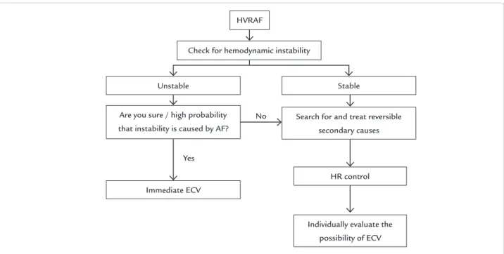
ONCLUSIONManagement of patients with high ventricular response AF in the emergency room is complex. Identifying factors that cause AF is critical to correct treatment. When pos-sible, heart rate control is the priority. At the time of hos-pital discharge, the patient should be evaluated for indica-tion and possibility of oral anticoagulaindica-tion. In a simpliied manner, the algorithm for conduct is shown in Figure 2.
R
ESUMOFibrilação atrial de alta resposta ventricular na sala de emergência: qual é a melhor estratégia de tratamento?
Palavras-chave: i brilação atrial, emergência, arritmia.
R
EFERENCES1. American College of Cardiology Foundation; American Heart Association; European Society of Cardiology; Heart Rhythm Society, Wann LS, Curtis AB, et al. Management of patients with atrial i brillation (compilation of 2006 ACCF/AHA/ESC and 2011 ACCF/AHA/HRS recommendations): a report of the American College of Cardiology/American Heart Association Task Force on practice guidelines. Circulation. 2013; 127(18):1916-26. 2. Magalhães LP, Figueiredo MJO, Cintra FD, Saad EB, Kuniyoshi RR, Teixeira
RA, et al. II Diretrizes Brasileiras de Fibrilação Atrial. Arq Bras Cardiol 2016; 106(4Supl. 2):1-22.
3. Camm AJ, Lip GY, De Caterina R, Savelieva I, Atar D, Hohnloser SH, et al.; ESC Committee for Practice Guidelines (CPG). 2012 focused update of the ESC Guidelines for the management of atrial i brillation: an update of the 2010 ESC Guidelines for the management of atrial i brillation. Developed with the special contribution of the European Heart Rhythm Association. Eur Heart J. 2012; 33(21):2719-47.
4. Chugh SS, Blackshear JL, Shen WK, Hammill SC, Gersh BJ. Epidemiology and natural history of atrial i brillation: clinical implications. J Am Coll Cardiol. 2001; 37(2):371-8.
5. Heeringa J, van der Kuip DA, Hofman A, Kors JA, van Herpen G, Stricker BH, et al. Prevalence, incidence and lifetime risk of atrial i brillation: the Rotterdam study. Eur Heart J 2006; 27(8):949-53.
6. Scheuermeyer FX, Pourvali R, Rowe BH, Grafstein E, Heslop C, MacPhee J, et al. Emergency department patients with atrial i brillation or l utter and an acute underlying medical illness may not benei t from attempts to control rate or rhythm. Ann Emerg Med. 2015; 65(5):511-22.e2.
7. Van Gelder IC, Groenveld HF, Crijns HJ, Tuininga YS, Tijssen JG, Alings AM, et al. Lenient versus strict rate control in patients with atrial i brillation. N Engl J Med. 2010; 362(15):1363-73.
8. Van Gelder IC, Hagens VE, Bosker HA, Kingma JH, Kamp O, Kingma T, et al. A comparison of rate control and rhythm control in patients with recurrent persistent atrial i brillation. N Engl J Med. 2002; 347(23):1834-40. 9. Siu CW, Lau CP, Lee WL, Lam KF, Tse HF. Intravenous diltiazem is superior
to intravenous amiodarone or digoxin for achieving ventricular rate control
in patients with acute uncomplicated atrial i brillation. Crit Care Med. 2009; 37(7):2174-9.
10. Demircan C, Cikriklar HI, Engindeniz Z, Cebicci H, Atar N, Guler V, et al. Comparison of the effectiveness of intravenous diltiazem and metoprolol in the management of rapid ventricular rate in atrial i brillation. Emerg Med J. 2005; 22(6):411-4.
11. Phillips BG, Gandhi AJ, Sanoski CA, Just VL, Bauman JL. Comparison of intravenous diltiazem and verapamil for the acute treatment of atrial i brillation and atrial l utter. Pharmacotherapy. 1997; 17(6):1238-45. 12. Ellenbogen KA, Dias VC, Plumb VJ, Heywood JT, Mirvis DM. A
placebo-controlled trial of continuous intravenous diltiazem infusion for 24-hour heart rate control during atrial i brillation and atrial l utter: a multicenter study. J Am Coll Cardiol. 1991; 18(4):891-7.
13. Platia EV, Michelson EL, Porteri eld JK, Das G. Esmolol versus verapamil in the acute treatment of atrial i brillation or atrial l utter. Am J Cardiol. 1989; 63(13):925-9.
14. Jordaens L, Trouerbach J, Calle P, Tavernier R, Derycke E, Vertongen P, et al. Conversion of atrial i brillation to sinus rhythm and rate control by digoxin in comparison to placebo. Eur Heart J. 1997; 18(4):643-8. 15. Hofmann R, Steinwender C, Kammler J, Kypta A, Leisch F. Effects of a high
dose intravenous bolus amiodarone in patients with atrial i brillation and a rapid ventricular rate. Int J Cardiol. 2006; 110(1):27-32.
16. Cappato R, Ezekowitz MD, Klein AL, Camm AJ, Ma CS, Le Heuzey JY, et al. Rivaroxaban vs. vitamin Kantagonists for cardioversion in atrial i brillation. Eur Heart J. 2014; 35(47):3346-55.
17. Nagarakanti R, Ezekowitz MD, Oldgren J, Yang S, Chernick M, Aikens TH, et al. Dabigatran versus warfarin in patients with atrial i brillation: an analysis of patients undergoing cardioversion. Circulation. 2011; 123(2):131-6. 18. Flaker G, Lopes RD, Al-Khatib SM, Hermosillo AG, Hohnloser SH, Tinga
B, et al.; ARISTOTLE Committees and Investigators. Efi cacy and safety of apixaban in patients after cardioversion for atrial i brillation: insights from the ARISTOTLE Trial (Apixaban for Reduction in Stroke and Other Thromboembolic Events in Atrial Fibrillation). J Am Coll Cardiol. 2014; 63(11):1082-7.
19. Friberg L, Rosenqvist M, Lip GY. Evaluation of risk stratii cation schemes for ischaemic stroke and bleeding in 182 678 patients with atrial i brillation: the Swedish Atrial Fibrillation cohort study. Eur Heart J. 2012; 33(12):1500-10.
FIGURE 2 General algorithm for approaching patients with high ventricular response AF in the emergency unit. HVRAF: high ventricular response atrial i brillation; ECV: synchronized electrical cardioversion; HR: heart rate.
HVRAF
Check for hemodynamic instability
Unstable
No
Yes Are you sure / high probability that instability is caused by AF?
Immediate ECV
Search for and treat reversible secondary causes
HR control
Individually evaluate the possibility of ECV
20. Connolly SJ, Ezekowitz MD, Yusuf S, Eikelboom J, Oldgren J, Parekh A, et al.; RE-LY Steering Committee and Investigators. Dabigatran versus warfarin in patients with atrial ibrillation. N Engl J Med. 2009; 361(12):1139-51. 21. Patel MR, Mahaffey KW, Garg J, Pan G, Singer DE, Hacke W, et al.; ROCKET
AF Investigators. Rivaroxaban versus warfarin in nonvalvular atrial ibrillation. N Engl J Med. 2011; 365(10):883-91.